. 2025 May 30;23(1):395.
doi: 10.1186/s12951-025-03423-w.

Immunogenicity and cellular response of a herpes zoster virus gEgI fusion protein adjuvanted with CpG-emulsion in mice

Affiliations

Immunogenicity and cellular response of a herpes zoster virus gEgI fusion protein adjuvanted with CpG-emulsion in mice

Sibo Zhang et al. J Nanobiotechnology. .

Abstract

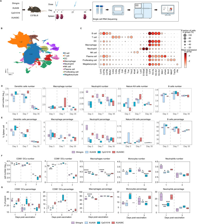
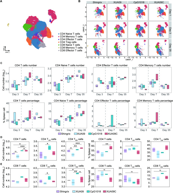
Herpes zoster (HZ), commonly known as shingles, arises from the reactivation of the latent varicella-zoster virus (VZV) when VZV-specific cellular immunity declines below a critical threshold necessary for viral suppression. The current leading vaccine, Shingrix, which incorporates the adjuvant AS01B with glycoprotein E (gE), has significantly contributed to HZ prevention but raises concerns regarding safety and accessibility. Addressing the need for safer and more accessible HZ vaccinations, we developed a vaccine comprising a fusion protein of glycoprotein E and I (gEgI), connected via a linker, targeting abundant B cell and CD4 T cell epitopes. Our study assessed the immunogenicity of the gE alone and the gEgI fusion protein in adult mice, revealing that gEgI prompts a more potent and comprehensive T cell response compared to gE alone. Furthermore, we introduced a composite adjuvant, an emulsion-type adjuvant combined with CpG1018 (XUA09C), which was shown to enhance both humoral and cellular immune responses beyond the capabilities of XUA09 with CpG alone. Comparative analyses demonstrated that the XUA09C-adjuvanted gEgI vaccine induces comparable antibody responses and significantly superior T cell responses relative to Shingrix in both adult, VZV-primed, and aged mice. Single-cell RNA sequencing highlighted that gEgI/XUA09C more effectively promotes early immune activation, B and T cell proliferation, and memory T cell augmentation compared to Shingrix. These findings position the XUA09C-adjuvanted gEgI as a promising candidate for further development in HZ vaccine strategies, potentially better serving the needs of the immunocompromised population.

Keywords: Adjuvant; CpG-emulsion; Glycoprotein E; Glycoprotein I; HZ vaccine; Single-cell RNA sequencing; Varicella-zoster virus.


Conflict of interest statement

Declarations. Competing interests: T.L., S.L., S.Z., Y.Z., L.C., Y.G., T.C. and N.X. are inventors on a patent application (PCT/CN2024/073289) filed by the Xiamen University that covers gEgI fusion protein design described in this work.

Figures

Fig. 1
Fig. 1
Design, purification, and characterization of purified gE and gEgI proteins. (A) Schematic representation of the primary structure and construct design for gE and gEgI proteins. SP denotes the signal peptide; ECD, the extracellular domain; TM, the transmembrane domain; and CT, the cytoplasmic tail. gEgI was engineered by fusing the ECDs of gE and gI. Predicted N-glycosylation and O-glycosylation sites are indicated above the domain bars. (B) SDS-PAGE and Western blot analysis of purified gE and gEgI proteins using anti-gE antibody 1B11 and anti-gI antibody 18B2. (C, D) High-pressure size-exclusion chromatography (HPSEC) profiles of gE and gEgI. (E, F) Size-exclusion chromatography-multi-angle light scattering (SEC-MALS) profiles showing the molar mass and size of gE and gEgI proteins. UV absorption at 280 nm and molar mass are represented by black and red lines, respectively. (G, H) Analytical ultracentrifugation (AUC) sedimentation velocity profiles of gE and gEgI. (I, J) Differential scanning calorimetry (DSC) profiles of purified gE and gEgI. (K, L) Analysis of gE and gEgI proteins treated with Endo-H and PNGase-F, or left untreated, visualized via SDS-PAGE and immunoblotting with anti-gE antibody 1B11 and anti-gI antibody 18B2. (M) LC-MS/MS analysis of N-glycans on specified Asn positions and O-glycans on Thr positions of purified gEgI. (N) ELISA reactivity of liquid and lyophilized gE and gEgI proteins against anti-gE mAbs and anti-gI mAbs. EC50 values were calculated using GraphPad Prism 8 software.
Fig. 2
Fig. 2
Immunogenicity of gEgI compared to gE in mice. (A) Immunization schedule for vaccines containing 5 µg-dose gE, 5 µg-dose gEgI, and 8.3 µg-dose gEgI in C57BL/6J mice, compared with Shingrix control. (B) Bi-weekly determination of gE-specific IgG titers to evaluate antigen-specific IgG durability. (C) gE-binding IgG titers measured by ELISA two weeks post-boost immunization. (D) Neutralizing antibody titers assessed by plaque reduction neutralization test post-boost. The detection limit is indicated by a horizontal dotted line. (E to G) ELISA-measured gE-binding IgG1 (E), IgG2b (F), and IgG2c (G) titers two weeks post-boost immunization. (H) Ratio of gE-specific IgG2c to IgG1 antibody titers. n = 6 per group. (I to P) Intracellular cytokine staining (ICS) assessing cytokine production by CD4+ and CD8+ T cells in response to gE or gEgI peptides. Box and whisker plots show the frequency of cytokine-positive CD4+ and CD8+ T cells for IFN-γ, IL-2, and TNF-α. (Q to T) Representative ELISPOT images and quantification of IFN-γ (R) and IL-2 (T) spot-forming cells (SFC) isolated from spleens post-stimulation with gE or gEgI peptide pools. Data are presented as means ± SD. Statistical differences were analyzed using one-way ANOVA, two-way ANOVA, and unpaired t-tests; significance denoted as *p < 0.05, **p < 0.01, ***p < 0.001, ****p < 0.0001, and “ns” for no significance.
Fig. 3
Fig. 3
Immunogenicity of gEgI formulated with XUA09C adjuvant in mice. (A) Immunization schedule for various vaccines containing 10 µg of gEgI with multiple adjuvants, using Shingrix as a control. (B) Weekly collection of serum samples to assess the durability of antigen-specific IgG titers. (C to G) Two weeks post-boost immunization, serum samples were analyzed for (C) anti-gEgI IgG titers, (D) neutralizing antibody titers, (E) IgG1 titers, (F) IgG2c titers, and (G) IgG2c/IgG1 ratio. n = 6 per group. (H to M) Intracellular cytokine staining (ICS) assessed cytokine production by CD4+ and CD8+ T cells in response to gEgI peptide pool stimulation. Histograms illustrate the frequency of cytokine-positive CD4+ and CD8+ T cells. Specifically, the frequency of CD4+ IFN-γ+ (H), CD4+ IL-2+ (I), CD4+ TNF-α+ (J), CD8+ IFN-γ+ (K), CD8+ IL-2+ (L), and CD8+ TNF-α+ (M) T cells. (N and O) ELISPOT quantification of IFN-γ (N) and IL-2 (O) spot-forming cells (SFC) isolated from spleens post-stimulation with gEgI peptide pools. Data are presented as means ± SD. Statistical analyses were performed using one-way ANOVA, Brown-Forsythe and Welch ANOVA tests, and the Kruskal-Wallis test for multiple comparisons; *p < 0.05, **p < 0.01, ***p < 0.001, ****p < 0.0001, and “ns” denotes no significant difference.
Fig. 4
Fig. 4
Comparative immunogenicity of gEgI formulated with XUA09C-1018 and Shingrix in mice. (A) Schematic representation of XUA09C compound adjuvant formulation, showing the naked eye appearance (left), morphology of XUA09/XUA09C (middle), and XUA09C (right) as observed by transmission electron microscopy and cryo-electron microscopy, respectively. (B to D) Characterization of particle sizes (B), polydispersity index (C), and zeta potentials (D) of XUA09 and its variants. (E) Immunization schedule for vaccine candidates containing a 10 µg-dose of gEgI formulated with various XUA09C adjuvants in 5-week-old C57BL/6J mice, compared to Shingrix control. (F) Weekly determination of gEgI-specific IgG titers to assess the durability of the antibody response. (G) Neutralizing antibody titers assessed by plaque reduction neutralization assay post-boost. Histograms depict the frequency of cytokine-positive CD4+ and CD8+ T cells. Specifically, frequencies of CD4+ IFN-γ+ (H), CD4+ IL-2+ (I), CD4+ TNF-α+ (J), CD8+ IFN-γ+ (K), CD8+ IL-2+ (L), and CD8+ TNF-α+ (M) T cells. (N and O) ELISPOT analysis of IFN-γ and IL-2 spot-forming cells (SFC) isolated from the spleen post-stimulation with gEgI peptide pools. Results are presented as means ± SD. Statistical differences were analyzed using one-way ANOVA with multiple comparisons; significance levels indicated by *p < 0.05, **p < 0.01, ***p < 0.001, ****p < 0.0001, and “ns” for no significance.
Fig. 5
Fig. 5
Toxicological profile of gEgI/XUA09C-1018 vaccine candidates in mice. (A to F) Body weight changes of mice during the first 10 days post-vaccination. (G to I) Liver and kidney functions were evaluated through blood biochemical parameters; Aspartate aminotransferase (AST) and Alanine aminotransferase (ALT) for liver function (G, H), and creatinine (CRE) for kidney function (I). (J) Representative histopathological sections (H&E staining) of liver, spleen, and kidney from naive and vaccine-immunized mice. Scale bars, 50 μm. Results are presented as means ± SD. Statistical differences between groups were analyzed using one-way ANOVA with multiple comparisons; *p < 0.05, **p < 0.01, ***p < 0.001, ****p < 0.0001, and “ns” for no significance.
Fig. 6
Fig. 6
Immunogenicity of gEgI/XUA09C-1018 compared to Shingrix in VZV-primed and aged mouse models. (A) Immunization schedule for vaccine candidate containing 10 µg-dose gEgI formulated with various XUA09C adjuvants in VZV-primed mouse models, compared with Shingrix control. (B, C) Antibody responses in mice, with gEgI-specific IgG (B) and neutralizing antibody titers (C) determined at week 5 post-vaccination. (D to I) Frequency of cytokine-producing CD4+ and CD8+ T cells assessed by flow cytometry: CD4+ IFN-γ+ (D), CD4+ IL-2+ (E), CD4+ TNF-α+ (F), CD8+ IFN-γ+ (G), CD8+ IL-2+ (H), and CD8+ TNF-α+ (I). (J, K) ELISPOT quantification of IFN-γ (J) and IL-2 (K) spot-forming cells isolated from spleens post-stimulation with gEgI peptide pools. (L) Immunization schedule for different vaccine candidates in aged C57BL/6J mice, compared with Shingrix control. (M, N) gEgI-specific IgG (M) and neutralizing antibody titers (N) measured at week 5. (O to T) Cytokine production frequencies in aged mice: CD4+ IFN-γ+ (O), CD4+ IL-2+ (P), CD4+ TNF-α+ (Q), CD8+ IFN-γ+ (R), CD8+ IL-2+ (S), and CD8+ TNF-α+ (T). (U, V) ELISPOT analysis of IFN-γ (U) and IL-2 (V) spot-forming cells from the spleen. Results are presented as means ± SD. Statistical differences were analyzed using one-way ANOVA with multiple comparisons; significance levels denoted as *p < 0.05, **p < 0.01, ***p < 0.001, ****p < 0.0001, and “ns” for no significance.
Fig. 7
Fig. 7
Cellular landscape of mice immunized with Shingrix, XUA09, CpG1018, and XUA09C as identified by scRNA-seq and flow cytometry. (A) Flowchart detailing the experimental design and schema for single-cell RNA sequencing. n = 3 biologically independent mice per group. (B) Spectral UMAP plot displaying cells from mice immunized with Shingrix, XUA09, CpG1018, XUA09C, and controls, identifying 9 major cell types annotated by cell-type identity. (C) Dot plots representing cells expressing selected canonical marker genes for the identification of 9 different cell types in the mouse spleen across all samples. Dot size and color correspond to the percentage of cells within the cell population expressing the gene and the average expression level of each marker gene, respectively. (D, E) Box plots illustrating the cell numbers and proportions of DCs, macrophages, neutrophils, NK cells, and B cells across all groups. n = 3 biologically independent mice per group. Box plots indicate the mean (center line) and the range from minimum to maximum values (bounds of the box). (F, G) Box plots showing the numbers and proportions of CD80+/CD86+ DCs, macrophages, monocytes, and neutrophils analyzed by flow cytometry. n = 3 per group.
Fig. 8
Fig. 8
T cell dynamics in mice immunized with Shingrix, XUA09, CpG1018, and XUA09C, analyzed by scRNA-seq and flow cytometry. (A) UMAP plot illustrating iterative clustering results of T cells, revealing subpopulations corresponding to different states of differentiation. (B) UMAP plots depicting the dynamics of T cells from mice immunized with Shingrix, XUA09, CpG1018, and XUA09C at various time points. (C) Box plots showing the cell numbers and proportions of all different CD4+ T cells across all groups. n = 3 biologically independent mice per group. Box plots show the mean (center line) and the range from minimum to maximum values (bounds of the box). (D) Box plots detailing the numbers and proportions of CD4+ T cells, CD4+ central memory (TCM)/effector memory (TEM) cells analyzed by flow cytometry at day 35. (E) Box plots detailing the numbers and proportions of CD8+ T cells, CD8+ TCM/TEM cells analyzed by flow cytometry at day 35. n = 3 per group.

Similar articles

Cited by

  • Research Progress on Varicella-Zoster Virus Vaccines.
    Liu H, Cui L, Zhang S, Wang H, Xue W, Li H, Zhang Y, Chen L, Gu Y, Li T, Xia N, Li S. Liu H, et al. Vaccines (Basel). 2025 Jul 4;13(7):730. doi: 10.3390/vaccines13070730. Vaccines (Basel). 2025. PMID: 40733707 Free PMC article. Review.

References

    1. Zerboni L, Sen N, Oliver SL, Arvin AM. Molecular mechanisms of varicella Zoster virus pathogenesis. Nat Rev Microbiol. 2014;12:197–210. - PMC - PubMed
    1. A AM: Varicella-Zoster virus. CLINMICROBIOLREV 1996:361–81.
    1. H. GD: Varicella-zoster virus DNA in human sensory ganglia. Nature 1983, 306. - PubMed
    1. Ku CC, Zerboni L, Ito H, Graham BS, Wallace M, Arvin AM. Varicella-zoster virus transfer to skin by T cells and modulation of viral replication by epidermal cell interferon-alpha. J Exp Med. 2004;200:917–25. - PMC - PubMed
    1. Laing KJ, Ouwendijk WJD, Koelle DM, Verjans G. Immunobiology of Varicella-Zoster virus infection. J Infect Dis. 2018;218:S68–74. - PMC - PubMed

MeSH terms

LinkOut - more resources