. 2025 May 30;32(1):54.
doi: 10.1186/s12929-025-01148-4.

USP24 upregulation stabilizes PKA-Cα to promote lipogenesis, inflammation, and fibrosis during MASH progression

Affiliations

USP24 upregulation stabilizes PKA-Cα to promote lipogenesis, inflammation, and fibrosis during MASH progression

Beh Ning et al. J Biomed Sci. .

Abstract

Background: Ubiquitin-specific peptidase 24 (USP24), a deubiquitinating enzyme, regulates protein stability by removing ubiquitin. This study investigates the role of UPS24 in lipid metabolism, inflammation, and fibrosis. It also explores the effect of targeting USP24 on metabolic disorders, focusing on high-fat diet (HFD)-induced obesity and liver diseases.

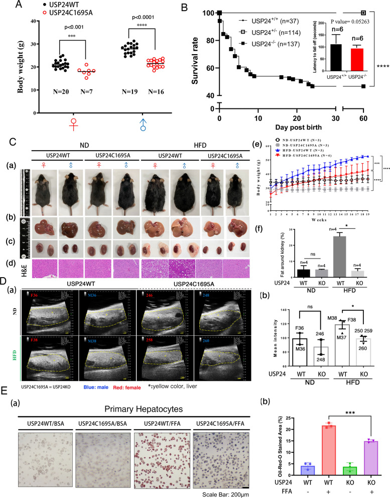
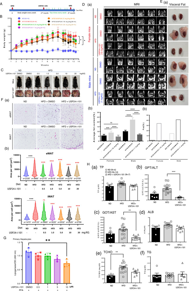
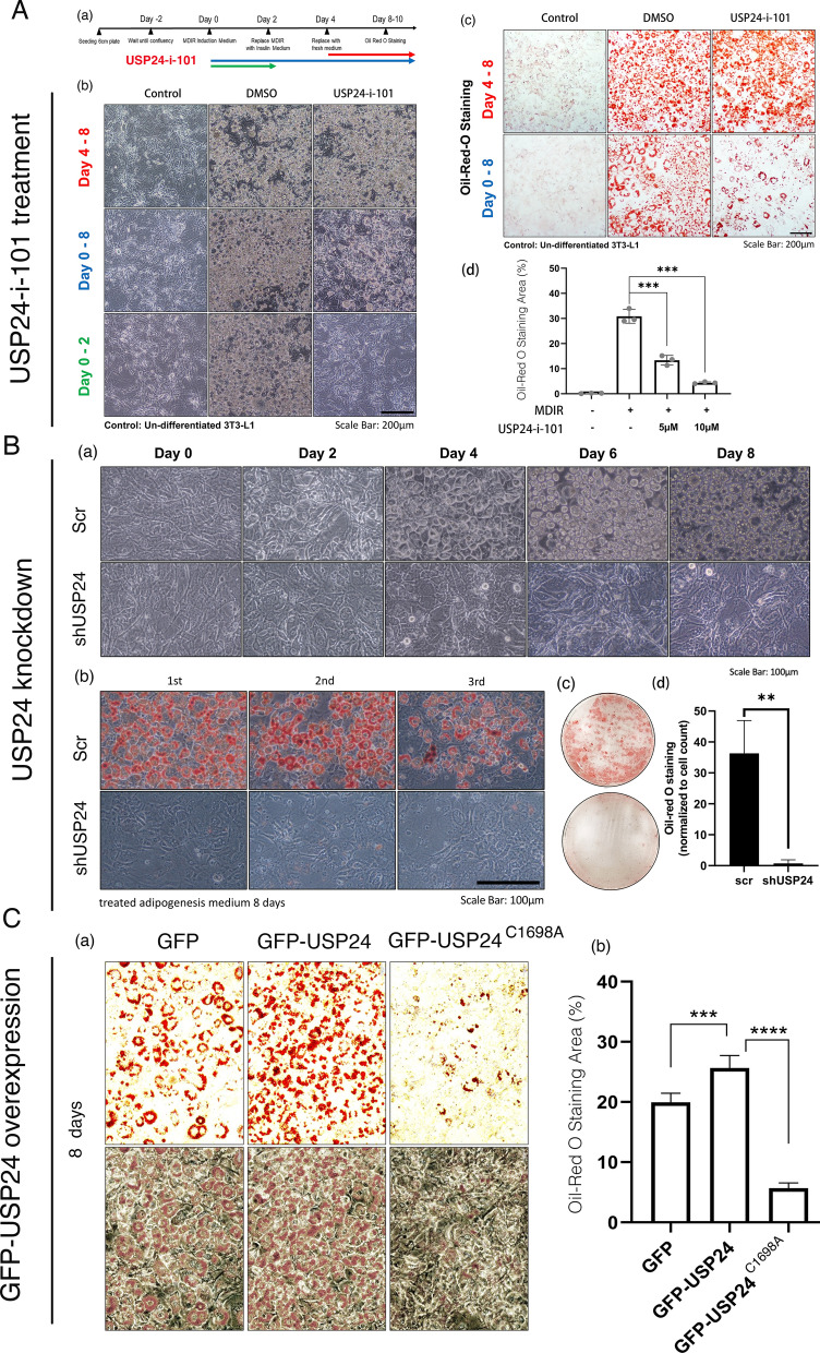
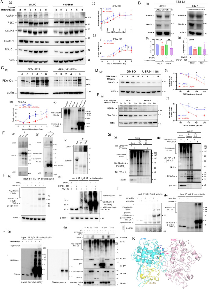
Methods: This study utilized CRISPR/Cas9 to create functional knockout mice (USP24C1695A) and treated HFD-fed mice with USP24 inhibitor (USP24-i-101). The effects of USP24 inhibition or knockout on 3T3-L1 derived adipocytes, primary hepatocytes, hepatic stellate cells, and murine hepatocyte cell line AML12 (alpha mouse liver 12) cells were assessed with RNA-sequencing. Molecular mechanisms and the interaction between USP24 and PKA-Cα were studied with co-immunoprecipitation. Downstream signaling pathways involving CREB, SREBP1, PPARγ, and C/EBPβ, as well as USP24 role in liver inflammation and fibrosis, were studied using western blot and real-time PCR. Clinical and animal tissue samples were examined with immunohistochemistry to identify the correlations between USP24 and metabolic-associated liver diseases.

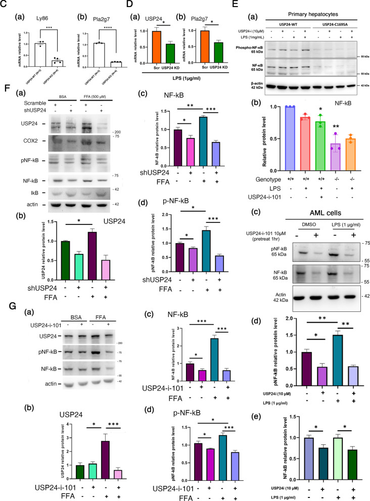
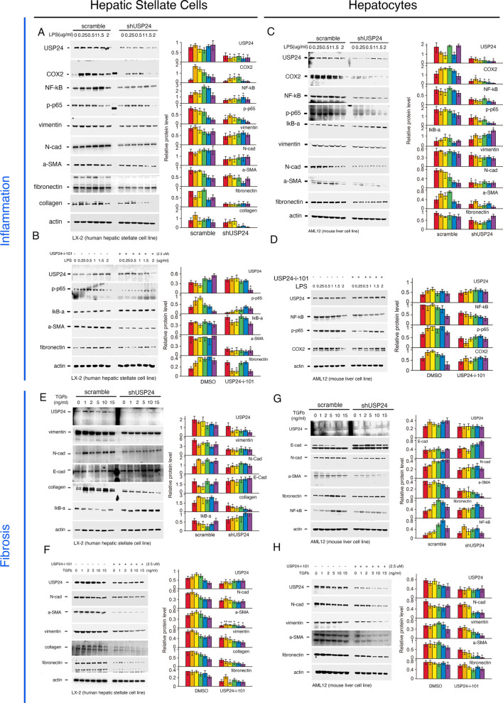
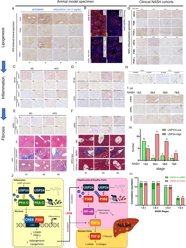
Results: Knockout or inhibition of USP24 reduced body weight, lipid accumulation, inflammation, and fibrosis in HFD-fed mice. The expression of genes related to lipogenesis, inflammation, and fibrosis was downregulated in USP24C1695A mice and those treated with USP24 inhibitor (USP24-i-101). USP24 inhibition decreased lipid droplet accumulation in adipocytes and hepatocytes, suppressed inflammation in hepatocytes and AML12 cells, and reduced fibrosis in hepatic stellate cells. Mechanistically, USP24 expression was upregulated by PKA activation during adipocyte differentiation, leading to increased PKA-Cα stability and CREB phosphorylation, which promoted lipogenic gene expression. Free fatty acids (FFA) increased USP24 expression, activating NF-κB and TGFβ pathways to induce inflammation (Cox2) and fibrosis (α-SMA). USP24 was highly expressed in patients with metabolic dysfunction-associated steatohepatitis (MASH) and correlated with Cox2 and α-SMA levels.

Keywords: Lipogenesis; MASH; PKA-Cα; USP24; p-CREB.


Conflict of interest statement

Declarations. Ethics approval and consent to participate: The IRB number is KMUHIRB-G(II)-20170013, which was approved by Institutional Review Board of Kaohsiung Medical University Hospital. Consent for publication: No applicable. Competing interests: The authors declare no potential competing interests.

Figures

Fig. 1
Fig. 1
Functional knockout of USP24 suppresses lipogenesis. A Body weights of USP24WT and USP24C1695A mice fed a normal diet were measured. B Survival rate of newborn mice (B, left panel) and physiological activities of mice (B, right panel) with or without USP24 knockout were assessed by tracing the survival rate and performing a rotarod test, respectively. The results of A and B were analyzed by statistical assay, t-test, ***p < 0.001, ****p < 0.0001. USP24WT (n = 3) and USP24C1695A (n = 3) mice and USP24WT (n = 3) and USP24C1695A (n = 4) mice were fed a normal diet (ND, 13.1% fat) or a high-fat-diet (HFD, 60% fat) for 4 months, respectively. Mouse body sizes (C, a), organs (liver and kidney) (C, b, c), and H&E staining (C, d) are shown. The body weight of mice (C, e) and fat around kidneys (C, f) were quantified, and statistical analysis was performed using a t-test, *p < 0.05, ***p < 0.001, ****p < 0.0001. Fat content inside ND- and HFD-fed mice with or without function knockout mice (USP24WT & USP24C1695A) was determined using an ultrasound machine (D, a), and the signal highlight with yellow dotted circle was quantified by Image J, and statistical analysis was conducted by t-test, *p < 0.05 (D, b). Lipid droplet formation in primary hepatocytes isolated from USP24WT and USP24C1695A mice treated with free fatty acid (500 μM FFA) was examined via oil red O staining assay (E, a). Subsequent quantification of lipid signaling levels followed three independent experiments, with statistical analysis performed using a t-test, ***p < 0.001 (E, b)
Fig. 2
Fig. 2
USP24-i-101, which targets USP24, inhibits lipogenesis. USP24WT and USP24C1695A mice were fed a normal diet (ND; n = 6) or a high-fat diet (HFD; n = 33) for 2 months and then treated with 0.5 mg/kg (n = 6), 1.0 mg/kg (n = 6), 5 mg/kg (n = 6), 10 mg/kg (n = 4) or 20 mg/kg (n = 5) USP24-i-101 twice a week for 1 month (A). Body weight changes in all mice were measured weekly (B). Mouse sizes and their lipid, SAT and VAT, contents, are presented (C). Lipid signals in USP24WT and USP24C1695A mice on either an ND or HFD with or without USP24-i-101 treatment were measured using MRI (D, a). The signal was quantified by Image J, and statistical analysis was performed by t-test, ***p < 0.001, ****p < 0.0005, ns: non-significant (D, b). Visceral adipose tissue (VAT) around the kidneys of female and male mice with or without USP24-i-101 treatment was shown (E, a), The area of VAT was quantified by Image J, and statistical analysis was conducted by t-test, ***p < 0.001 (E, b). iWAT and eWAT in male mice on either an ND or HFD with or without 1 mg/kg USP24-i-101 treatment are depicted (F, a). Adipocyte sizes were measured using Image J, and statistical analysis was performed by t-test, **p < 0.01, ***p < 0.001 (F, b). Primary hepatocytes isolated from the mice were treated with various doses of USP24-i-101 and FFA for 24 h, after which lipid droplet formation was studied by oil red O staining. After three independent experiments, lipid droplets contents were quantified, and statistical analysis was performed via a t-test, **p < 0.01 (G). Serum collected from the mice was used to study the levels of several lipogenesis-related markers, such as total protein (TP), GPT/ALT, GOT/AST, ALB, total cholesterol (TCHO) and triglyceride (TG) levels, via ELISA, followed by statistical analysis via a t-test, **p < 0.01, ***p < 0.001 (H)
Fig. 3
Fig. 3
Upregulation of USP24 promotes adipogenesis. The schedule for adipocyte differentiation from 3T3-L1 cells and the cellular morphology with or without USP24-i-101 treatment at different time points, days 0–2, days 0–8 and days 4–8, are shown (A, a, b). Lipid droplet content during adipogenesis with or without 10 μM USP24-i-101 treatment were measured via oil red O staining (A, c), and the number of lipid droplets (day 0–8) was quantitated using Image J (A, d). 3T3-L1 cells with or without USP24 expression knockdown by shUSP24 lentivirus were observed (B). The morphology of the cells was observed (B, a), and fat accumulation was determined by oil red O staining (B, b and c). The levels of fat in adipocytes were quantified after three independent experiments (B, d). GFP, GFP-USP24WT, and GFP-USP24.C1698A were overexpressed in 3T3-L1 cells during adipogenesis. Lipid droplet formation was measured by oil red O staining (C, a). After three independent experiments, the contents of lipid droplets were quantified via Image J (C, b). Statistical analysis was performed via a t-test, **p < 0.01, ***p < 0.001, ****p < 0.0001
Fig. 4
Fig. 4
The USP24/PKA/p-CREB/CEBPβ/PPARγ axis regulates adipocyte differentiation. Lipogenesis-related proteins expressed in 3T3-L1 cells treated with 10 μM USP24-i-101 (A) were knocked down with shRNA-USP24 (B) or overexpressed with GFP/GFP-USP24/GFP-USP24C1698A (C) during adipogenesis, and then samples were harvested on the -2nd, 0, 2nd, 4th, 6th and 8th days. The expression of lipogenesis-related proteins was measured via IB with antibodies agonist the indicated proteins (AC). Primary hepatocytes isolated from mice with or without USP24 knockout, USP24WT or USP24.C1695A, were treated with 500 μM FFA, and then, the cells were lysed to study the expression of USP24 and PLIN2 via IB (D). 3T3-L1 cells were treated with MDIR and the PKA inhibitor 10 μM H89 for 24 h, and then the samples were collected in sample buffer. The expression of USP24 was measured via IB with anti-USP24 antibodies (E). 3T3-L1 cells with or without 10 μM USP24-i-101 treatment were lysed to study the expression of CREB and pCREB via IB (F, a). After three independent experiments, the expression of pCREB (F, b) and CREB (F, c) were quantified. 3T3-L1 cells were treated with cycloheximide (CHX), and then, cell lysates were collected at the indicated times to study the expression of pCREB/CREB (G, a). After three independent experiments, the expression of pCREB/CREB (D, b) and PKA-Cα (G, b) was quantified. After three independent experiments, various protein expressions were quantified. The statistical analysis was performed via a t-test, *p < 0.05, **p < 0.01, ***p < 0.001
Fig. 5
Fig. 5
USP24 stabilizes PKA-Cα and p300 during adipogenesis. The expression of USP24 in 3T3-L1 cells was knocked down using shRNA-USP24 for three days, after which the cells were differentiated into adipocytes. Samples were collected at different time points to study the expression of the indicated proteins via IB with antibodies against the indicated proteins (A, a). After three independent experiments, the expression of CaMKII (A, b) and PKA-Cα (A, c) was quantified. 3T3-L1 cells were treated with or without 10 μM USP24-i-101, and samples were collected at day 2 and day 4 (B, a) to measure the expression of PKA-Cα and CaMKII via IB. After three independent experiments, the expression of PKA-Cα (B, b) and CaMKII (B, c) was quantified. GFP-USP24 and GFP-USP24C1698A were overexpressed in 3T3-L1 cells, which were then induced for adipocyte differentiation via MDIR. Samples were collected at different time points to measure the expression of PKA-Cα via IB with anti-PKA-Cα antibodies (C). 3T3-L1 cells were treated with cycloheximide (CHX) with or without 10 μM USP24-i-101 treatment (D) and USP24 knockdown (E), and then cell lysates were collected at the indicated times to study the expression of PKA-Cα (D and E). After three independent experiments, the expression of PKA-Cα (D and E, b) was quantified. 3T3-L1 cells were differentiated, and samples were collected on the 2nd day for IP with anti-USP24 and IgG (F, a, c) or anti-PKA-Cα and IgG (F, b), and then IB with antibodies against indicated proteins. GFP, GFP-USP24WT and GFP-USP24C1698A were transfected into 3T3-L1 cells for differentiation, and samples were for IP with anti-GFP antibodies or anti-USP24 antibodies and IB with antibodies against USP24, PKA-Cα, ubiquitin and actin (G). 3T3-L1 cells with or without USP24-i-101 (H), or USP24 knockdown (I) were differentiated with MG132 treatment, and samples were collected on the 2nd day for IP with anti-ubiquitin and IgG and IB with antibodies against PKA-Cα. GFP and GFP-PKA-Cα were overexpressed in 3T3-L1 cells for 2 days. Polyubiquitinated PKA-Cα was pulldown by IP with anti-ubiquitin antibodies (J, a), anti-PKA-Cα and anti-GFP antibodies (J, b), followed by the addition of myc-USP24 (1 μg recombinant protein) for 1 h. The levels of various proteins were measured by IB with indicated antibodies (J). The generation of the USP24-ubiquitin-PKA-Cα complex may indicate its function. An irreversible bond is formed between ubiquitin G76 and PKA-Cα K310. The catalytic site of USP24 envelops the ubiquitin-PKA-Cα bond and can be near the USP24 catalytic residues C10, H282, and N302. The modelled complex suggests potential functions for further cellular processing of the complex (K)
Fig. 6
Fig. 6
Numerous genes related to lipogenesis are regulated by USP24. Total RNA samples were isolated from the liver tissues of USP24WT, USP24C1695A or USP24-i-101-treated mice fed a ND or HFD for RNA-Seq and then analyzed via the bioinformatics tools DEG Down GO TERMs – BP Enrichment (A, a; USP24C1695A/USP24WT), DEG All Top 20 GO Barplot (A, b; USP24C1695A/USP24WT), DEG Down ALL GO Dotplot (A, c; 1 mg/KG USP24-i-101, i.p., two times per week) and Volcano Plot (A, d; 1 mg/KG USP24-i-101, i.p., two times per week). Inflammation-related genes whose expression was regulated by USP24C1695A were studied via RNA-seq and analyzed with a heatmap (B, USP24C1695A/USP24WT). Total RNA was extracted from mice with or without USP24 expression knockout to study the mRNA expression of Ly86 (C, a) and pLa2g7 (C, b) via qPCR. AML12 cells with or without USP24 knockdown were treated with LPS for 2 h, and the mRNA expression of USP24 (D, a) and Pla2g7 (D, b) were measured via qPCR. Primary hepatocytes isolated from mice with or without USP24 knockout (E, a, b) or hepatocyte AML cells (E, c, d, e) were treated with LPS or USP24-i-101. The expression of USP24, p-NF-κB and NF-κB were measured via IB (E). AML12 cells with or without USP24 knockdown (F) or 10 μM USP24-i-101 treatment (G) were treated with 500 μM free fatty acid (FFA) or BSA for 24 h, after which the samples were harvested to measure the expression of USP24,COX2, IκB, p-NF-κB and NF-κB via IB. After three independent experiments, the quantification and statistical analysis were performed via t-test: *p < 0.05, **p < 0.01, ***p < 0.001
Fig. 6
Fig. 6
Numerous genes related to lipogenesis are regulated by USP24. Total RNA samples were isolated from the liver tissues of USP24WT, USP24C1695A or USP24-i-101-treated mice fed a ND or HFD for RNA-Seq and then analyzed via the bioinformatics tools DEG Down GO TERMs – BP Enrichment (A, a; USP24C1695A/USP24WT), DEG All Top 20 GO Barplot (A, b; USP24C1695A/USP24WT), DEG Down ALL GO Dotplot (A, c; 1 mg/KG USP24-i-101, i.p., two times per week) and Volcano Plot (A, d; 1 mg/KG USP24-i-101, i.p., two times per week). Inflammation-related genes whose expression was regulated by USP24C1695A were studied via RNA-seq and analyzed with a heatmap (B, USP24C1695A/USP24WT). Total RNA was extracted from mice with or without USP24 expression knockout to study the mRNA expression of Ly86 (C, a) and pLa2g7 (C, b) via qPCR. AML12 cells with or without USP24 knockdown were treated with LPS for 2 h, and the mRNA expression of USP24 (D, a) and Pla2g7 (D, b) were measured via qPCR. Primary hepatocytes isolated from mice with or without USP24 knockout (E, a, b) or hepatocyte AML cells (E, c, d, e) were treated with LPS or USP24-i-101. The expression of USP24, p-NF-κB and NF-κB were measured via IB (E). AML12 cells with or without USP24 knockdown (F) or 10 μM USP24-i-101 treatment (G) were treated with 500 μM free fatty acid (FFA) or BSA for 24 h, after which the samples were harvested to measure the expression of USP24,COX2, IκB, p-NF-κB and NF-κB via IB. After three independent experiments, the quantification and statistical analysis were performed via t-test: *p < 0.05, **p < 0.01, ***p < 0.001
Fig. 7
Fig. 7
USP24 is involved in the expression of MASH-related proteins. USP24 expression was knocked down (A) or treated with 2.5 μM USP24-i-101 (E) in LX-2 cells (B) with or without LPS treatment, and samples were harvested to study the expression of the indicated proteins via IB. USP24 was knocked down (C) or treated with 2.5 μM USP24-i-101 (D) in AML12 cells with or without LPS treatment, and samples were harvested to study the expression of the indicated proteins via IB. USP24 was knocked down (E) or treated with 2.5 μM USP24-i-101 (F) in AML12 cells and LX-2 cells (G, H) with or without TGFβ treatment, and samples were harvested to study the expression of the indicated proteins via IB. After three independent experiments, the quantification and statistical analysis were performed via t-test: *p < 0.05, **p < 0.01
Fig. 8
Fig. 8
Positive correlation between USP24 expression and that of MAFLD-related markers in animal samples and clinical specimens. The protein expressions of the indicated proteins in the livers of HFD-fed mice with or without USP24-i-101 treatment were measured by IHC (A). Primary hepatocytes were isolated from mice treated with free fatty acid (FFA) and USP24-i-101 for 24 h showed lipid level measurements via immunofluorescence with anti-PLIN2 antibodies (B). The expression of the inflammation- and fibrosis-related proteins, COX-2, and NF-κB in HFD-fed USP24WT and USP24.C1695A mice (C), and HFD-fed mice treated with USP24-i-101 (D) was studied via IHC. The level of α-SMG and collagen in ND- and HFD-fed mice with or without USP24 functional knockout (E) or USP24-i-101 treatment (F) were determined using IHC and Masson’s trichome staining. The expression of USP24, p300, PPARγ in MAFL clinical cohorts was determined via IHC (G). The expression of USP24, COX2 and αSMA in the MASH patients was measured via IHC, and the signals were divided into low and high expression. The relationship between USP24 expression and different stages of MASH is shown (H, a, b), and the correlations among the expression of USP24, COX2 and αSMA in the different stages of MASH are shown (H, a, c). Working model—PKA-mediated upregulation of USP24 during lipogenesis increases CREB1 phosphorylation and stabilizes p300. This promotes PPARγ and C/EBPβ gene expression, leading to adipogenesis and fatty liver. FFA treatment increases USP24 expression in the liver, promoting inflammation and fibrosis through activating the NF-κB and TGF-β signaling pathways (I)

References

    1. Young M-J, Hsu K-C, Lin TE, Chang W-C, Hung J-J. The role of ubiquitin-specific peptidases in cancer progression. J Biomed Sci. 2019;26:42. - PMC - PubMed
    1. Cruz L, Soares P, Correia M. Ubiquitin-specific proteases: players in cancer cellular processes. Pharmaceuticals (Basel). 2021;14(9):848. - PMC - PubMed
    1. Dewson G, Eichhorn PJA, Komander D. Deubiquitinases in cancer. Nat Rev Cancer. 2023;23:842–62. - PubMed
    1. Thayer JA, et al. The PARK10 gene USP24 is a negative regulator of autophagy and ULK1 protein stability. Autophagy. 2020;16:140–53. - PMC - PubMed
    1. Zhao B, et al. Association analysis of single-nucleotide polymorphisms of USP24 and USP40 with Parkinson’s disease in the Han Chinese population. Eur Neurol. 2012;68:181–4. - PubMed

Substances