. 2025 May 30;23(1):397.
doi: 10.1186/s12951-025-03494-9.

Carbon dots from purple sweet potato as a promising anti-inflammatory biomaterial for alleviating the LPS-induced inflammation in macrophages

Affiliations

Carbon dots from purple sweet potato as a promising anti-inflammatory biomaterial for alleviating the LPS-induced inflammation in macrophages

Jiebang Jiang et al. J Nanobiotechnology. .

Abstract

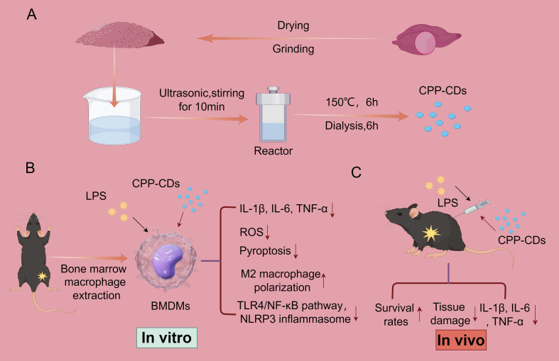
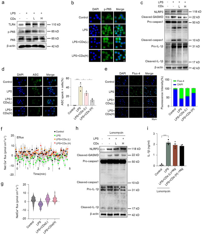
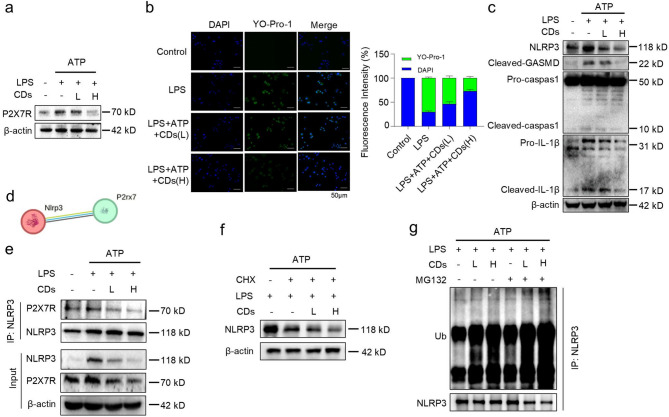
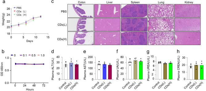
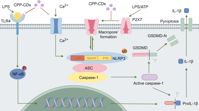
This study synthesizes carbon dots derived from crude extracts of purple sweet potato (CPP-CDs) and evaluates its anti-inflammatory effects in a lipopolysaccharide (LPS) -induced acute inflammation model. Characterization revealed that CPP-CDs possess a uniform spherical structure and excellent photoluminescent properties. In vitro, CPP-CDs significantly inhibited the expression of pro-inflammatory cytokines (IL-1β, IL-6, TNF-α), reduced the accumulation of reactive oxygen species (ROS), suppressed pyroptosis, and facilitated the polarization of macrophages from the M1 phenotype to the M2 phenotype. In vivo, CPP-CDs significantly improved the survival rates of LPS-treated mice, mitigated tissue damage, and suppressed the levels of pro-inflammatory cytokines. Mechanistic studies indicated that CPP-CDs exert anti-inflammatory effects through the inhibition of the TLR4/NF-κB signaling pathway and the modulation of the NLRP3 inflammasome. Additionally, CPP-CDs exhibited excellent biocompatibility, with no significant toxicity observed in mice. This study provides strong evidence supporting the application of CPP-CDs as a novel anti-inflammatory material, highlighting their potential for acute inflammation treatment and expanding the possibilities for the development of carbon-dot-based anti-inflammatory therapies.

Keywords: Anti-inflammatory material; Carbon dots; NLRP3 inflammasome; Purple sweet potato; Reactive oxygen species; TLR4/NF-κB signaling pathway.


Conflict of interest statement

Declarations. Ethical approval: The Ethics Committee of the Affiliated Hospital of Nantong University granted approval for this study. All animal procedures were reviewed and authorized by the Animal Care and Use Committee of Nantong University and were carried out in compliance with its guidelines (No: B20240420011). Consent for publication: All authors provided their consent for publication. Competing interests: The authors declare no competing interests.

Figures

Scheme 1
Scheme 1
Synthesis of CPP-CDs and their application in acute inflammation treatment. a. Purple sweet potato tubers were utilized to synthesize CPP-CDs via a one-step hydrothermal method. b. Mouse bone marrow-derived macrophages were isolated and cultured to establish an LPS-induced inflammation model; CPP-CDs effectively ameliorated the inflammatory response through multiple anti-inflammatory pathways. c. Following intraperitoneal administration, CPP-CDs effectively alleviated systemic inflammation, reduced tissue damage, and improved survival in mice
Fig. 1
Fig. 1
Effects of three carbon dots on inhibiting LPS-induced inflammation. a-c. Left: Images of the raw material extracts for the three types of carbon dots. Right: Images of the three types of carbon dots under UV light. CDs: CPP-CDs, CDs1:CPSPSL-CDs, CDs2: SPR-CDs. d-f. Relative gene expression levels of IL-1β, IL-6, and TNF-α measured by RT-qPCR. BMDMs were treated with LPS (1 µg/mL, 4 h), low-concentration CPP-CDs (L, 0.5 mg/mL, 8 h), and high-concentration CPP-CDs (H, 1 mg/mL, 8 h) (n = 3 biological replicates). g. Relative gene expression levels of IL-1β, IL-6, and TNF-α measured by RT-qPCR. BMDMs were treated with LPS (1 µg/mL, 4 h), PSPCE and CPP-CDs (both 1 mg/mL, 8 h) (n = 3 biological replicates). d-g. Data were analyzed by One-way ANOVA for multiple group comparisons. *p < 0.05, **p < 0.01, ***p < 0.001
Fig. 2
Fig. 2
Synthesis and characterization of CPP-CDs. a. TEM images of CPP-CDs showing an overall size of 50 nm and individual particles measuring 5 nm. b. Size distribution analysis of CPP-CDs. c. Fluorescence excitation and emission spectra of CPP-CDs. d. FTIR spectra of CPP-CDs. e. XRD patterns of CPP-CDs. f. Full survey XPS spectra of CPP-CDs. g. High-resolution XPS spectrum of C 1s for CPP-CDs. h. High-resolution XPS spectrum of O 1s for CPP-CDs
Fig. 3
Fig. 3
Effects of CPP-CDs on LPS-induced inflammation. a. ELISA measurements of IL-1β, IL-6, and TNF-α protein levels in BMDMs culture supernatants. Experimental groups were treated with LPS (1 µg/mL, 4 h), Nigericin (Nig, 5µM, 45 min), and various concentrations of CPP-CDs (n = 4 biological replicates). b-c. ATP levels were measured using an ATP assay kit to assess intracellular and extracellular ATP levels in BMDMs (n = 4 biological replicates). d. ROS levels in BMDMs were measured using a ROS detection probe (n = 3 biological replicates). e. Morphological changes in BMDMs were observed under an optical microscope for each treatment group. Red arrows indicate pyroptotic cells (n = 3 biological replicates). Scale bar: 20 μm. f. LDH release levels in BMDMs for each treatment group, represented as the percentage of LDH activity in the cell lysate (n = 4 biological replicates). g. The relative expression of M2 macrophage markers ARG1 and CD206 was measured by RT-qPCR in BMDMs treated with various concentrations of CPP-CDs (n = 3 biological replicates). a-g. Data are expressed as mean ± standard error of the mean (SEM). One-way or Two-way ANOVA was used for multiple group comparisons. *p < 0.05, **p < 0.01, ***p < 0.001
Fig. 4
Fig. 4
In vivo anti-inflammatory effects of CPP-CDs in an LPS-induced mouse model. a. Mouse model construction process: Mice were divided into groups and given intraperitoneal injections of low (L: 15 mg/kg) or high (H: 30 mg/kg) concentrations of CPP-CDs, followed by LPS injection (5 mg/kg) 2 h later. b. Survival rate of mice monitored for 72 h after LPS and CPP-CDs treatment (n = 6, two independent experiments). c. Colon and liver tissues were collected 24 h after LPS and CPP-CDs treatment and stained with H&E to observe tissue damage (magnification: 15×). d. Pathological scoring of colonic lesions and liver inflammation. Colon score was based on weight loss, rectal bleeding, and stool consistency; liver score was based on the number and area of inflammatory foci around the central vein (n = 6, two independent experiments). e. ELISA measurements of IL-1β, IL-6, and TNF-α levels in mouse serum (n = 4 biological replicates). b, d-e. All data are expressed as mean ± standard error of the mean (SEM). One-way or Two-way ANOVA was used for multiple group comparisons. *p < 0.05, **p < 0.01, ***p < 0.001
Fig. 5
Fig. 5
CPP-CDs modulate inflammatory signaling pathways. a. Western blot analysis of TLR4, p-P65, and P65 protein expression levels. BMDMs were treated with LPS (1 µg/mL, 4 h), low-concentration CPP-CDs (L, 0.5 mg/mL, 8 h), and high-concentration CPP-CDs (H, 1 mg/mL, 8 h). b. Immunofluorescence staining showing nuclear translocation of p-P65 in BMDMs (green) and DAPI-stained nuclei (blue) (n = 3 biological replicates). c. Western blot analysis of NLRP3, Cleaved-GSDMD, Pro-caspase-1, Cleaved-caspase-1, Pro-IL-1β, and Cleaved-IL-1β protein expression levels in BMDMs was performed using the same treatment as described in Fig. 5a. d. Immunofluorescence staining showing ASC speck formation and DAPI-stained nuclei (blue). BMDMs were treated with LPS (1 µg/mL, 4 h), Nig (5 µM, 45 min), and various concentrations of CPP-CDs (n = 3 biological replicates). e. Calcium ion influx in BMDMs measured using fluo-4 after LPS treatment for 10 min, with or without CPP-CDs pre-treatment for 6 h (n = 3 biological replicates). f, g. f (Upper panel): Net calcium flux measurements for different treatment groups using non-invasive microtest technology (NMT) in BMDMs, with each group tested for 5 min. g (Lower panel): Data are displayed as violin plots (n = 6 biological replicates). h. Western blot analysis of NLRP3, Cleaved-GSDMD, Pro-caspase-1, Cleaved-caspase-1, Pro-IL-1β, and Cleaved-IL-1β protein expression in BMDMs treated with LPS (1 µg/mL, 4 h), CPP-CDs (1 mg/mL, 8 h), and Ionomycin (5 µM, 1 h). i. ELISA measurements of IL-1β levels in BMDMs culture supernatants (n = 4 biological replicates). g, h, i. Data are expressed as mean ± standard error of the mean (SEM). One-way ANOVA was used for multiple group comparisons. *p < 0.05, **p < 0.01, ***p < 0.001
Fig. 6
Fig. 6
Regulation of P2X  7R/NLRP3 interaction by CPP-CDs. a. Western blot analysis of P2 X 7R protein expression in different treatment groups. BMDMs were treated with LPS (1 µg/mL, 4 h), ATP (5 mM, 30 min), low -concentration CPP-CDs (L: 0.5 mg/mL), and high-concentration CPP-CDs (H: 1 mg/mL) (n = 3 biological replicates). b. Left: YO-Pro-1 staining showing changes in BMDMs membrane permeability. Green represents YO-Pro-1 staining, and blue represents DAPI-stained nuclei. Scale bar: 50 μm. Right: Relative fluorescence intensity of YO-Pro-1 and DAPI (n = 3 biological replicates). c. Western blot analysis of NLRP3, Cleaved-GSDMD, Pro-caspase-1, Cleaved-caspase-1, Pro-IL-1β, and Cleaved-IL-1β protein expression levels in BMDMs treated as indicated in Fig. 6a (n = 3 biological replicates). d. Interaction network prediction of P2 X 7R and NLRP3 proteins using the STRING database, showing potential direct binding relationships. e. Immunoprecipitation assays detecting the interaction between P2 X 7R and NLRP3 in different treatment groups. BMDMs were treated with LPS (1 µg/mL, 6 h), ATP (5 mM, 30 min), and different concentrations of CPP-CDs (L and H) (n = 3 biological replicates). f. Western blot analysis of NLRP3 protein stability after treatment with Cycloheximide in BMDMs (Chx, 20 µg/mL, 8 h) (n = 3 biological replicates). g. Western blot analysis of NLRP3 ubiquitination levels after immunoprecipitation of NLRP3. Treatment conditions were LPS (1 µg/mL, 6 h), ATP (5 mM, 30 min), and different concentrations of CPP-CDs, with MG132 (10 µM, 6 h) used to block protein degradation pathways in BMDMs (n = 3 biological replicates)
Fig. 7
Fig. 7
Safety Assessment of CPP-CDs. a. Assessment of the impact of CPP-CDs administration on mouse body weight throughout the treatment period (n = 5, two independent experiments). b. Cytotoxicity evaluation of CPP-CDs on BMDMs using the CCK-8 assay. Cell viability was quantified at 0, 24, 48, and 72 h under different concentrations of CPP-CDs (0, 0.1, 0.5, and 1.0 mg/mL) based on OD450 values (n = 3, biological replicates). c. Morphological examination of the colon, liver, spleen, lung, and kidney tissues of mice from each group through H&E staining (magnification: 15×). d-h. Measurement of serum biomarkers, including alanine aminotransferase (ALT), aspartate aminotransferase (AST), uric acid (UA), urea (UREA), and creatinine (CREA). This analysis assessed the effects of CPP-CDs on key physiological indicators (n = 4, biological replicates). a-b, d-h. Data are expressed as mean ± standard error of the mean (SEM). One-way or Two-way ANOVA was used for multiple group comparisons. *p < 0.05, **p < 0.01, ***p < 0.001
Fig. 8
Fig. 8
Schematic representation of the mechanism through which CPP-CDs regulate LPS-induced inflammatory responses in macrophages

Similar articles

Cited by

References

    1. Breijyeh Z, Jubeh B, Karaman R. Resistance of Gram-Negative Bacteria to current antibacterial agents and approaches to resolve it. Molecules. 2020;25(6). - PMC - PubMed
    1. Silhavy TJ, Kahne D, Walker S. The bacterial cell envelope. Cold Spring Harb Perspect Biol. 2010;2(5):a000414. - PMC - PubMed
    1. Raetz CR, Whitfield C. Lipopolysaccharide endotoxins. Annu Rev Biochem. 2002;71:635–700. - PMC - PubMed
    1. Delcour AH. Outer membrane permeability and antibiotic resistance. Biochim Biophys Acta. 2009;1794(5):808–16. - PMC - PubMed
    1. Medzhitov R. The spectrum of inflammatory responses. Science. 2021;374(6571):1070–5. - PubMed

MeSH terms

LinkOut - more resources