. 2025 May 30;20(1):552.
doi: 10.1186/s13018-025-05837-6.

Effects of dynamic stabilization and fusion on postoperative paraspinal muscle degeneration and lumbar function recovery

Affiliations

Effects of dynamic stabilization and fusion on postoperative paraspinal muscle degeneration and lumbar function recovery

Jiawei Song et al. J Orthop Surg Res. .

Abstract

Objective: To investigate the relationship between segmental motion and paraspinal muscle degeneration at the surgical level in patients with single-level lumbar degenerative disease (LDD) who have undergone either fusion or dynamic stabilization surgery.

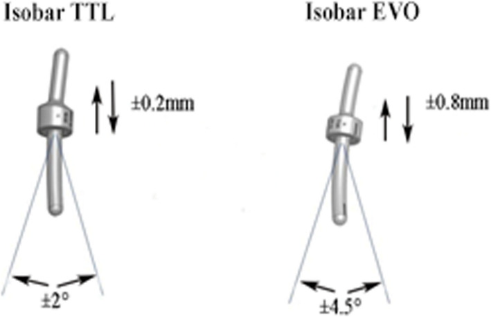
Methods: Patients treated with posterolateral intertransverse fusion (PITF group), Isobar TTL dynamic internal fixation (TTL group), or Isobar EVO dynamic internal fixation (EVO group) for single-level lumbar degenerative disease (LDD) between March 2012 and September 2020 were included in the study. Cases were selected with complete records and follow-up times ranging from 2 to 5 years, taking into account the effects of postoperative muscle edema and age-related muscle degeneration. The study compared operative time, intraoperative blood loss, preoperative and postoperative Oswestry Disability Index (ODI) scores, Lumbar Stiffness Disability Index (LSDI) scores, range of motion (ROM) at the surgical segment, cross-sectional area (CSA) of the paraspinal muscles, and functional cross-sectional area (FCSA) of the paraspinal muscles before and after surgery across all groups.

Results: A total of 73 patients were included in this study: 23 in the PITF group, 26 in the TTL group, and 24 in the EVO group. There were no statistically significant differences among the three groups in terms of age, gender, follow-up duration, body mass index (BMI), preoperative lumbar VAS score, preoperative ODI score, and preoperative LSDI score (P > 0.05). Postoperative ODI scores were significantly higher in the PITF group compared to the TTL and EVO groups, with ODI scores demonstrating a moderate negative correlation with postoperative range of motion (ROM) of the surgical segment (Pearson's r = -0.333, P < 0.004). A strong negative monotonic relationship was observed between ROM of the surgical segment and the rate of change in functional cross-sectional area (FCSA) of the paraspinal muscles across all groups, as evidenced by Spearman's correlation coefficients (PITF: r = -0.766, P < 0.001; TTL: r = -0.818, P < 0.001; EVO: r = -0.865, P < 0.001) (Fig. 7). Multiple linear regression models confirmed that age, BMI, and gender had no significant effect on the rate of FCSA change. For the PITF, TTL, and EVO groups, the regression coefficients for ROM's association with FCSA change were β = -0.653 (P < 0.005), β = -0.956 (P < 0.001), and β = -0.908 (P < 0.001), respectively. Similarly, postoperative LSDI scores were significantly elevated in the PITF group compared to the TTL and EVO groups, with LSDI scores exhibiting a strong negative correlation with postoperative ROM (r = -0.802, P < 0.001).

Conclusion: Compared to traditional decompression combined with rigid fusion surgery, decompression coupled with dynamic stabilization techniques can more effectively alleviate postoperative lumbar stiffness and functional impairment in patients. Moderately enhancing the range of motion at the surgical level facilitates the remodeling of paraspinal muscle tissue following surgery.

Keywords: Isobar; Lumbar degenerative disease; Paraspinal muscles fat infiltration; Posterior pedicle screw fixation; Posterolateral intertransverse lumbar fusion.


Conflict of interest statement

Declarations. Competing interests: The authors declare no competing interests.

Figures

Fig. 1
Fig. 1
Imaging data for a male patient before and after surgery. (A) Preoperative lateral X-ray in flexion shows an angle of 7.1° at the surgical level. (B) Preoperative lateral X-ray in extension shows an angle of 18.6° at the surgical level. (C) Preoperative sagittal MRI demonstrates lumbar canal stenosis at L4-L5, with a white line indicating the inferior endplate of the upper vertebra at the surgical level, the plane selected for measuring CSA and FCSA. (C1) Preoperative axial MRI, with green lines outlining the psoas muscle (PS), blue lines outlining the erector spinae (ES), and red lines outlining the multifidus muscle (MM). (C2) Yellow lines outline the ES and MM preoperatively, with the red area within this range representing the functional cross-sectional area (FCSA) of the paraspinal muscles. (D) Postoperative lateral X-ray in flexion shows an angle of 19.9° at the surgical level. (E) Postoperative lateral X-ray in extension shows an angle of 24.9° at the surgical level. (F) Postoperative sagittal MRI reveals resolution of spinal canal stenosis at the surgical level. White outlines visible on L4 and L5 vertebrae indicate the presence of implanted screws. The white line indicates the inferior endplate of the upper vertebra at the surgical level, the plane selected for measuring postoperative CSA and FCSA. (F1) Postoperative axial MRI, with green lines outlining the PS, blue lines outlining the ES, and red lines outlining the MM. (F2) Yellow lines outline the ES and MM postoperatively, with the red area within this range representing the functional cross-sectional area (FCSA) of the paraspinal muscles
Fig. 2
Fig. 2
(A) Preoperative axial MRI of the surgical segment for a patient in the PITF group. (a) Axial MRI of the surgical segment during postoperative follow-up for the corresponding patient. (B, b) Images processed using ImageJ from (A, a); the red portion adjacent to the spine represents the pure muscle area, which is noticeably reduced postoperatively compared to preoperatively. (C) Preoperative axial MRI of the surgical segment for a patient in the TTL group. (c) Axial MRI of the surgical segment during postoperative follow-up for the corresponding patient. (D, d) Images processed using ImageJ from (C, c); the red portion adjacent to the spine represents the pure muscle area, which shows a mild reduction postoperatively compared to preoperatively. (E) Preoperative axial MRI of the surgical segment for a patient in the EVO group. (e) Axial MRI of the surgical segment during postoperative follow-up for the corresponding patient. (F, f) Images processed using ImageJ from (E, e); the red portion adjacent to the spine represents the pure muscle area, which does not show a significant reduction postoperatively compared to preoperatively
Fig. 3
Fig. 3
Intergroup comparison of ODI scores for the PITF, TTL, and EVO groups preoperatively and at the final follow-up
Fig. 4
Fig. 4
Correlation between the range of motion (ROM) at the surgical segment and the ODI
Fig. 5
Fig. 5
Intergroup comparison of the ROM at the surgical segment for the PITF, TTL, and EVO groups preoperatively and at the final follow-up
Fig. 6
Fig. 6
Intergroup comparison of the change in Functional Cross-Sectional Area (FCSA) for the PITF, TTL, and EVO groups before and after surgery
Fig. 7
Fig. 7
Correlation between the rate of change in FCSA and the range of motion (ROM) at the surgical segment for each of the PITF, TTL, and EVO groups
Fig. 8
Fig. 8
Isobar dynamic stabilization devices. (Left) Isobar TTL and (Right) Isobar EVO

Similar articles

References

    1. Suwa H, Hanakita J, Ohshita N, et al. Postoperative changes in paraspinal muscle thickness after various lumbar back surgery procedures. Neurol Med Chir (Tokyo). 2000;40(3):151–5. - PubMed
    1. Lee D, Cha B, Kim J, et al. Paraspinal muscles atrophy on both sides and at multiple levels after unilateral lumbar partial discectomy. Medicine (Baltimore). 2023;102(3):e32688. - PMC - PubMed
    1. Stevens S, Agten A, Timmermans A, et al. Unilateral changes of the multifidus in persons with lumbar disc herniation: a systematic review and meta-analysis. Spine J. 2020;20(10):1573–85. - PubMed
    1. Gengyu H, Jinyue D, Chunjie G, et al. The predictive value of preoperative paraspinal muscle morphometry on complications after lumbar surgery: a systematic review. Eur Spine J. 2022;31(2):364–79. - PubMed
    1. Noonan AM, Brown SHM. Paraspinal muscle pathophysiology associated with low back pain and spine degenerative disorders. JOR Spine. 2021;4(3):e1171. - PMC - PubMed

MeSH terms

LinkOut - more resources