. 2025 Jun 15;39(11):e70679.
doi: 10.1096/fj.202500086R.

A Novel Interaction of Staphylococcal Protein A With Human Fibronectin and Its Implications in Host Cell Adhesion

Affiliations

A Novel Interaction of Staphylococcal Protein A With Human Fibronectin and Its Implications in Host Cell Adhesion

S Camaione et al. FASEB J. .

Abstract

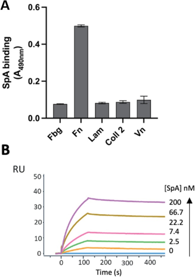
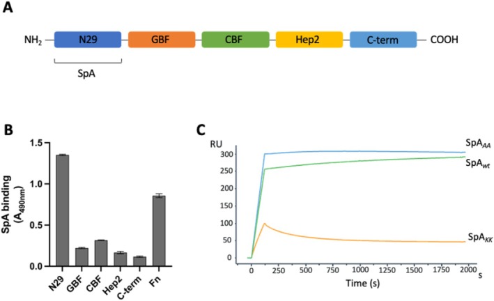
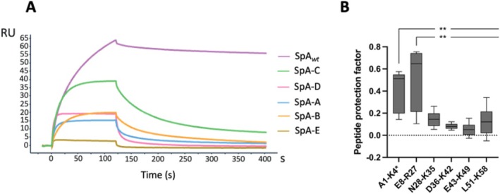
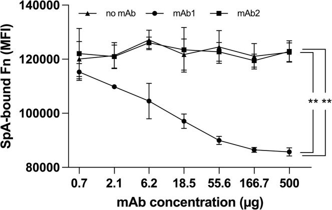
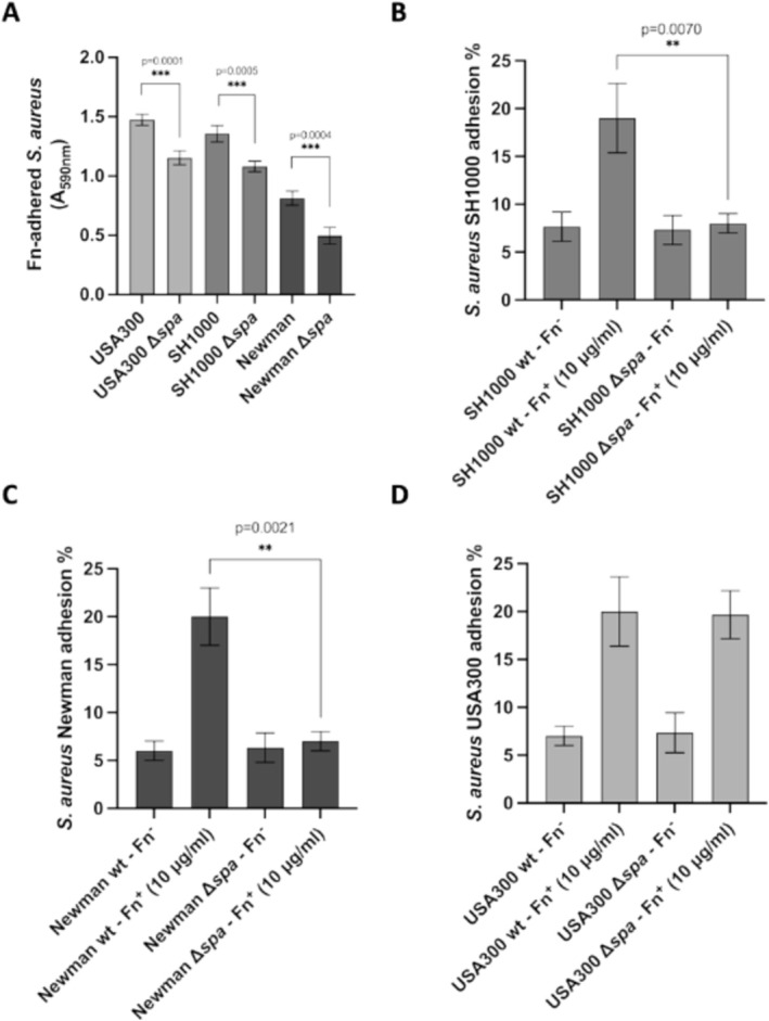
Staphylococcus aureus is the causative agent of serious human health conditions, such as sepsis, endocarditis, and necrotizing pneumonia, as well as less severe clinical manifestations including epithelial and mucosal infections. This pathogen expresses a wide range of surface virulence factors, among which fibronectin-binding proteins play a crucial role in both bacterial adhesion and infection of host cells. Fibronectin is utilized by S. aureus during the early stages of infection to form a protective coating that shields the bacterium from host defenses, facilitating adhesion to the extracellular matrix of host cells and promoting immune evasion and subsequent invasion. S. aureus protein A (SpA) is a key multi-domain cell wall-anchored and secreted molecule that functions to evade the human immune response by non-specifically interacting with Fc and the Fab VH3 domains of immunoglobulins. Other known human ligands of SpA include the von Willebrand factor, the tumor necrosis factor receptor 1, and a platelet surface protein, all of which contribute to immune evasion and pathogenesis. The present study reveals that SpA can also bind human fibronectin with high affinity, adding a new function to this already multifunctional virulence factor. We show that the N-terminus of fibronectin is involved in the interaction and demonstrate by carbene footprinting experiments that the SpA fibronectin binding site spans the interdomain linker region and helix 1 of the domains D, A, B, and C, partially overlapping with the Fc binding site. In the presence of fibronectin, SpA knock-out mutant strains showed reduced adhesion to human endothelial cells compared to wild-type bacteria, suggesting that this interaction may play a significant role in the attachment to host tissues by S. aureus.

Keywords: Staphylococcus aureus; SpA; bacterial adhesion; fibronectin; monoclonal antibody; protein–protein interaction.


Conflict of interest statement

At the time of the study, all authors, excluding G. Pietrocola and A. Pellegrini , were employees of the GSK group of companies. S. Camaione was the recipient of a GSK fellowship from the PhD program of the University of Pavia. I. Margarit, N. Norais, I. Ferlenghi, and S. Savino hold shares of GSK.

Figures

FIGURE 1
FIGURE 1
Schematic depiction of the SpA structure, highlighting its five Ig‐binding domains labeled by their respective letters. The Xr and Xc linker regions are also indicated: The Xr region exhibits variable length, while the Xc linker region contains motifs essential for bacterial cell wall binding. Additionally, the IgG‐binding regions within each domain are visualized, along with the mutants generated by knocking out these binding sites [14, 15]. The square brackets on top of the figure indicate the SpA wt region used for the experiments reported in this study.
FIGURE 2
FIGURE 2
Specific binding of SpA to Fn with high affinity. (A) Binding of SpA to various immobilized ECM analyzed by ELISA. Complex formation was detected using HRP‐conjugated IgG bound by SpA. Data represent the means ± SD of three independent experiments, each performed in triplicate; statistically significant differences are indicated (***p < 0.001). (B) Representative sensorgrams from three different experiments showing the binding of increasing concentrations of SpA to immobilized Fn, analyzed by SPR. Fn was immobilized on a CM5 sensor chip and SpA was injected at increasing concentrations in the mobile phase.
FIGURE 3
FIGURE 3
The Fc binding site of SpA is responsible for the SpA‐Fibronectin interaction. Binding of recombinant SpAwt or its mutant variants SpA AA and SpA KK to immobilized Fn analyzed by ELISA. Complex formation was detected using an HRP‐conjugated IgG. Data represent the means ± SD of three independent experiments, each performed in triplicate. Statistically significant differences are indicated (***p < 0.001).
FIGURE 4
FIGURE 4
The SpA binding site is localized at the N‐terminus of Fn. (A) Schematic representation of Fn highlighting the N‐terminal domain (N29), gelatin‐binding domain (GBF), cell‐binding domain (CBF), heparin‐binding domain (Hep2), and C‐terminal domain (C‐term). Domains are named following established nomenclature [21]. The SpA binding site is indicated on the N‐terminal domain (NTD). (B) Binding of recombinant SpA wt to immobilized Fn or its fragments analyzed by ELISA. Complex formation was detected using a HRP‐conjugated anti‐IgG antibody. Data represent means ± SD of three independent experiments, each performed in triplicate. Statistically significant differences are indicated (***p < 0.001). (C) Representative sensograms from three independent experiments showing the binding of SpA wt and its mutants, SpA AA and SpA KK to immobilized N29 fragment, analyzed by SPR. N29 was immobilized on a CM5 sensor chip, and the recombinant protein was injected at a single concentration in the mobile phase.
FIGURE 5
FIGURE 5
The Fn binding site spans the inter‐domain linker region and helix 1 of SpA. (A) Representative sensorgrams showing the binding of SpA wt and its individual domains (C‐D‐A‐B and E) to immobilized N29 fragment, analyzed by SPR. N29 was immobilized on a CM5 sensor chip, and each recombinant protein was injected at a single concentration in the mobile phase. (B) Box plot depicting the carbene footprinting‐derived protection factor for each of the analyzed peptides. Significance of the result was calculated via two‐samples Wilcoxon non‐parametric test. All peptides amino acid localization has been normalized on the Uniprot sequence for SpA‐C domain (entry A0A0H3K686 • SPA_STAAE) to account for the His‐tag insert used for affinity purification. Data derived from three independent experiments, each performed in triplicate and statistically significant differences are indicated (**p < 0.01).
FIGURE 6
FIGURE 6
Luminex displacement of bFn from immobilized SpA by a SpA‐specific mAb. SpA‐coated magnetic beads were pre‐incubated with bFn under saturating conditions, followed by incubation with SpA‐specific mAb1 or non‐SpA‐specific mAb2 and subsequently with PE‐conjugated streptavidin. Data derived from three independent experiments, each performed in triplicate and statistically significant differences are indicated (**p < 0.01).
FIGURE 7
FIGURE 7
SpA mediates Fn‐dependent host cell adhesion. (A) Adhesion of the indicated S. aureus strains to immobilized Fn. Bound bacteria were detected by crystal‐violet staining. The data points are the means ± SD from three independent experiments, each performed in triplicate (***p < 0.001). B–D) Confluent HUVEC cell monolayers, grown in Fn‐free medium preincubated or not with exogenously added Fn, were infected with the indicated bacterial strains and SpA knockout mutants. The percentage of adhesion for each strain was calculated relative to the initial inoculum. The data points are the means ± SD from three independent experiments, each performed in triplicate. Statistically significant differences are indicated (**p < 0.01).

References

    1. Que Y. A., Haefliger J. A., Piroth L., et al., “Fibrinogen and Fibronectin Binding Cooperate for Valve Infection and Invasion in Staphylococcus aureus Experimental Endocarditis,” Journal of Experimental Medicine 201 (2005): 1627–1635. - PMC - PubMed
    1. Que Y. A. and Moreillon P., “Infective Endocarditis,” Nature Reviews. Cardiology 8, no. 6 (2011): 322–336, 10.1038/nrcardio.2011.43. - DOI - PubMed
    1. DeLeo F. R. and Chambers H. F., “Reemergence of Antibiotic‐Resistant Staphylococcus aureus in the Genomics Era,” Journal of Clinical Investigation 119, no. 9 (2009): 2464–2474. - PMC - PubMed
    1. Foster T. J., Geoghegan J. A., Ganesh V. K., and Höök M., “Adhesion, Invasion and Evasion: The Many Functions of the Surface Proteins of Staphylococcus aureus ,” Nature Reviews. Microbiology 12, no. 1 (2014): 49–62, 10.1038/nrmicro3161. - DOI - PMC - PubMed
    1. Schneewind O., Fowler A., and Faull K. F., “Structure of the Cell Wall Anchor of Surface Proteins in Staphylococcus aureus ,” Science 268, no. 5207 (1995): 103–106, 10.1126/science.7701329. - DOI - PubMed

MeSH terms

Grants and funding

LinkOut - more resources