. 2025 Aug;12(32):e14956.
doi: 10.1002/advs.202414956. Epub 2025 May 31.

Listerin Alleviates Alzheimer's Disease through IRE1-mediated Decay of TLR4 mRNA

Affiliations

Listerin Alleviates Alzheimer's Disease through IRE1-mediated Decay of TLR4 mRNA

Fei Qin et al. Adv Sci (Weinh). 2025 Aug.

Abstract

Alzheimer's disease (AD) is the most prevalent neurodegenerative disorder, accounting for ≈60-70% of all dementia cases worldwide. Microglial-mediated brain inflammation is thought to play key roles in AD progression. Clinical evidence and animal models have indicated that the ribosome-associated quality control (RQC) component Listerin is involved in the development of AD. How Listerin regulates the development and progression of AD is unknown. Here, it is demonstrated that Listerin can decrease brain inflammation and alleviate AD-related cognitive impairments. Microglial-specific knockout of Listerin exhibits deteriorative cognitive symptoms based on the extracellular Amyloid-β (Aβ) or Lipopolysaccharide (LPS) injection. Mechanistically, Listerin directly binds to Toll-like receptor 4 (TLR4) mRNA and facilitates the IRE1α-mediated cleavage and degradation of TLR4 mRNA, leading to the alleviation of TLR4-induced brain inflammation. Adenovirus-mediated overexpression of Listerin decelerates the disease progression in the mouse model of Aβ-mediated neurodegeneration. Thus, Listerin is an important suppressor of microglia-induced brain inflammation and may be a potential therapeutic target for AD treatment.

Keywords: Alzheimer's disease; IRE1α‐dependent decay (RIDD); Listerin; TLR4; inflammation.


Conflict of interest statement

The authors declare no conflict of interest.

Figures
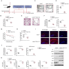
Figure 1
Figure 1
Deletion of Listerin aggravate disease in an AD mouse model. A) Schematic diagram of the experimental design. PBS or Aβ were injected into the bilateral entorhinal cortices of 2‐month‐old Listerin fl/fl and Listerin fl/fl Cx3cr1 Cre mice. After 12 days, the behavior tests were performed and the sampling were collected at last. B–F) 12 days after Aβ injection in Listerin fl/fl and Listerin fl/fl Cx3cr1 Cre mice, Y maze (B), Novel object recognition (C, D) or Open field test (E, F) was performed. G,H) Time to reach hidden platform in Morris water maze and time in critical quadrant (first quadrant) was tested. I,J) Immunolabeling of hippocampi for Iba1 (microglia), AT8 (phospho‐Tau), and NeuN (neurons) of Listerin fl/fl and Listerin fl/fl Cx3cr1 Cre mice after Aβ injection. K) TNF‐α, IL‐6, IL‐12p40, and IL‐1β concentrations in the hippocampi of Listerin fl/fl and Listerin fl/fl Cx3cr1 Cre mice after Aβ injection measured by ELISA. L) Representative immunoblots of phospho‐IKKβ and phospho‐P65 in the hippocampi of Listerin fl/fl and Listerin fl/fl Cx3cr1 Cre mice after Aβ injection. Data shown as mean ± SD and are representative of two independent experiments with similar results. Statistical analysis was performed using unpaired Student's t‐test. ** P < 0.01.
Figure 2
Figure 2
Listerin inhibits AD‐related neuroinflammation. A) Primary microglia prepared from Listerin fl/fl and Listerin fl/fl Cx3cr1 Cre mice were stimulated with LPS or Aβ for 24 h, cell lysates were collected and TNFα, IL6, IL12b, and IL1β mRNA were quantified by qPCR. B) Supernatant in (A) were collected and TNF‐α, IL‐6 and IL‐12p40 were quantified by ELISA. C) HEK293‐TLR4 cells transfected with Vector or Flag‐Listerin constructs followed by stimulated with LPS for 8 h, cell lysates were collected and TNFα, IL6, IL12b, and IL1β mRNA were quantified by qPCR. Bars represent mean ± SD. D) Pathway activation in primary microglia prepared from Listerin fl/fl and Listerin fl/fl Cx3cr1 Cre mice under LPS for the indicating times. E) Pathway activation in primary microglia prepared from Listerin fl/fl and Listerin fl/fl Cx3cr1 Cre mice under Aβ for the indicating times. F) Confocal images of nuclear translocation of NF‐κB p65 (red) after Aβ treatment in primary microglia prepared from Listerin fl/fl and Listerin fl/fl Cx3cr1 Cre mice. G) Quantitative analyses of % nuclear NF‐κB‐positive microglia in (F). H) Schematic representation of co‐culture model: primary microglia prepared from Listerin fl/fl and Listerin fl/fl Cx3cr1 Cre mice were plated on transwells. Primary hippocampal neurons from wild type mice were plated in 24‐well plates. I) Representative images of Hoechst and propidium iodide (PI) staining from primary hippocampal neurons co‐incubated with primary microglia prepared from Listerin fl/fl and Listerin fl/fl Cx3cr1 Cre mice, and further incubated with Aβ for 3 days. Scale bar, 20 µm. J) Quantification of cell death in (I). K) The cytotoxicity was detected by the TUNEL assay from primary hippocampal neurons co‐incubated with primary microglia prepared from Listerin fl/fl and Listerin fl/fl Cx3cr1 Cre mice, and further incubated with Aβ for 3 days. L) LDH assay from primary hippocampal neurons co‐incubated with primary microglia prepared from Listerin fl/fl and Listerin fl/fl Cx3cr1 Cre mice, and further incubated with Aβ for 3 days. Data shown as mean ± SD and are representative of two independent experiments with similar results. Statistical analysis was performed using unpaired Student's t‐test. ** P < 0.01.
Figure 3
Figure 3
Listerin decreases the expression of TLR4 protein. A) Representative immunoblots of primary microglia prepared from Listerin fl/fl and Listerin fl/fl Cx3cr1 Cre mice under LPS stimulation for the indicating times. B) Representative immunoblots of primary microglia prepared from Listerin fl/fl and Listerin fl/fl Cx3cr1 Cre mice under Aβ stimulation for the indicating times. C) Representative immunoblots of HEK293T cells transfected with His‐TLR4 together with gradient amount of Flag‐Listerin constructs. D) Representative immunoblots of HEK293‐TLR4 cells transfected with gradient amount of Flag‐Listerin constructs and stimulated with LPS for 8 h. E) Representative immunoblots of primary microglia prepared from Listerin fl/fl and Listerin fl/fl Cx3cr1 Cre mice followed by stimulated with LPS for 4 h and then treated with the protein synthesis inhibitor CHX for the indicated times. F) Quantification analysis of proteins degradation kinetics in (E). G) Representative immunoblots of HEK293‐TLR4 cells transfected with Vector or Flag‐Listerin constructs followed by stimulated with LPS for 4 h and then treated with the protein synthesis inhibitor CHX for the indicated times. H) Quantification analysis of proteins degradation kinetics in (G). I) Representative immunoblots of HEK293T cells transfected with Vector or Flag‐Listerin constructs followed by treatment with Mg132 (10 µm), Chloroquine (10 µm), Bafilomycin A1 (0.5 µm) or 3‐Methyladenine (10 mm) for 10 h.
Figure 4
Figure 4
Listerin decreased TLR4 mRNA transcript levels. A) Primary microglia prepared from Listerin fl/fl and Listerin fl/fl Cx3cr1 Cre mice were stimulated with LPS for 0–4 h, cell lysates were collected and TLR4 mRNA was quantified by qPCR. B) HEK293T cells transfected with His‐TLR4 together with gradient amount of Flag‐Listerin constructs, cell lysates were collected and TLR4 mRNA was quantified by qPCR. C) HEK293‐TLR4 cells transfected with Vector or Flag‐Listerin constructs followed by stimulated with PBS or LPS for 4 h, cell lysates were collected and TLR4 mRNA was quantified by qPCR. D) Representative images of FISH for TLR4 mRNA in HEK293T cells transfected with His‐TLR4 together with Vector or Flag‐Listerin constructs. Images are representative of 3 independent experiments. Scale bars, 20 µm. E) Primary microglia prepared from Listerin fl/fl and Listerin fl/fl Cx3cr1 Cre mice were stimulated with LPS for 4 h and then treated with actinomycin D for the indicated times, cell lysates were collected and TLR4 mRNA (left) and TLR4 mRNA degradation (right) was quantified by qPCR. F) HEK293‐TLR4 cells were transfected with Vector or Flag‐Listerin constructs followed by stimulated with LPS for 4 h and then treated with actinomycin D for the indicated times, cell lysates were collected and TLR4 mRNA (left) and TLR4 mRNA degradation (right) was quantified by qPCR. G) Primary microglia prepared from Listerin fl/fl and Listerin fl/fl Cx3cr1 Cre mice were stimulated with LPS for 4 h and then exposed to a 30‐min EU pulse, nascent RNA was extracted and TLR4 mRNA was quantified by qPCR. H) RIP assay for TLR4 mRNA in HEK293‐TLR4 cells transfected with Vector or Flag‐Listerin constructs. Normalized data were shown as relative fold enrichment to the control group. I) Representative images of FISH for TLR4 mRNA and fluorescence immunostaining for Flag‐Listerin in HEK293T cells transfected with His‐TLR4 together with Flag‐Listerin constructs. J) Intensity profiles in (I). K) Representative immunoblots of HEK293‐TLR4 cells transfected with vector, Flag‐Listerin, Flag‐Listerin (C/A) or Flag‐Listerin (△R) constructs. Results are representative of 3 independent experiments. L) Cells treated as in (K) were collected and TLR4 mRNA were quantified by qPCR. M) Representative immunoprecipitation (IP) of HEK293T cells transfected with Myc‐IRE1α together with vector, Flag‐Listerin, Flag‐Listerin (C/A) or Flag‐Listerin (△R) constructs. Data shown as mean ± SD and are representative of three independent experiments with similar results. Statistical analysis was performed using unpaired Student's t‐test. ** P < 0.01.
Figure 5
Figure 5
IRE1α is required for Listerin mediated TLR4 degradation. A) Representative immunoblots of HEK293‐TLR4 cells transfected with vector or Flag‐Listerin constructs for 20 h and then treated with 4µ8C (10 µm) or STF‐083010 (10 µm) for 10 h. B) Cells treated as in (A) were collected and TLR4 mRNA were quantified by qPCR. C) Representative immunoblots of HEK293T cells silenced for control (siCtrl) or IRE1α (siIRE1α) for 24 h followed by transfected with His‐TLR4 together with vector or Flag‐Listerin constructs for 24 h. D) Cells treated as in (C) were collected and TLR4 mRNA were quantified by qPCR. E) Representative immunoblots of IRE1α knockout cells transfected with His‐TLR4 together with gradient amount of Flag‐Listerin constructs. F) Co‐IP analysis of the interaction in HEK293‐TLR4 cells transfected with Myc‐IRE1α together with vector, Flag‐Listerin, Flag‐Listerin (C/A) or Flag‐Listerin (△R) constructs. Normalized data were shown as relative fold enrichment to the control group. G) Representative images of FISH for TLR4 mRNA and fluorescence immunostaining for Flag‐Listerin and Myc‐IRE1α in HEK293T cells transfected with His‐TLR4, Myc‐IRE1α together with Flag‐Listerin constructs. H) Intensity profiles in (G).Data shown as mean ± SD and are representative of three independent experiments with similar results. Statistical analysis was performed using unpaired Student's t‐test. ** P < 0.01.
Figure 6
Figure 6
Listerin is essential for IRE1α to cleavage TLR4 mRNA. A) RNA cleavage assay of Xbp‐1 or TLR4 transcripts incubated with recombinant protein IRE1α together with PBS, Listerin or Listerin (△R) for 30 min, followed by resolved for a 2.5% agarose gel and visualized by a BioRad Molecular Imager. B) RNA cleavage assay of TLR3 transcripts incubated with recombinant protein IRE1α together with PBS or Listerin for 30 min, followed by resolved for a 2.5% agarose gel and visualized by a BioRad Molecular Imager. C) Illustration of predicted stem‐loop secondary structure shown for human wild type (WT) TLR4 mRNA and the mutant (Mut, GC to CA) TLR4 mRNA. The potential IRE1α cleavage site is indicated by an arrow. D) RNA cleavage assay of WT or Mut TLR4 transcripts incubated with recombinant protein IRE1α together with PBS or Listerin for 30 min, followed by resolved for a 2.5% agarose gel and visualized by a BioRad Molecular Imager. E) Representative immunoblots of HEK293T cells transfected with WT or Mut His‐TLR4 together with vector or Flag‐Listerin constructs. F) Fluorescence‐based analysis of IRE1α cleavage of the synthetic WT‐1 or mutant (mut, GC to CA) Mut‐1 mRNA. G) HEK293T cells were transfected with WT or Mut‐1 His‐TLR4 together vector or Flag‐Listerin constructs followed by stimulated with LPS for 6 h, cell supernatant was collected and TNF‐α (left) or IL‐6 (right) were quantified by ELISA. H) Representative immunoblots of phospho‐P65 in HEK293T cells treated as in (G). Data shown as mean ± SD and are representative of three independent experiments with similar results. Statistical analysis was performed using unpaired Student's t‐test. ** P < 0.01.
Figure 7
Figure 7
Listerin ameliorates the inflammation of AD progression in vivo. A) Schematic diagram of the experimental design. Aβ were injected into the bilateral entorhinal cortices of 2‐month‐old WT mice. After 12 days, Adv‐GFP or Adv‐Listerin‐GFP were injected into the bilateral entorhinal cortices for 3 days, the behavior tests were performed and the sampling were collected at last. B) Representative immunoblots of Listerin expression in hippocampi from mice treated in (A). Results are representative of 3 mice. C–G) Mice were treated as in (A), Y maze (C), Novel object recognition (D, E) or Open field test (F, G) was performed (n = 5 mice per group). H) Immunolabeling of hippocampi for Iba1 (microglia), AT8 (phospho‐Tau), and NeuN (neurons) of mice treated as in (A). I) TNF‐α, IL‐6, IL‐12p40, and IL‐1β concentrations in the hippocampi of mice treated as in (A) measured by ELISA (n = 5 mice per group). J) Representative immunoblots of phospho‐IKKβ and phospho‐P65 in the hippocampi of mice treated as in (A). Data shown as mean ± SD and are representative of three independent experiments with similar results. Statistical analysis was performed using unpaired Student's t‐test. ** P < 0.01.

References

    1. Dubois B., Villain N., Frisoni G. B., Rabinovici G. D., Sabbagh M., Cappa S., Bejanin A., Bombois S., Epelbaum S., Teichmann M., Habert M. O., Nordberg A., Blennow K., Galasko D., Stern Y., Rowe C. C., Salloway S., Schneider L. S., Cummings J. L., Feldman H. H., Lancet Neurol. 2021, 20, 484. - PMC - PubMed
    1. a) Chhatwal J. P., Schultz S. A., McDade E., Schultz A. P., Liu L., Hanseeuw B. J., Joseph‐Mathurin N., Feldman R., Fitzpatrick C. D., Sparks K. P., Levin J., Berman S. B., Renton A. E., Esposito B. T., Fernandez M. V., Sung Y. J., Lee J. H., Klunk W. E., Hofmann A., Noble J. M., Graff‐Radford N., Mori H., Salloway S. M., Masters C. L., Martins R., Karch C. M., Xiong C., Cruchaga C., Perrin R. J., Gordon B. A., et al., Lancet Neurol. 2022, 21, 140; - PMC - PubMed
    2. b) Ossenkoppele R., van der Kant R., Hansson O., Lancet Neurol. 2022, 21, 726; - PubMed
    3. c) Tijms B. M., Gobom J., Reus L., Jansen I., Hong S., Dobricic V., Kilpert F., Ten Kate M., Barkhof F., Tsolaki M., Verhey F. R. J., Popp J., Martinez‐Lage P., Vandenberghe R., Lleó A., Molinuevo J. L., Engelborghs S., Bertram L., Lovestone S., Streffer J., Vos S., Bos I., Blennow K., Scheltens P., Teunissen C. E., Zetterberg H., Visser P. J., Brain 2020, 143, 3776. - PMC - PubMed
    1. a) Heneka M. T., Carson M. J., El Khoury J., Landreth G. E., Brosseron F., Feinstein D. L., Jacobs A. H., Wyss‐Coray T., Vitorica J., Ransohoff R. M., Herrup K., Frautschy S. A., Finsen B., Brown G. C., Verkhratsky A., Yamanaka K., Koistinaho J., Latz E., Halle A., Petzold G. C., Town T., Morgan D., Shinohara M. L., Perry V. H., Holmes C., Bazan N. G., Brooks D. J., Hunot S., Joseph B., Deigendesch N., et al., Lancet Neurol. 2015, 14, 388; - PMC - PubMed
    2. b) De Strooper B., Karran E., Cell 2016, 164, 603. - PubMed
    1. a) Castro‐Gomez S., Heneka M. T., Immunity 2024, 57, 790; - PubMed
    2. b) Gao C., Jiang J., Tan Y., Chen S., Signal Transduction Targeted Ther. 2023, 8, 359. - PMC - PubMed
    1. Heneka M. T., van der Flier W. M., Jessen F., Hoozemanns J., Thal D. R., Boche D., Brosseron F., Teunissen C., Zetterberg H., Jacobs A. H., Edison P., Ramirez A., Cruchaga C., Lambert J. C., Laza A. R., Sanchez‐Mut J. V., Fischer A., Castro‐Gomez S., Stein T. D., Kleineidam L., Wagner M., Neher J. J., Cunningham C., Singhrao S. K., Prinz M., Glass C. K., Schlachetzki J. C. M., Butovsky O., Kleemann K., De Jaeger P. L., et al., Nat. Rev. Immunol. 2025, 25, 321. - PubMed

LinkOut - more resources