. 2025 Aug;44(32):2831-2849.
doi: 10.1038/s41388-025-03461-6. Epub 2025 May 31.

Drug tolerance and persistence to EGFR inhibitor treatment are mediated by an ILK-SFK-YAP signaling axis in lung adenocarcinoma

Affiliations

Drug tolerance and persistence to EGFR inhibitor treatment are mediated by an ILK-SFK-YAP signaling axis in lung adenocarcinoma

Rocky Shi et al. Oncogene. 2025 Aug.

Abstract

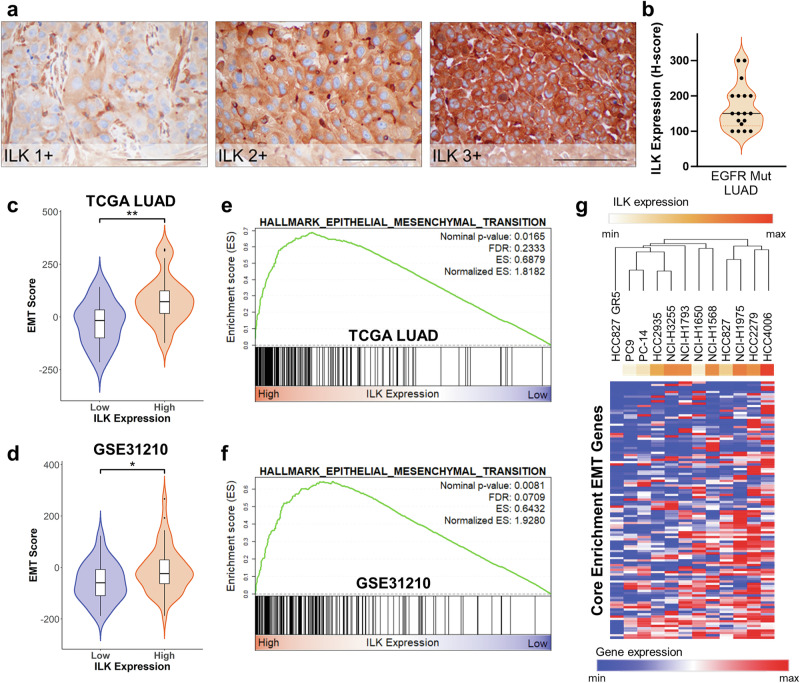
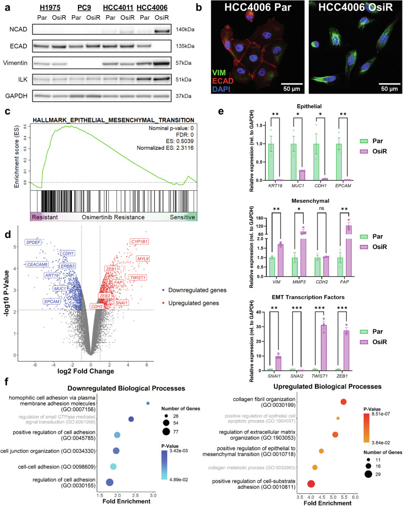
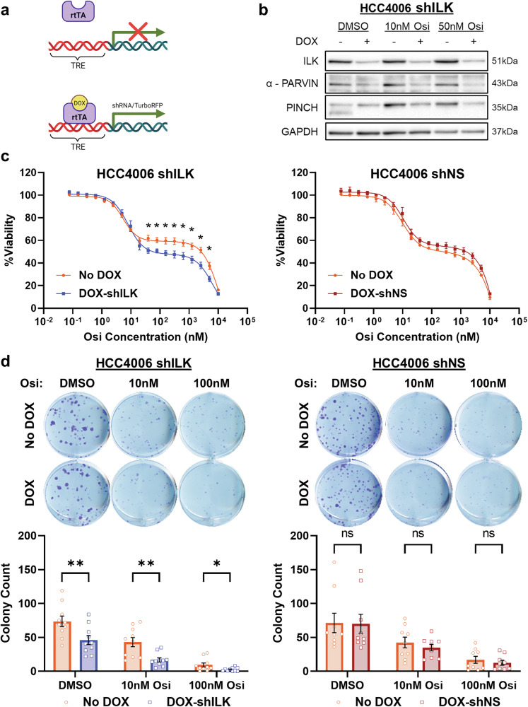
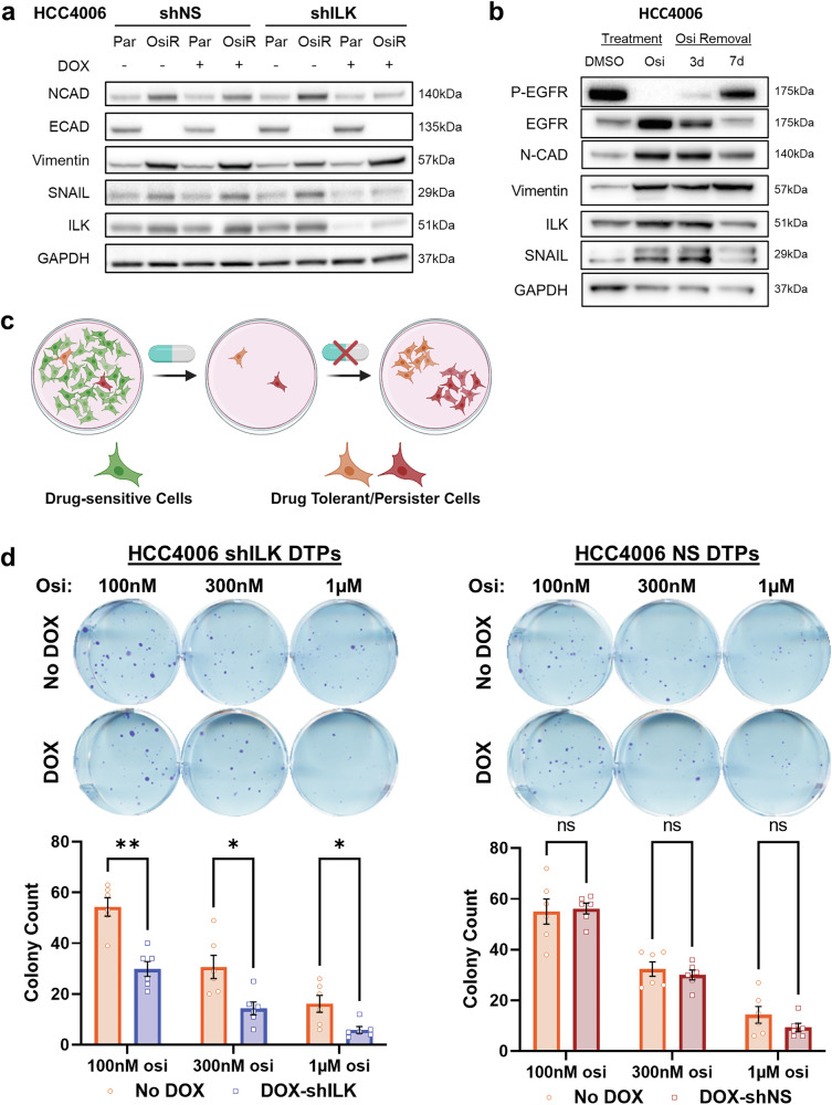
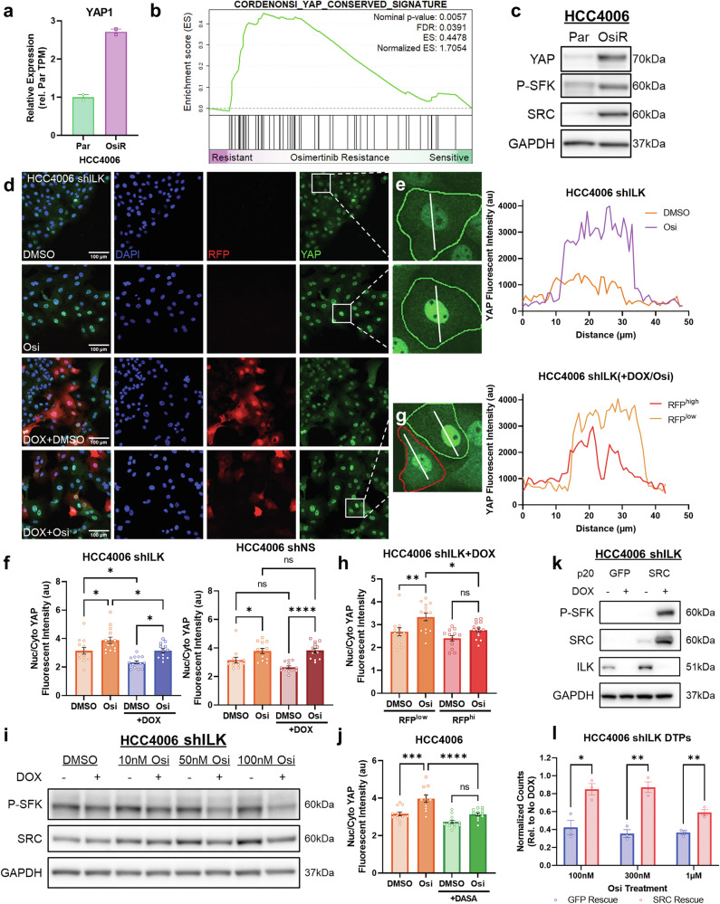
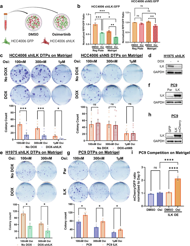
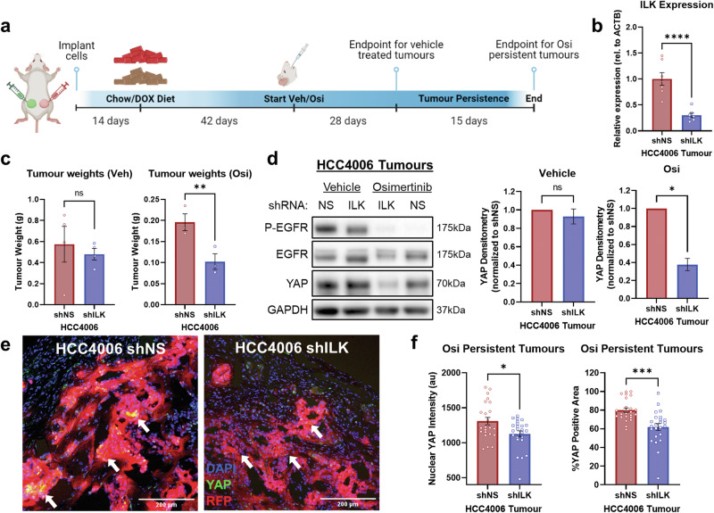
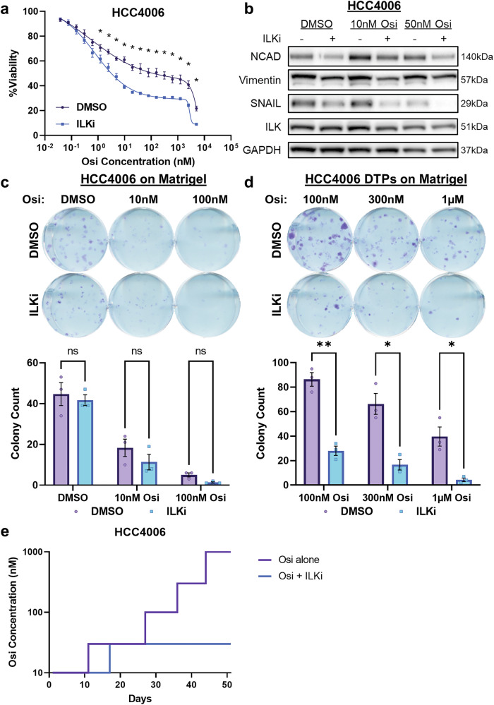
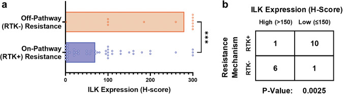
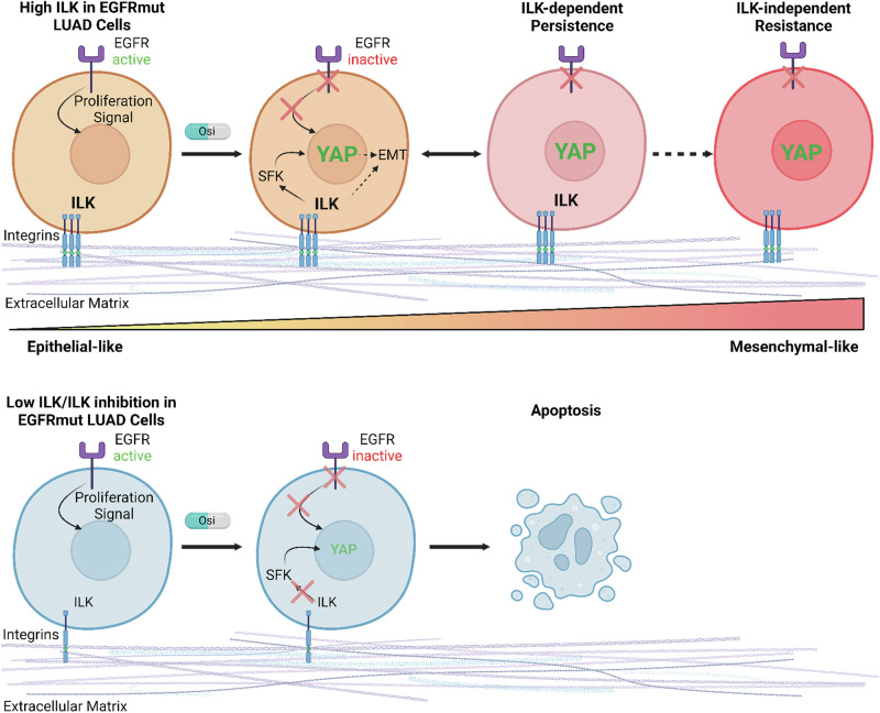
Combating resistance to targeted therapy remains a major challenge to improving lung cancer care. Epithelial-mesenchymal transition (EMT) in tumour cells is an established non-genetic resistance mechanism to EGFR tyrosine kinase inhibitors (TKI) that is also associated with worse outcome in patients. Here we demonstrate that integrin-linked kinase (ILK) is an important driver of EMT-mediated TKI resistance in lung adenocarcinoma (LUAD) by promoting a drug-tolerant persister (DTP) cell phenotype. Our results indicate that high ILK expression is associated with EMT in LUAD patients and that genetic suppression of ILK can limit EMT progression and reduce the viability of DTP cells by impairing YAP activation, ultimately improving osimertinib (Osi) sensitivity in LUAD cells. Importantly, LUAD cells with high ILK expression are able to persist during EGFR-TKI treatment, acquiring additional genetic and phenotypic alterations to develop EGFR-TKI resistance. To improve clinical translatability of our findings, we showed that pharmacological inhibition of ILK can suppress EMT and improve Osi response in LUAD cells. Lastly, we found that strong immunohistochemistry staining of ILK in patient biopsies was significantly associated with and may be used to predict receptor tyrosine kinase-independent mechanisms of EGFR-TKI resistance. Overall, our results suggest that ILK is an important regulator of EGFR-TKI response and may be exploited as a predictor for acquired resistance, providing evidence for co-targeting ILK with EGFR to better control minimal residual disease and EGFR-TKI resistance in lung cancer.


Conflict of interest statement

Competing interests: The authors declare no competing interests. Ethical approvals: Patient tissue samples were collected under MSKCC IRB-approved biospecimen collection protocols, and informed consent was obtained. All housing, care, and experimental conditions and protocols for mice were followed in accordance with the guidelines of the University of British Columbia Animal Care Committee and the Canadian Council for Animal Care.

Figures

Fig. 1
Fig. 1. ILK is associated with EMT in EGFR-mutant LUAD.
a IHC staining of ILK in EGFR-mutant LUAD patient tumours. Representative images of staining scores 1, 2, or 3 are shown. Black scale bar = 100 µm. b Summary of ILK staining in pre-treated EGFR-mutant LUAD tumours (n = 18 from 10 patients). c, d Comparison of EMT Score between high-expressing vs low-expressing ILK EGFR-mutant LUAD patient tumour samples in c TCGA PanCan Atlas LUAD (n = 24 each) and d Japanese LUAD microarray cohort (n = 42 each; GSE31210). Wilcoxon rank sum Test were used to statistically compare high vs low ILK samples. e, f Enrichment plots of the top Hallmark gene set from GSEA comparing high to low ILK-expressing EGFR Mut LUAD tumours from indicated datasets. g Heatmap of DepMap EGFR Mut LUAD cell lines indicating expression of core enrichment genes from the EMT gene set of patient tumours in (e, f).
Fig. 2
Fig. 2. EGFR-TKI resistance in high ILK-expressing HCC4006 cells is associated with EMT.
a Western blot of ILK and EMT marker levels in parental (Par) and osimertinib (Osi) resistant EGFR-mutant cell lines. b Immunofluorescence images of HCC4006 Par and OsiR cells. c Enrichment plot of the top enriched Hallmark gene set from GSEA comparing HCC4006 OsiR (n = 2 experimental replicates) to Par (n = 2 experimental replicates) cells. d Volcano plot of significant differentially expressed genes. Genes were considered differentially expressed if adjusted P-value (by the Benjamini-Hochberg method) was less than 0.05, which corresponds to a raw P-value of ~0.008. e RT-qPCR validation of representative epithelial, mesenchymal and EMT transcription factor genes (n = 3 each). Student’s t-test were used to statistically compare Par vs OsiR (*p < 0.05; **p < 0.01; ***p < 0.001). Bar graphs with error bars represent the mean ± SEM. f Top 6 downregulated and upregulated biological processes from Gene Ontology analysis (PANTHER Over Representation test) of differentially expressed genes in HCC4006 OsiR versus Par cells. Bolded are known biological processes that ILK regulates. Osi was maintained at 1 µM in OsiR cells in these experiments.
Fig. 3
Fig. 3. ILK knockdown improves response to Osi treatment.
a Schematic of pTRIPZ shRNA constructs. b Validation of knockdown in HCC4006 shILK cells; western blot of proteins in the IPP complex in HCC4006. c Three day Osi dose-response in HCC4006 shILK (n = 3) and shNS (n = 3) ± DOX. Multiple t-tests with Holm-Sidak correction were used to statistically compare DOX vs No DOX at each Osi concentration (*p < 0.05). d Clonogenic assay of HCC4006 shILK (n = 10) and shNS (n = 9) cells treated with Osi for 7 days ± DOX. Quantification of colony count is below the representative plates. Multiple paired t-tests with Holm-Sidak correction were used to statistically compare DOX vs No DOX at each Osi concentration (*p < 0.05; **p < 0.01). Error bars represent the mean ± SEM. DOX concentration = 100 ng/mL.
Fig. 4
Fig. 4. ILK promotes DTP survival and the progression towards EMT-mediated Osi resistance.
a Western blot of EMT markers in HCC4006 shNS, and shILK cells ± DOX Par and OsiR. b Western blot of ILK and EMT markers following treatment with DMSO or 100 nM Osi for 7 days and subsequent removal of Osi. c Schematic of persister cell assay. d Persister clonogenic assay of HCC4006 shILK (n = 6) and shNS (n = 6) cells treated with Osi for 7 days ± DOX. Quantification of colony count is below the representative plates. Multiple paired t-tests with Holm-Sidak correction were used to statistically compare DOX vs No DOX at each Osi concentration (*p < 0.05; **p < 0.01). Error bars represent the mean ± SEM. DOX concentration = 100 ng/mL.
Fig. 5
Fig. 5. Cytoprotective effects of ILK are mediated through an ILK-SFK-YAP axis.
a YAP1 expression in HCC4006 Par and OsiR cells (n = 2 experimental replicates each). b Enrichment plot of the YAP expression signature from the Oncogenic Signature gene sets from GSEA comparing HCC4006 OsiR to Par cells. c Western blot of YAP, P-SFK and SRC in HCC4006 Par and OsiR cells. d Representative immunofluorescent confocal imaging of Osi/DMSO-treated HCC4006 shILK cells ± DOX. e Insets of example HCC4006 cells treated with DMSO or Osi. YAP intensity profile across the white line is plotted on the right. f Quantification of nuclear/cytoplasmic YAP intensity in Osi (100 nM) treated HCC4006 shILK (n = 12–14 images in each condition across 3 experimental replicates) and shNS (n = 12 images in each condition across 3 experimental replicates) cells. g Insets of 2 example Osi treated HCC4006 shILK+DOX cells with high and low RFP expression neighbouring each other. YAP intensity profile across the white line is plotted on the right. h Quantification of nuclear/cytoplasmic YAP intensity in Osi/DMSO-treated HCC4006 shILK cells +DOX (n = 12–14 images in each condition across 3 experimental replicates). i Representative western blot of P-SFK and SRC in Osi-treated HCC4006 shILK cells ± DOX. j Quantification of nuclear/cytoplasmic YAP intensity in Osi (50 nM) treated HCC4006 cells ± dasatinib (30 nM DASA) (n = 12 images in each condition across 3 experimental replicates). Statistical analyses were performed with One-way ANOVA with Sidak post-hoc test (*p < 0.05; **p < 0.01; ***p < 0.001; ****p < 0.0001). k Validation of DOX-inducible SRC overexpression HCC4006 shILK p20-GFP/SRC cells ± DOX. l Quantification of GFP or SRC rescue in Osi-treated HCC4006 shILK DTP cells (n = 3 each). Colony counts were normalized to the No DOX condition for each Osi concentration. Multiple paired t-tests with Holm-Sidak correction were used to statistically compare DOX vs No DOX at each Osi concentration (*p < 0.05; **p < 0.01). Error bars represent the mean ± SEM. DOX concentration = 100 ng/mL.
Fig. 6
Fig. 6. ECM increases dependences on ILK-mediated cytoprotective effects during Osi treatment.
a Schematic of competition assay. b Quantification of normalized RFP/GFP ratio in Osi (100 nM) treated HCC4006 shILK/GFP or HCC4006 shNS/GFP cells plated on either regular or Matrigel-coated plates (n = 3 experimental replicates in each condition). One-way ANOVA was used to statistically compare across these various conditions (*p < 0.05; **p < 0.01; ***p < 0.001). c Persister clonogenic assay of HCC4006 shILK (n = 9) and shNS (n = 9) cells plated on Matrigel-coated plates and treated with Osi for 7 days ± DOX. Quantification of colony count is below the representative plates. d Validation of ILK knockdown in H1975 shILK cells. e Persister clonogenic assays of H1975 shILK cells cultured on Matrigel-coated plates treated with Osi for 7 days ± DOX. Multiple paired t-tests with Holm-Sidak correction were used to statistically compare DOX vs No DOX at each Osi concentration (*p < 0.05; ***p < 0.001). DOX concentration = 100 ng/mL. f Validation of ILK overexpression in PC9 ILK cells. g Persister clonogenic assays of PC9 and PC9 ILK cells on Matrigel-coated plates treated with Osi for 7 days. Quantifications of colony count are below the representative plates. Multiple t-tests were used to statistically compare PC9 vs PC9 ILK at each Osi concentration (*p < 0.05). h Validation of ILK overexpression in PC9 ILK mCherry cells vs GFP cells. i Quantification of mCherry/GFP ratio in Osi/DMSO-treated PC9 mCherry/GFP or PC9 ILK mCherry/GFP cells plated on Matrigel-coated plates (n = 3 experimental replicates in each condition). One-way ANOVA was used to statistically compare across these various conditions (****p < 0.0001). Error bars represent the mean ± SEM.
Fig. 7
Fig. 7. ILK preserves residual tumours and promotes YAP activation during Osi treatment in vivo.
a Schematic of HCC4006 shNS/shILK tumour xenograft experiment. b Validation of ILK knockdown by RT-qPCR. Ratio paired t-test was used to statistically compare between HCC4006 shNS vs shILK tumours (n = 7) in the same mouse (***p < 0.001). c Tumour weights of implanted HCC4006 shNS and shILK tumours extracted from mice gavaged with Vehicle (Veh; n = 4) or Osi (n = 3) at endpoint. Ratio paired t-test was used to statistically compare between HCC4006 shNS vs shILK tumours (n = 3) in the same mouse (**p < 0.01). d Representative western blot of HCC4006 shNS and shILK tumours treated with Veh or Osi. Quantification of YAP band density are plotted on the right. Ratio paired t-test was used to statistically compare between HCC4006 shNS vs shILK tumours (n = 3) in the same mouse (*p < 0.05). e Representative images of YAP staining in HCC4006 shNS and shILK tumours. White arrows indicate regions of YAP staining. f Quantification of nuclear YAP intensity and %YAP positive area between shNS vs shILK tumours (n = 4 images/section × 2 section/tumour × 3 tumours). Student’s t-tests were used to statistically compare HCC4006 shNS vs shILK persistent tumours (*p < 0.05, ***p < 0.001). Error bars represent the mean ± SEM.
Fig. 8
Fig. 8. Pharmacological inhibition of ILK synergizes with Osi to reduce viability of DTP cells.
a Three days Osi dose-response in HCC4006 cells (n = 3 experimental replicates) ± ILKi (5 µM QLT0267). Multiple t-tests with Holm-Sidak correction were used to statistically compare DOX vs No DOX at each Osi concentration (*p < 0.05). b Representative western blot of mesenchymal markers in Osi-treated HCC4006 cells ± ILKi (5 µM QLT0267). c Clonogenic and d persister clonogenic assays of HCC4006 cells cultured on Matrigel-coated plates treated with Osi for 7 days ± ILKi (5 µM QLT0267). Quantification of colony count is below the representative plates. Multiple paired t-tests with Holm–Sidak correction were used to statistically compare ILKi vs DMSO at each Osi concentration (*p < 0.05; **p < 0.01). e Onset of resistance of HCC4006 cells dose-escalated with either Osi alone or Osi + ILKi.
Fig. 9
Fig. 9. ILK expression is associated with RTK-independent resistance mechanisms to EGFR-TKIs in clinical biopsies.
a ILK expression (by H-Score) in EGFR-TKI resistant patient tumour samples with RTK pathway-independent (RTK-) mechanisms (n = 12) vs RTK pathway-dependent (RTK+) mechanisms (n = 48). Mann–Whitney Test was used to statistically compare RTK- vs RTK+ resistant samples. b Coincidence of high vs low ILK expression and RTK+ vs RTK- resistance mechanisms in 18 LUAD patients post-treatment with EGFR-TKI. P values were calculated by a two-tailed 2 × 2 Fisher’s Exact Test to assess contingency across the two variables.
Fig. 10
Fig. 10
Graphical summary of ILK’s role during EGFR-TKI treatment.

Similar articles

References

    1. Sung H, Ferlay J, Siegel RL, Laversanne M, Soerjomataram I, Jemal A, et al. Global Cancer Statistics 2020: GLOBOCAN Estimates of Incidence and Mortality Worldwide for 36 Cancers in 185 Countries. CA Cancer J Clin. 2021;71:209–49. - PubMed
    1. Herbst RS, Morgensztern D, Boshoff C. The biology and management of non-small cell lung cancer. Nature. 2018;553:446–54. - PubMed
    1. Soria JC, Ohe Y, Vansteenkiste J, Reungwetwattana T, Chewaskulyong B, Lee KH, et al. Osimertinib in untreated EGFR-mutated advanced non-small-cell lung cancer. N Engl J Med. 2018;378:113–25. - PubMed
    1. Leonetti A, Sharma S, Minari R, Perego P, Giovannetti E, Tiseo M. Resistance mechanisms to osimertinib in EGFR-mutated non-small cell lung cancer. Br J cancer. 2019;121:725–37. - PMC - PubMed
    1. Fukuda K, Takeuchi S, Arai S, Kita K, Tanimoto A, Nishiyama A, et al. Glycogen synthase kinase-3 inhibition overcomes epithelial-mesenchymal transition-associated resistance to osimertinib in EGFR-mutant lung cancer. Cancer Sci. 2020;111:2374–84. - PMC - PubMed

MeSH terms