. 2025 May 31;15(1):19168.
doi: 10.1038/s41598-025-03790-2.

Decreased expression of insulin-degrading enzyme increases gluconeogenesis and glucose production in cultured hepatocytes administered with glucagon

Affiliations

Decreased expression of insulin-degrading enzyme increases gluconeogenesis and glucose production in cultured hepatocytes administered with glucagon

Carlos M González-Casimiro et al. Sci Rep. .

Abstract

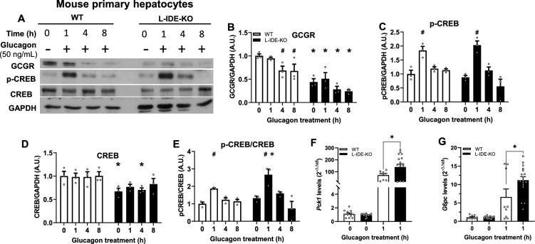
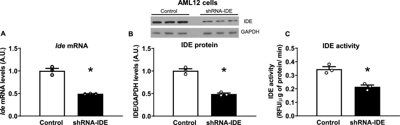
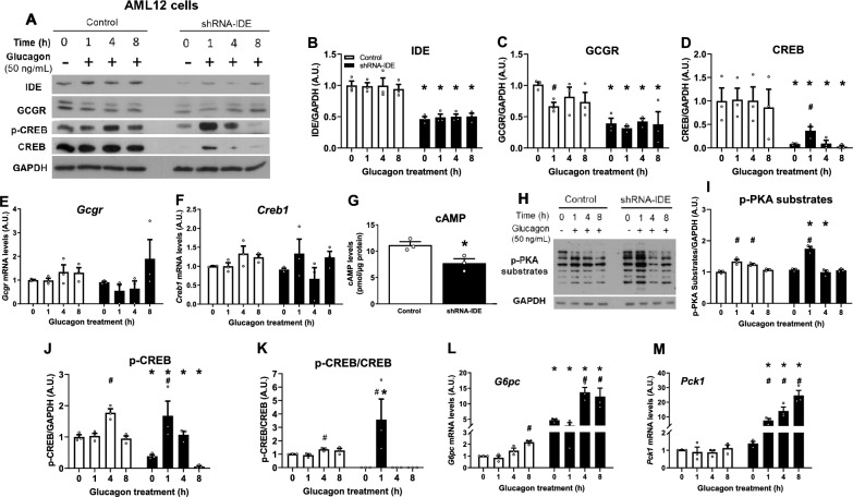
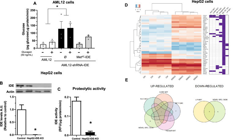
Insulin-degrading enzyme (IDE) is a protein with proteolytic and non-proteolytic functions that regulates glucose homeostasis. In the fasted state, glucagon regulates glycemia through induction of hepatic gluconeogenesis. The rate of hepatic gluconeogenesis is elevated in subjects with type 2 diabetes (T2D) compared with healthy subjects. Interestingly, subjects with T2D show decreased expression of hepatic IDE. However, the role of IDE on the regulation of hepatic gluconeogenesis is completely unknow. We hypothesize that IDE deficiency alters glucagon signaling and thereby gluconeogenesis. To test this hypothesis, we used mouse liver tissues and cultured hepatocytes with total or partial IDE deficiency. The glucagon signaling pathway, expression of gluconeogenic genes, glucose production, and transcriptomic analysis were performed in control and IDE-KO hepatocytes. Total or partial loss of IDE in liver tissues or cultured mouse hepatocytes resulted in lower levels of the glucagon receptor (GCGR) and the cAMP-response element binding protein (CREB). However, glucagon stimulation increased the phosphorylation of CREB, despite lower levels of cAMP in IDE-deficient mouse hepatocytes. The activation of CREB was associated with an upregulation of the gluconeogenic genes Pck1 and G6pc (~ 200% and ~ 70% respectively) and higher glucose production in IDE-deficient mouse hepatocytes. Finally, genetic depletion of IDE in HepG2 hepatocytes led to upregulation of genes involved in cellular functions related to membranes, organelles and signaling receptors. These findings may be of relevance to better understand the regulation of hepatic gluconeogenesis and the use of IDE as a potential therapeutic target for the treatment of T2D.

Keywords: Glucagon; Gluconeogenesis; Hepatocytes; Insulin-degrading enzyme.


Conflict of interest statement

Declarations. Competing interests: The authors declare no competing interests.

Figures

Fig. 1
Fig. 1
Effects of liver-specific depletion of IDE on the glucagon signaling pathway. Livers from fasted 3-month-old WT and L-IDE-KO mice were excised and protein lysates were prepared by homogenizing the tissues in lyses buffer. (A) Representative western blot of liver lysates (40 µg protein/sample) from WT (white bars) or L-IDEKO (black bars). (B) Densitometric analyses of the data in panel A of the glucagon receptor (GCGR), (C) p-CREB, (D) CREB and (E) the ratio p-CREB versus total CREB protein. Data are expressed relative to WT. Mean ± SEM for n = 4 independent experiments per genotype. *p value < 0.05 versus WT by Students´ T-test.
Fig. 2
Fig. 2
Effects of genetic depletion of IDE on glucagon signaling and expression of gluconeogenic genes in primary mouse hepatocytes. Primary hepatocytes isolated from fasted 3-month-old WT and L-IDE-KO mice were treated with glucagon (50 ng/mL) at the indicated times followed by quantification of protein levels of the glucagon signaling pathway. (A) Representative western blot (40 µg protein/sample) depicting WT (white bars) and L-IDE-KO (black bars) mouse primary hepatocytes treated with glucagon. Densitometric analyses of the data in panel A of the GCGR (B), p-CREB (C), CREB (D) and the ratio p-CREB versus total CREB protein (E). Data are expressed relative to WT. Mean ± SEM for n = 3 independent experiments per genotype. *p value < 0.05 versus WT by two-way ANOVA. #p value < 0.05 versus untreated cells (time 0) by two-way ANOVA. Primary hepatocytes isolated from WT and L-IDE-KO mice were treated with glucagon as above followed extraction and quantification of mRNA levels of Pck1 (F) and G6pc (G). Data are mean ± SEM (relative to control) for n = 3–6 independent experiments in triplicate per genotype and condition. *p value < 0.05 versus WT by Students’ T-test.
Fig. 3
Fig. 3
Development and characterization of a mouse IDE knockdown cell line. Mouse hepatocytes (AML12) were transfected with an shRNA to knockdown Ide. (A) mRNA levels, (B) representative western blot image and quantification of the protein levels, and (C) assessment of the proteolytic activity of IDE. Results are mean ± SEM. n = 3 per group. *p value < 0.05 versus control by Students’ T-test.
Fig. 4
Fig. 4
Effects of IDE deficiency on glucagon signaling and expression of gluconeogenic genes in a cell line of hepatocytes. AML12 cells were serum-starved for 18 h followed by incubation with glucagon (50 ng/mL) at the indicated times and the effects of Ide deficiency on glucagon signaling and expression of gluconeogenic genes were examined. (A) Representative western blots depicting control (white bars) and shRNA-IDE (black bars) hepatocytes treated with glucagon. Densitometric analysis of data in panel A for IDE (B), GCGR (C) and CREB (D). Data are mean ± SEM. n = 3 per group. #p value < 0.05 versus untreated cells (time 0) or *p value < 0.05 versus control cells by two-way ANOVA. Gene expression levels of Gcgr (E) or Creb1 (F). Data are mean ± SEM. n = 3 per group. #p value < 0.05 versus untreated cells (time 0) or *p value < 0.05 versus control cells by two-way ANOVA. (G) cAMP levels after 30 min of glucagon stimulation in control and IDE-deficient cells. Data are mean ± SEM. n = 3 per group. *p value < 0.05 versus control cells by Students´ T-test. (H) Representative western blots of p-PKA substrates for control and shRNA-IDE cells treated with glucagon (50 ng/mL) at the indicated times. (I) Densitometric analysis of data in panel H for p-PKA substrates. Data are mean ± SEM. n = 3 per group. #p value < 0.05 versus untreated cells (time 0) or *p value < 0.05 versus control cells by two-way ANOVA. (J) Densitometric analysis of data in panel A for p-CREB and the ratio p-CREB/CREB (K). Data are mean ± SEM. n = 3 per group. #p value < 0.05 versus untreated cells (time 0) or *p value < 0.05 versus control cells by two-way ANOVA. Gene expression levels of G6pc (L) or Pck1 (M). Data are mean ± SEM. n = 3 per group. #p value < 0.05 versus untreated cells (time 0) or *p value < 0.05 versus control cells by two-way ANOVA.
Fig. 5
Fig. 5
Effect of the cytoplasmic IDE on glucose production and transcriptomic analysis in IDE-deficient hepatocytes. The cytoplasmic isoform of IDE regulates glucose production in hepatocytes. (A) AML12-shRNA-IDE cells were transfected with a plasmid containing the cytoplasmic isoform of IDE (Met42-IDE) and stimulated with glucagon (50 ng/mL) for 1 h. Afterwards, glucose production was quantified. Results are mean ± SEM. n = 3 per condition. *p value < 0.05 versus control by two-way ANOVA. Characterization of HepG2 cells with genetic depletion of IDE. HepG2-IDE-KO cells were generated using the CRISPR/Cas9 as described in the Methods section. (B) IDE protein levels. Upper panel, representative western blot image. Lower panel, quantification of IDE in control and HepG2-IDE-KO cells. Results are mean ± SEM. n = 4 per group. *p value < 0.05 versus control by Students’ T-test. (C) Assessment of the proteolytic activity of IDE in control and HepG2-IDE-KO cells. Results are mean ± SEM. n = 3 (control) and n = 5 (HepG2-IDE-KO). *p value < 0.05 versus control by Students´ T-test. Transcriptomic analyses in HepG2 cells. (D) Colored heatmap (left) shows gene expression (blue: low expression, red: high expression) across control and HepG2-IDE-KO samples. Right panel shows a functional profile of differentially expressed genes (NUC MET: nucleotides metabolism; CARB MET: carbohydrates metabolism; MIT FUNC: mitochondrial function; ME, OR, SIG: Membrane, organelles, signaling receptor; LIP MET: lipids metabolism). (E) Venn diagram. Number of up-regulated (left) and down-regulated (right) genes sharing one or more functional profiles. Genes with an adjusted p-value < 0.05 and a log2(Fold Change) > 1 were considered significantly differently expressed.

Similar articles

References

    1. Gonzalez-Casimiro, C. M. et al. Modulation of insulin sensitivity by insulin-degrading enzyme. Biomedicines9, 86. 10.3390/biomedicines9010086 (2021). - PMC - PubMed
    1. Authier, F. & Desbuquois, B. Glucagon receptors. Cell. Mol. Life Sci.65, 1880–1899. 10.1007/s00018-008-7479-6 (2008). - PMC - PubMed
    1. Leissring, M. A., Gonzalez-Casimiro, C. M., Merino, B., Suire, C. N. & Perdomo, G. Targeting insulin-degrading enzyme in insulin clearance. Int. J. Mol. Sci.22, 2235. 10.3390/ijms22052235 (2021). - PMC - PubMed
    1. Kullenberg, H. et al. Increased levels of insulin-degrading enzyme in patients with type 2 diabetes mellitus. Endocrine77, 561–565. 10.1007/s12020-022-03123-7 (2022). - PubMed
    1. Sofer, Y. et al. Insulin-degrading enzyme higher in subjects with metabolic syndrome. Endocrine71, 357–364. 10.1007/s12020-020-02548-2 (2021). - PubMed

MeSH terms

LinkOut - more resources