. 2025 May 31;25(1):198.
doi: 10.1186/s12935-025-03831-w.

NSC632839 suppresses esophageal squamous cell carcinoma cell proliferation in vitro by triggering spindle assembly checkpoint-mediated mitotic arrest and CREB-Noxa-dependent apoptosis

Affiliations

NSC632839 suppresses esophageal squamous cell carcinoma cell proliferation in vitro by triggering spindle assembly checkpoint-mediated mitotic arrest and CREB-Noxa-dependent apoptosis

Shan Zhao et al. Cancer Cell Int. .

Abstract

Objective: Esophageal cancer is one of the most common digestive cancers in the world. Because of the limitation and resistence of the traditional chemotherapy drugs, it is important to explore new therapeutic targets and strategies for this refractory cancer. Recently, targeting deubiquitinases has emerged as a promising avenue for the development of anti-tumor drugs. However, the role and underlying mechanism of NSC632839, a broad-spectrum deubiquitinases inhibitor, in esophageal squamous cell carcinoma in vitro remain elusive.

Methods: Cell Counting Kit-8 assay, colony formation assay, EdU proliferation experiment and cell morphology observation were used to detect the effect of NSC632839 on cell growth. Flow cytometry was employed to detect cell apoptosis and cell cycle arrest. Immunoblot and immunofluorescence was used to evaluate the expression level of cell cycle-, apoptosis-, and autophagy-related proteins.

Results: NSC632839 inhibited the proliferation of Kyse30 and Kyse450 cells. Mechanistically, NSC632839 induced the formation of multipolar spindles, and its concomitant spindle assembly checkpoint-dependent mitotic arrest, followed by CREB-Noxa-mediated apoptosis. Reversine, a classical MPS1 kinase inhibitor known for its ability to inhibit the spindle assembly checkpoint, could rescue NSC632839-induced cell cycle arrest and apoptosis. Additionally, NSC632839 could trigger pro-survival autophagy. Combination of autophagy inhibitor, CQ and BafA1, with NSC632839 could induce stronger cell proliferation inhibition and apoptosis than NSC632839 alone.

Conclusions: These findings provided a novel anti-cancer mechanism of NSC632839 and highlighted it as a potential anti-tumor agent for the treatment of esophageal cancer.

Keywords: Apoptosis; Esophageal squamous cell carcinoma; Mitotic arrest; NSC632839.


Conflict of interest statement

Declarations. Ethics approvals: Not applicable. Competing interests: The authors declare no competing interests.

Figures
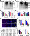
Fig. 1
Fig. 1
Effect of NSC632839 on cell growth. (A, B) NSC632839 accumulated ubiquitinated proteins in Kyse30 and Kyse450 cells. Cells were incubated with different dose of NSC632839 for 24 h (A) or for different time at 2.5 µM (B). Then cell proteins were analyzed with Ub antibody. GAPDH as a control. (C, D) Effect of NSC632839 on cell viability. Cells were incubated with NSC632839 or DMSO at the indicated concentrations for 48 h (C) or with NSC632839 (2.5µM and 3.5µM) for different times (D). Cell viability was measured with the CCK-8 kit. (E, F) Effect of NSC632839 on cell proliferation. Cells were incubated with NSC632839 or vehicle for 24 h and the effect of NSC632839 on Kyse30 and Kyse450 cell proliferation was evaluated by EdU incorporation assay (E). Scale bars = 200 μm. The percentage of EdU+ cells were counted and analyzed (F). (G, H) Effect of NSC632839 on cell colony formation. Cells were incubated with NSC632839 or vehicle. The cell colonies were stained with crystal violet, images were taken (G) and the numbers of the colonies were statistically analyzed (H)
Fig. 2
Fig. 2
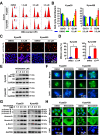
NSC632839 causes cells arrest at M phase. (A, B) Cell cycle was analyzed by flow cytometry. NSC632839 treatment led to a concentration-dependent G2-M phase arrests. DMSO was used as control. Representative histograms for DMSO and NSC632839 were shown (A). Statistical analysis was shown in panel B. (C, D) Immunofluorescence microscopy of p-H3 expression. Cells were incubated with DMSO or NSC632839 and stained with phospho-histone H3 antibodies (red). DAPI (blue) was used to stain the nucleus. The DAPI and p-H3 staining were merged. Representative images were shown in panel C. Scale bar = 50 μm. Statistical analysis of p-H3-positive (mitotic) cells were shown in panel D. (E) The expression of p-H3 (Ser10) and H3 were evaluated by immunoblot assay. (F) NSC632839 disrupts the mitotic spindle organization. Cells were incubated with DMSO or NSC632839 and stained with anti-α-tubulin antibodies (green). DAPI (blue) was used to detect DNA. Cells were analyzed and representative immunofluorescence images are shown. Scale bar = 15 μm. (G) The expression of p-Aurora A/B/C, Aurora A/B, p-PLK1 and PLK1 were detected by immunoblot assay. (H) Nuclear morphologies of cells were stained with α-tubulin (green) antibody and DAPI (blue). Scale bar = 50 μm
Fig. 3
Fig. 3
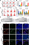
NSC632839 treatment induces intrinsic apoptosis. (A) AnnexinV-FITC/PI double stained kit was used to detect NSC632839-induced apoptosis. Cells were incubated with NSC632839 for 48 h, collected and stained with AnnexinV-FITC and PI for apoptosis detection using FACS. The cells in the upper and lower right quadrant represent Annexin V+ cells, which were defined as apoptotic cells. Representitive images were shown in left panel. Statistical analysis was shown in the right panel. (B) Detecting the effect of NSC632839 on caspase3 activity. Cells were incubated with NSC632839 for 48 h. Then cells were collected and caspase3 activity was evaluated by CaspGLOW Fluorescein Active Caspase3 Staining kit. Representitive images were shown in left panel. The cell numbers with positive caspase3 activity were calculated, statistical analysis and shown in the right panel. (C) Evaluation of mitochondrial membrane depolarization. Mitochondrial membrane depolarization was analyzed by flow cytometry and displayed as in left panel. Cells that had lost mitochondrial membrane potential (MMP) appeared in the right lower quadrant, which were calculated, analyzed and shown in right panel. (D) Detection of apoptosis-related proteins using immunoblot assay
Fig. 4
Fig. 4
CREB-Noxa axis is important for NSC632839-induce apoptosis. (A-C) Noxa is involved in NSC632839-induce apoptosis. siControl and siNoxa were transfected into cells and treated with DMSO or NSC632839, respectively. Apoptosis was detected by AnnexinV-FITC/PI double stain Kit and analyzed by FACS as described in Fig. 3A and B. The silencing efficiency and the effect on apoptosis were evaluated by immunoblot assay using specific antibodies against Noxa and cleaved PARP (C). (D) The expression of Noxa-related transcription factors was evaluated using immunoblot assay. (E-G) Activation of CREB is responsible for NSC632839-induce apoptosis by transcriptionally regulating the expression of Noxa. Cells were incubated with transcriptional activity inhibitor 666 − 15 for CREB and DMSO or NSC632839 (NSC). 48 h later, apoptotic rate was measured by FACS using AnnexinV-FITC/PI double stain Kit and analyzed as described in Fig. 4E and F. Cell proteins were collected and analyzed (G)
Fig. 5
Fig. 5
NSC632839-induced M phase arrest happens prior to apoptosis. (A-E) Cells were incubated with NSC632839 and collected at indicated time for detecting apoptosis (A-B) or cell cycle (C-D) by FACS. Cell proteins were collected to evaluate the expression of p-H3 (Ser 10) and cleaved-PARP (E). (F) Apoptotic cells come from M-phase-arrested cell in NSC632839-treated cells. After treated with NSC632839 or DMSO, the expression of p-H3 and the activation of caspase3 was evaluated by immunofluorescence as described in Material and Methods. Scale bar = 100 μm
Fig. 6
Fig. 6
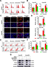
Activation of spindle assembly checkpoint is responsible for M phase arrest and subsequent CREB-Noxa-mediated apoptosis. After incubated with DMSO, Reversine, NSC632839, or Reversine + NSC632839 (NSC), cells cycles were detected by FACS (A) and statistically analyzed (B). The number of mitotic cells were examined by evaluating the p-H3 (Ser 10)-positive cells using immunofluorescence (C, D). Apoptosis was measured by FACS (E, F). The effect of Reversine on NSC632839-induced activation of CREB and the protein level of Noxa and cleaved PARP was evaluated using immunoblot (G)
Fig. 7
Fig. 7
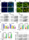
NSC632839 treatment triggers protective autophagy. (A-C) NSC632839 induced autophagy. After treated with DMSO or NSC632839, LC3 puncta was detected using immunofluorescence (A) and autolysosomes were examined by acridine orange (AO) staining (B). The expression of LC3 I/II was evaluated using immunoblot assay (C). (D, E) NSC632839 treatment enhanced autophagic flux. Cells were treated with NSC632839 combined with CQ or BafA1. Then cell proteins were collected and the protein level of LC3 I/II was analyzed. (F-I) NSC632839 treatment triggers protective autophagy. Cells were incubated with NSC632839 and CQ or BafA1. Cell viability was detected by CCK-8 Kit (F), apoptotic rate was detected by FACS using AnnexinV-FITC/PI double staining kit (G-H), the protein level of cleaved-PARP was evaluated by immunoblot assay (I)
Fig. 8
Fig. 8
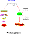
Working model of NSC632839-induced cell growth inhibition in esophageal cancer

References

    1. Thrift AP. Global burden and epidemiology of Barrett oesophagus and oesophageal cancer. Nat Rev Gastro Hepat. 2021;18(6):432–43. - PubMed
    1. Morgan E, Soerjomataram I, Rumgay H, Coleman HG, Thrift AP, Vignat J, et al. The global landscape of esophageal squamous cell carcinoma and esophageal adenocarcinoma incidence and mortality in 2020 and projections to 2040: new estimates from GLOBOCAN 2020. Gastroenterology. 2022;163(3):649–58.e2. - PubMed
    1. Mazidimoradi A, Amiri S, Khani Y, Allahqoli L, Salehiniya H. Burden of esophageal cancer between 2010 and 2019 in Asian countries by geographical region and sociodemographic index: A comparison with global data. Thorac Cancer. 2023. - PMC - PubMed
    1. Sheikh M, Roshandel G, McCormack V, Malekzadeh R. Current status and future prospects for esophageal Cancer. Cancers. 2023; 15(3). - PMC - PubMed
    1. McHugh A, Fernandes K, Chinner N, Ibrahim AFM, Garg AK, Boag G, et al. The identification of potential therapeutic targets for cutaneous squamous cell carcinoma. J Invest Dermatol. 2020;140(6):1154–65. e5. - PMC - PubMed

LinkOut - more resources