Review
. 2025 May 31;21(1):14.
doi: 10.1186/s12993-025-00279-w.

Neurodegeneration models in Parkinson's disease: cellular and molecular paths to neuron death

Affiliations
Review

Neurodegeneration models in Parkinson's disease: cellular and molecular paths to neuron death

Diana D Álvarez-Luquín et al. Behav Brain Funct. .

Abstract

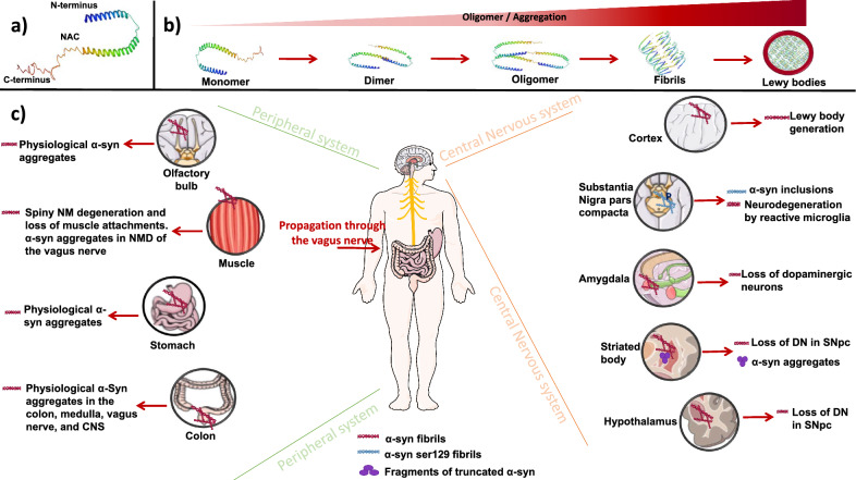
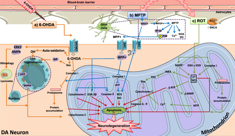
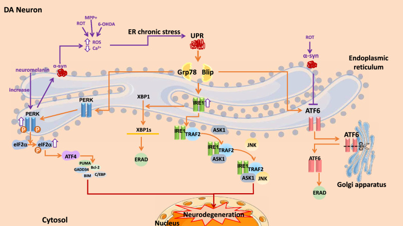
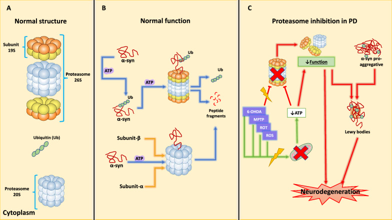
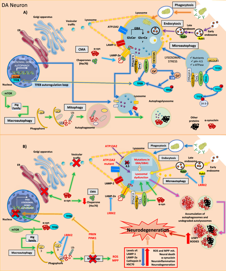
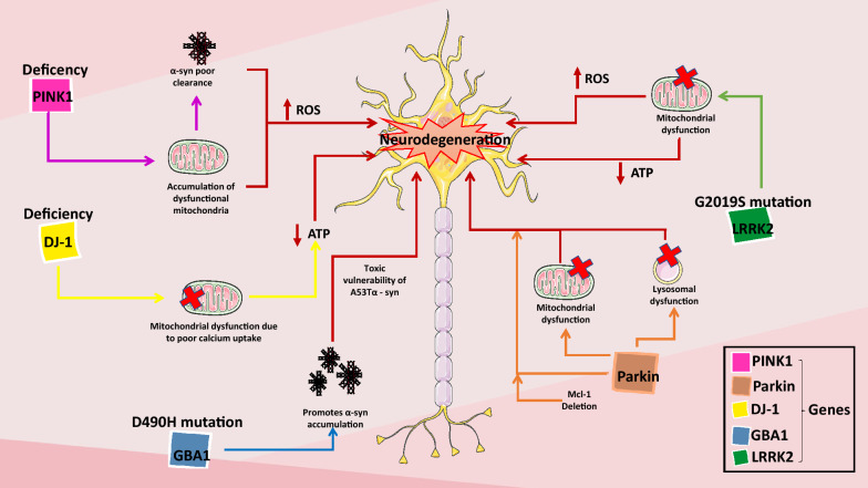
Parkinson's disease (PD) is a progressive neurodegenerative disorder that affects dopaminergic neurons in the substantia nigra pars compacta. It is a complex disease that is strongly influenced by environmental and genetic factors. While the exact causes of PD are not well understood, research on the effects of toxic substances that induce neuronal death has shed some light on the etiology of the disease. In addition, studies have implicated protein aggregation and impaired mitochondrial, endoplasmic reticulum (ER), proteasome, and/or lysosomal function in the pathogenesis of PD. This review focuses on the alterations in intraneuronal organelles and the role of toxic agents that lead to organelle damage and neurodegeneration that characterize PD. We describe in vivo and in vitro models that have been used to elucidate the factors that lead to the death of dopaminergic neurons and summarize the molecular mechanisms that may underlie the changes that promote neurodegeneration. A deeper understanding of the mechanisms of neuronal death may help us to develop new therapies and interventions to delay or prevent the progression of PD.

Keywords: Dopaminergic neuron; Molecular mechanism; Neurodegeneration; Neuronal death; Parkinson disease.


Conflict of interest statement

Declarations. Ethics approval and consent to participate: Not applicable. Consent for publication: Not applicable. Competing interests: The authors declare no competing interests.

Figures

Fig. 1
Fig. 1
α-syn accumulation. A Three-dimensional structure of 140-aa α-syn, composed by three regions: N-terminus, non-amyloid beta component (NAC), and C-terminus. PDB ID: 1QX8. Ulmer, T. S., Bax, A., Cole, N. B., & Nussbaum, R. L. (2005). Structure and dynamics of micelle-bound human α-syn. Journal of Biological Chemistry, 280 (10), 9595–9603. https://doi.org/10.1074/jbc.M411805200. B In PD, the aggregation of α-syn leads to the formation of Lewy bodies. C Regions of the human body where α-syn aggregates can occur. Alpha-synuclein (α-syn), Central Nervus System (CNS); Dopaminergic Neurons (DN); Substantia Nigra pars compacta (SNpc); Neuromelanine (NM); Nerve Motor Dorsal (NMD)
Fig. 2
Fig. 2
Molecular mechanisms of mitochondrial damage involved in Parkinson's disease. A Pathways of 6-OHDA-mediated neurodegeneration. 6-OHDA affects the mitochondrial complex I in neurons and promotes ROS production and DNA fragmentation, leading to neuron death. B Pathways of MPTP-mediated neurodegeneration. MPTP is transformed into MPP+ by glial cells and enters the neuron through DAT. MPTP affects the mitochondrial complex I by promoting ROS production and the activation of apoptosis pathways that lead to neurodegeneration. C Pathways of ROT-mediated neurodegeneration. Damage mechanisms including oxidative and nitrosative stress, mitochondrial damage, and ATP depletion, leading to apoptosis. 6-hydroxydopamine (6-OHDA); 1-methyl-4-phenyl-1,2,3,6-tetrahydropyridine (MPTP); 1-methyl-1,4-phenylpyridinium (MPP +); 1-methyl-4-phenyl-2,3-dihydropyridinium (MPDP+); Rotenone (ROT); dopamine transporter (DAT); norepinephrine transporter (NET); reactive oxygen species (ROS); glutathione (GSH); superoxide dismutase (SOD)
Fig. 3
Fig. 3
Molecular mechanisms of ER damage. The UPR activates three proteins: PERK, IRE1, and the ATF6. ER chronic stress causes an accumulation of unfolded proteins, which participate in neuronal death induction. Unfolded Protein Response (UPR); PRKR-like endoplasmic reticulum kinase (PERK), Inositol-requiring kinase 1 (IRE1); Transcription Activating Factor 6 (ATF6); Endoplasmic Reticulum (ER); 6-hydroxydopamine (6-OHDA); 1-methyl-1,4-phenylpyridinium (MPP +); Alpha-synuclein (α-syn)
Fig. 4
Fig. 4
Proteasome inhibition. A The proteasome has a stacked cylinder morphology. 19S subunits occupy the outer layer, and the 20S subunit, with beta and alpha rings, is located in the center. The assembly of these subunits constitutes the 26S proteasome. The 20S proteasome works independently and does not need ubiquitin for protein process. B The 26S and 20S proteasome are ATP-dependent. The 26S requires target protein polyubiquitination to be recognized by the 19S subunit, internalized, and degraded into smaller peptides. The 20S subunit only internalizes the protein and degrades it. C Mitochondrial damage and oxidative stress in PD cause proteasomal disassembly, impair its protein degradation function, and cause protein and subunit accumulation; this results in the production of Lewy bodies and further neurodegeneration. Alpha-synuclein (α-syn); Ubiquitin (Ub); adenosine triphosphate (ATP); 6-hydroxydopamine (6-OHDA); 1-methyl-1,4-phenylpyridinium (MPP +); Rotenone (ROT); reactive oxygen species (ROS)
Fig. 5
Fig. 5
Lysosome. A Main lysosomal degradation pathways activated during PD. Lysosomes are a common end for endocytosis, phagocytosis, and autophagy. Macroautophagy and CMA are the primary degradation pathways of α-syn during PD. mTOR modulates macroautophagy by regulating the Atg family and TFEB via the mTORC1 complex. mTORC1 inhibits TFEB nuclear translocation. Lysosomal stress causes Rag GTPases to deactivate, releasing and inactivating mTORC1 from the lysosomal surface. Unphosphorylated TFEB translocates to the nucleus and binds the CLEAR sequence of its target genes, inducing the expression of genes involved in lysosome function and autophagy. TFEB can regulate its own expression either by a positive feedback loop or through MCOLN1. B Alterations in the ALP pathways in PD: Mutations in ALPs and α-syn (GBA1, ATP13A2, LRRK2, PRKN, and PINK1) impair their lysosomal degradation and decrease autophagy, favoring the accumulation of autophagosomes and autolysosomes, and thus the formation of Lewy bodies. The accumulation of α-syn blocks ER-Golgi vesicular trafficking, which can impair autophagy. Decreased levels of cathepsin D, LAMP-1, LAMP-2a, and Hsc70 have been reported. Finally, PD causes alterations in the TFEB signaling pathway. TFEB co-locates with α-syn transcription factor in the cytoplasm, preventing its activation. PRKN and PINK1 inhibit TFEB translocation to the nucleus, prevent mitochondrial turnover, and increase oxidative stress. Chaperone-mediated autophagy (CMA); Autophagy-lysosome (ALP); Transcription factor EB (TFEB); Parkinson’s disease (PD); Coordinated lysosomal expression and regulation network (CLEAR); Alpha-synuclein (α-syn); Endoplasmic Reticulum (ER); Autophagy-related genes (Atg); Ubiquitin (Ub); mammalian target of rapamycin (mTOR); Reactive Oxygen Species (ROS); 1-methyl-1,4-phenylpyridinium (MPP +)
Fig. 6
Fig. 6
Genetic models of Parkinson's disease. PINK1: its deficiency decreases the clearance of dysfunctional mitochondria, reduces ATP levels and α-syn clearance, leading to ROS production and neurodegeneration. DJ-1: its deficit causes poor calcium uptake and dysfunction in the mitochondrial membrane, resulting in decreased ATP levels. LRRK2: The G2019S mutation of this gene causes mitochondrial damage, leading to lower ATP and increased ROS levels due to its low clearance. GBA1: The D490H mutation induces α-synuclein to form an aggregating state; its accumulation creates A53T toxic vulnerability, which leads to neurodegeneration. Parkin: Its deficiency causes mitochondrial damage and dysregulation, and the deletion of Mcl-1; acting together, they lead to dopaminergic neuron death. Alpha-synuclein (α-syn); Reactive Oxygen Species (ROS); adenosine triphosphate (ATP); PTEN-induced putative kinase 1 (PINK1); protein deglycase (DJ-1); Glucocerebrosidase 1 (GBA1); E3 ubiquitin ligase Parkin (Parkin); leucine-rich repeated kinase (LRRK2)

Similar articles

References

    1. Kin K, Yasuhara T, Kameda M, Date I. Animal models for Parkinson’s disease research: trends in the 2000s. Int J Mol Sci. 2019;20:5402. 10.3390/ijms20215402. - PMC - PubMed
    1. Le W, Sayana P, Jankovic J. Animal models of Parkinson’s disease: a gateway to therapeutics? Neurotherapeutics. 2014;11:92–110. 10.1007/s13311-013-0234-1. - PMC - PubMed
    1. Shibasaki Y, Baillie DAM, Clair D, Brookes AJ. High-resolution mapping of SNCA encoding α-synuclein, the non-Aβ component of Alzheimer’s disease amyloid precursor, to human chromosome 4q21.3→q22 by fluorescence in situ hybridization. Cytogenet Genome Res. 1995;71:54–5. 10.1159/000134061. - PubMed
    1. Gilmozzi V, Gentile G, Rueda MPC, Hicks AA, Pramstaller PP, Zanon A, et al. Interaction of alpha-synuclein with lipids: mitochondrial cardiolipin as a critical player in the pathogenesis of Parkinson’s disease. Front Neurosci. 2020. 10.3389/fnins.2020.578993. - PMC - PubMed
    1. Knecht L, Folke J, Dodel R, Ross JA, Albus A. Alpha-synuclein immunization strategies for synucleinopathies in clinical studies: a biological perspective. Neurotherapeutics. 2022;19:1489–502. 10.1007/s13311-022-01288-7. - PMC - PubMed

LinkOut - more resources