. 2025 Jul;52(7):e17904.
doi: 10.1002/mp.17904. Epub 2025 May 31.

Characterization of a practically designed plastic scintillation plate dosimeter

Affiliations

Characterization of a practically designed plastic scintillation plate dosimeter

Takeshi Ohta et al. Med Phys. 2025 Jul.

Abstract

Background: Advancements in radiotherapy have enabled the use of high-definition irradiation, leading to more precise and finely adjusted treatments in clinical settings. Nevertheless, the attainment of high resolution, an extensive measurement area, and the repeatability of dose distribution measurements persist as challenges in clinical practice, thereby often requiring multiple dosimetry systems to overcome measurement constraints. Consequently, there is a significant need to develop a dosimeter that offers both a high resolution and a capability for repeated use.

Purpose: A practical scintillation plate dosimeter was designed and its dosimetric characteristics were evaluated using x-ray beams from a linear accelerator.

Methods: A practical scintillation plate dosimeter comprised a 0.2 cm-thick scintillation plate sandwiched between a pair of 2.0 cm-thick Polymethyl methacrylate (PMMA) plates. A Complementary Metal Oxide Semiconductor (CMOS) camera was used to detect the scintillation light emitted from the scintillation plate when the x-ray beams were delivered to the plate. Measurements were made at 6 MV to test the dose linearity, reproducibility, and dependencies on the camera temperature and angles of incidence. The dose-rate dependency was also measured using 6 and 10 MV flattening filter-free (FFF) beams. The x-ray energy dependency was further tested using 4 MV, 6 MV, 10 MV, 6 MV FFF, and 10 MV FFF beams.

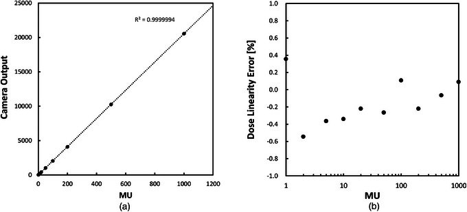
Results: A maximum linearity error of 0.4% was observed for doses ranging from 10 to 1000 MU. The coefficient of variation for the dose reproducibility was ± 0.062%, the temperature dependency was 0.07%/°C, and the angular variations were within ± 1.3% after the removal of Cherenkov light. The dose output decreased by 5.0% at 45 MU/min, compared with that at 1300 MU/min with the 6 MV FFF beams, and by 2.0% at 160 MU/min, compared to 1900 MU/min with the 10 MV FFF beams. The dependency of x-ray energy ranged from -2.1% to +1.4%.

Conclusions: The practical scintillation plate dosimeter showed favorable dose characteristics that can be applied in patient-specific quality assurance for volumetric modulated arc therapy.

Keywords: CMOS camera; characteristics; plastic scintillator; scintillator plate; x‐ray beams.


Conflict of interest statement

The authors declare no conflicts of interest.

Figures

FIGURE 1
FIGURE 1
(a) Structure of the plastic scintillation plate dosimeter for the measurement and an example of light distribution obtained by camera on an axial plane. (b) That is for the measurement on a coronal plane. The measurement plane can be changed by swapping the scintillation and camera units.
FIGURE 2
FIGURE 2
(a) An example dose distribution of an x‐ray beam measured on the axial plane. (b) An example dose distribution of an x‐ray beam planned on the axial plane. (c) X‐axis dose profile across the center of the scintillation plate. (d) Z‐axis dose profile across the center of the scintillation plate.
FIGURE 3
FIGURE 3
(a) An example dose distribution of an x‐ray beam measured on the coronal plane. (b) An example dose distribution of an x‐ray beam planned on the coronal plane. (c) X‐axis dose profile across the center. (d) Y‐axis dose profile across the center.
FIGURE 4
FIGURE 4
(a) Plot of the camera output signal as a function of the delivered dose for a field size of 10 × 10 cm2 with an x‐ray energy of 6 MV. (b) Plot of the resulting dose linearity error as a function of the delivered dose. The scintillation plate was set on the coronal plane at the isocenter, and the pixel intensities on the 10 × 10 pixels at the image center were averaged for this test.
FIGURE 5
FIGURE 5
Plot of the reproducibility of camera outputs by delivering 100 MU 10 times for a field size of 10 × 10 cm2 with an x‐ray energy of 6 MV. The scintillation plate was set on the coronal plane at the isocenter, and the pixel intensities of the 10 × 10 pixels at the image center were averaged. A Semiflex chamber was also placed for comparison.
FIGURE 6
FIGURE 6
Plot of the normalized camera outputs as a function of the actual dose rates ranging from 45 to 1900 MU/min. The normalization was made against the black‐bordered points. A dose of 100 MU was delivered with 6 and 10 MV flattening‐filter‐free (FFF) x‐ray beams with a field size of 10 × 10 cm2. The scintillation plate was set on the coronal plane at the isocenter, and the pixel intensities on the 10 × 10 pixels at the image center were averaged for this test.
FIGURE 7
FIGURE 7
Plot of the normalized camera output as a function of the camera temperature. The normalization was made at 24.1°C, which is one of the measurement points closest to room temperature. This is illustrated by the red circle. The camera temperature was monitored by a temperature sensor integrated inside the camera. During this test, a dose of 100 MU was repeatedly delivered to the scintillation plate with an x‐ray energy of 6 MV. The scintillation plate was set on the coronal plane at the isocenter, and the pixel intensities of the 10 × 10 pixels at the image center were averaged.
FIGURE 8
FIGURE 8
Plot of the camera output deviations as a function of the angle of incident x‐ray beams before and after Cherenkov background subtraction. The measured value was normalized by dividing it by the planned dose on the scintillation plate at each angle to compensate for different absorption values in the 2 cm‐thick PMMA plate. Then, the deviations were plotted so that the mean deviation of each plot was zero. A dose of 100 MU was delivered with a photon energy of 6 MV and a field size of 10 × 10 cm2. The scintillation plate was set on the coronal plane at the isocenter, and the pixel intensities on the 10 × 10 pixels at the image center were averaged for this test.
FIGURE 9
FIGURE 9
The relationship between TPR20,10 and normalized camera outputs. TPR20,10 is measured value in the water phantom.
FIGURE 10
FIGURE 10
(a) A dose distribution measured for a clinical stereotactic lung VMAT case in the axial plane. (b) A dose distribution planned for a clinical stereotactic lung VMAT case in the axial plane. (c) X‐axis dose profile comparison between measurement and calculation. (d) Z‐axis dose profile comparison between measurement and calculation.
FIGURE 11
FIGURE 11
(a) A dose distribution measured for a clinical stereotactic lung VMAT case in the coronal plane. (b) A dose distribution planned for a clinical stereotactic lung VMAT case in the coronal plane. (c) X‐axis dose profile comparison between measurement and calculation. (d) Y‐axis dose profile comparison between measurement and calculation.

References

    1. Beddar AS, Mackie TR, Attix FH. Water‐equivalent plastic scintillation detectors for high‐energy beam dosimetry: I. Physical characteristics and theoretical consideration. Phys Med Biol. 1992;37(10):1883‐1900. doi: 10.1088/0031-9155/37/10/006 - DOI - PubMed
    1. Beddar AS, Mackie TR, Attix FH. Water‐equivalent plastic scintillation detectors for high‐energy beam dosimetry: II. Properties and measurements. Phys Med Biol. 1992;37(10):1901‐1913. doi: 10.1088/0031-9155/37/10/007 - DOI - PubMed
    1. Beddar AS. Plastic scintillation dosimetry and its application to radiotherapy. Radiat Meas. 2006;41:S124‐S133. doi: 10.1016/j.radmeas.2007.01.002 - DOI
    1. Beaulieu L, Beddar S. Review of plastic and liquid scintillation dosimetry for photon, electron, and proton therapy. Phys Med Biol. 2016;61(20):R305‐R343. doi: 10.1088/0031-9155/61/20/R305 - DOI - PubMed
    1. Petric MP, Robar JL, Clark BG. Development and characterization of a tissue equivalent plastic scintillator based dosimetry system. Med Phys. 2006;33:96‐105. doi: 10.1118/1.2140118 - DOI - PubMed

MeSH terms

LinkOut - more resources