. 2025 Jul;301(7):110322.
doi: 10.1016/j.jbc.2025.110322. Epub 2025 May 30.

Myristoylation of TMEM106B by NMT1/2 regulates TMEM106B trafficking and turnover

Affiliations

Myristoylation of TMEM106B by NMT1/2 regulates TMEM106B trafficking and turnover

Alexander Lacrampe et al. J Biol Chem. 2025 Jul.

Abstract

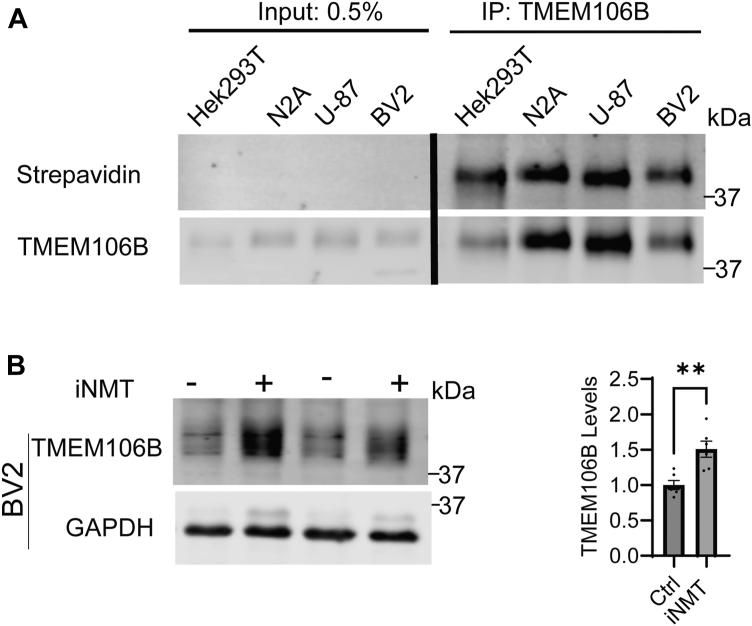
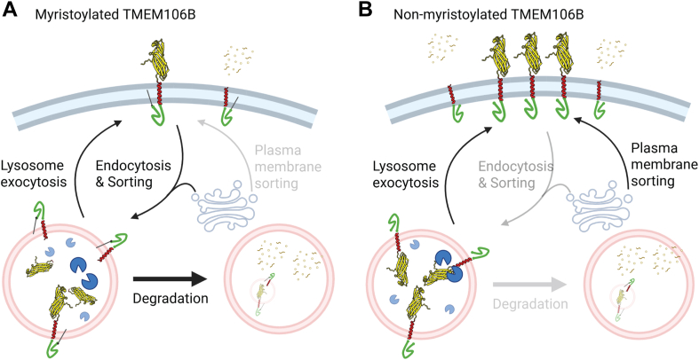
TMEM106B, a type II transmembrane protein localized on the lysosomal membrane, has been identified as a central player in neurodegeneration and brain aging during the past decade. TMEM106B variants that increase TMEM106B expression levels are linked to several neurodegenerative diseases, including frontotemporal lobar degeneration (FTLD). Additionally, the C-terminal lumenal fragment of TMEM106B was recently shown to form amyloid fibrils during aging and neurodegeneration. However, the mechanisms regulating TMEM106B levels are not well understood. Here we show that TMEM106B is myristoylated by NMT1/2 enzymes at its glycine 2 α-amino group and its lysine 3 ε-amino group. Myristoylation decreases TMEM106B levels by promoting its lysosomal degradation. Furthermore, we demonstrate that TMEM106B C-terminal fragments (CTFs) can be detected under physiological conditions, and the levels of CTFs are regulated by myristoylation and lysosomal activities. In addition, we show that non-myristoylated TMEM106B accumulates on the cell surface, indicating that myristoylation affects TMEM106B trafficking within the cell. Taken together, these findings suggest that TMEM106B myristoylation is an important mechanism regulating its function, trafficking, and turnover.

Keywords: NMT1/2; TMEM106B; lysosome; myristoylation; neurodegeneration; protein processing; protein trafficking.


Conflict of interest statement

Conflict of Interest The authors declare the following financial interests/personal relationships which may be considered as potential competing interests: H. L. is a founder and consultant for Sedec Therapeutics. AY, BG, IP, MAYC, and NFH are current or former employees of Alector, LLC and may have an equity interest in Alector, Inc. Several authors have patents related to TMEM106B-specific antibodies.

Figures

Figure 1
Figure 1
TMEM106B is myristoylated by NMT enzymes. A, HEK293T cells were transfected with WT TMEM106B and either empty vector, HA-NMT1 or HA-NMT2 as indicated. 48 h after transfection, lysates were immunoprecipitated (IP) using anti-HA antibodies, and the IP samples were analyzed by Western blot. B, HEK293T cells were incubated with myristic acid analog with or without NMT inhibitor. TMEM106B was then immunoprecipitated and biotin click chemistry was performed. Proteins were separated using SDS-PAGE and blotted with anti-TMEM106B antibodies or fluorescently labeled streptavidin. The intensity of biotinylated TMEM106B band was quantified and normalized to total TMEM106B levels probed by anti-TMEM106B antibodies (n = 3, one sample t test). C, HEK293T cells were transfected with WT or TMEM106B mutants as indicated and myristoylation assays were performed as in (B) (n = 3, one-way ANOVA with post hoc tests). D, HEK239T cells were transfected HA-NMT1 And HA-NMT2 along with empty vector, WT, or G2AK3R TMEM106B as indicated. 48 h after transfection the cells were lysed and immunoprecipitated using anti-TMEM106B antibodies. Samples were analyzed via western blotting. Data represent mean ± SEM; ns, not-significant; ∗, p < 0.05; ∗∗, p < 0.01; ∗∗∗, p < 0.001; ∗∗∗∗, p < 0.0001.
Figure 2
Figure 2
NMT1 myristoylates TMEM106B peptide in vitro. A–B, purified NMT1 enzyme was incubated with WT TMEM106BN-terminal peptide and myristoyl-CoA. The reaction products were detected using LC-MS. The ion chromatograms for the substrate and single (A) or double (B) myristoyl product ions are shown. C and D, purified NMT1 enzyme was incubated TMEM106B G2A (C) or K3R (D) peptide and myristoyl-CoA. The reaction products were detected using LC-MS. The ion chromatograms for the substrate and single lysine myristoylated product ions are shown.
Figure 3
Figure 3
Ablating TMEM106B myristoylation protects it from lysosomal degradation.A, HEK293T cells were transfected with WT, G2A, K3R, or G2AK3R mutant TMEM106B for 48 h. The levels of TMEM106B in the lysates were quantified and normalized to GAPDH (n = 6, one-sample t test). B, HEK293T cells transfected with WT or G2AK3R TMEM106B for 48 h were either untreated or treated with Bafilomycin A1 (BafA1) for 16 h and then lysed and analyzed with Western blot. The levels of TMEM106B in the lysates were quantified and normalized to GAPDH (n = 12, two-way ANOVA with post hoc tests). C, HEK293T cells were either treated untreated or treated with iNMT for 24 h before being lysed and analyzed via Western blot. The levels of TMEM106B in the lysates were quantified and normalized to GAPDH (n = 5, one-sample t test). D, HEK293T cells were treated with or without NMT inhibitor (iNMT) overnight and then treated with cycloheximide for 10 h before being lysed and analyzed via western blot. The relative levels of TMEM106B to GAPDH were normalized to its levels without cycloheximide treatment in each condition (n = 5, unpaired t test). E, HEK293T cells transfected with WT or G2AK3R TMEM106B. 24 h post-transfection cells were treated with either iNMT or DMSO control treatment for another 24 h. The levels of TMEM106B in the lysates were quantified and normalized to GAPDH (n = 6, unpaired t test) Data represent mean ± SEM; ns, not-significant; ∗, p < 0.05; ∗∗, p < 0.01; ∗∗∗, p < 0.001; ∗∗∗∗, p < 0.0001.
Figure 4
Figure 4
TMEM106B myristoylation regulates its levels in BV2 cells.A, HEK293T, N2A, U-87, and BV2 cells were incubated with myristic acid analog. TMEM106B was then immunoprecipitated and biotin click chemistry was performed. Proteins were separated using SDS-PAGE and blotted with anti-TMEM106B antibodies or fluorescently labeled streptavidin. B, BV2 cells were either treated untreated or treated with iNMT for 24 h before being lysed and analyzed via Western blot. As previously reported, when kept on ice TMEM106B runs as a monomer in microglia cells (48). The levels of TMEM106B in the lysates were quantified and normalized to GAPDH (n = 6, unpaired t test. Data represent mean ± SEM; ns, not-significant; ∗, p < 0.05; ∗∗, p < 0.01; ∗∗∗, p < 0.001; ∗∗∗∗, p < 0.0001.
Figure 5
Figure 5
Myristoylation is dispensable for TMEM106B lysosome trafficking and its ability to induce lysosomal enlargement. A, Neuro-2a cells transfected with empty vector, wild-type TMEM106B, or G2AK3R TMEM106B were fixed and stained with anti-TMEM106B and anti-LAMP1 antibodies. Samples were then imaged using confocal microscopy. Scale bar = 10 μm. B, quantification of lysosome enlargement and TMEM106B/LAMP1 co-localization for the experiment in (A). For Lysosome diameter, Image J was used to measure the diameter of the five largest lysosomes in each transfected cell. (n = 4, at least 20 cells were quantified per condition per experiment, one-way ANOVA with post hoc tests). Image J was used to measure the Pearsons coefficient between the TMEM106B signal and LAMP1 signal (n = 3, at least 20 cells were quantified per condition per experiment, unpaired t test =) Data represent mean ± SEM; ns, not-significant; ∗, p < 0.05; ∗∗, p < 0.01; ∗∗∗, p < 0.001; ∗∗∗∗, p < 0.0001.
Figure 6
Figure 6
TMEM106B myristoylation and lysosome pH affect CTF levels.A, HEK293T cells were transfected with either WT or G2AK3R mutant TMEM106B. The levels of full-length TMEM06B (FL), N-terminal fragment (NTF), or cytosolic domain (ICD) were analyzed by Western blot using rabbit anti-TMEM106B ICD antibodies 48 h after transfection. TMEM106B levels were quantified by normalizing FL TMEM106B to GAPDH, NTF and ICD levels were normalized to the levels of FL TMEM106B (n = 6, unpaired t test). B, HEK293T cells were transfected with WT or G2AK3R TMEM106B and were either untreated or treated with bafilomycin A1 (BafA1) for 16 h. The levels of full-length TMEM06B (FL) and C-terminal fragments (CTF) were analyzed by Western blot using human anti-TMEM106B CTF antibodies and quantified (n = 6, two-way ANOVA with post hoc tests). C, HEK293T cells were treated with BafA1 for 16 h. The levels of full-length TMEM06B (FL) and C-terminal fragment (CTF) were analyzed by Western blot using anti-TMEM106B CTF antibodies and quantified (n = 5, one-sample t test). D, HEK293T cells were treated with NMT inhibitor (iNMT) for 24 h or untreated. The levels of full-length TMEM06B (FL) and C-terminal fragment (CTF) were analyzed by Western blot using anti-TMEM106B CTF (Clone Ab78) antibodies and quantified (n = 5, unpaired t test). E, HEK293T cells were transfected with either WT or G2AK3R TMEM106B. 24 h post-transfection cells were treated with either iNMT or DMSO control treatment for another 24 h. The cells were then lysed and analyzed via Western blot using CTF antibodies (n = 6, two-way ANOVA with post hoc tests). The levels of FL and CTF were quantified and tha ratio between CTF to FL is calculatedData represent mean ± SEM; ns, not-significant; ∗, p < 0.05; ∗∗, p < 0.01; ∗∗∗, p < 0.001; ∗∗∗∗, p < 0.0001.
Figure 7
Figure 7
TMEM106B myristoylationregulates its trafficking to the cell surface.A, HEK293T cells were transfected with either WT or G2AK3R mutant TMEM106B. 24 h post-transfection cells were either treated with NMT inhibitor (iNMT) or DMSO control for an additional 24 h before immunostaining. Live staining was performed human anti-TMEM106B CTF antibodies. Cells were then fixed and permeabilzed and immunostained using rabbit anti-TMEM106B ICD antibodies. The ratio of the intensity of cell-surface TMEM106B (T106B CTF) and internal TMEM106B (T106B ICD) was measured per cell using Image J. (n = 3, at least 50 cells were quantified per condition per experiment. Matched two-way ANOVA with post hoc tests. Scale bar = 10 μm). B, Neuro2A cells were transfected with either WT or G2AK3R mutant TMEM106B and live staining and quantification was performed as in (A) (n = 3, at least 30 cells were quantified per condition per experiment. Ratio paired t test. Scale bar = 10 μm).
Figure 8
Figure 8
Model of the role of TMEM106B myristoylation in modulating TMEM106B trafficking and levels. A, when TMEM106B is myristoylated, its levels on the plasma membrane are reduced due to proper sorting to the lysosome at the Golgi or efficient endocytosis from the plasma membrane. Localization on the lysosome membrane renders TMEM106B more likely to be degraded by the lysosome. B, when TMEM106B is not myristoylated, it accumulates on the plasma membrane due to miss-sorting to the plasma membrane, increased lysosome exocytosis or defects in its endocytosis. This might lead to decreased CTF production or secretion of CTF into the extracellular space, and less degradation of full-length TMEM106B and NTF by the lysosome. Created with Biorender.com.

References

    1. Van Deerlin V.M., Sleiman P.M., Martinez-Lage M., Chen-Plotkin A., Wang L.S., Graff-Radford N.R., et al. Common variants at 7p21 are associated with frontotemporal lobar degeneration with TDP-43 inclusions. Nat. Genet. 2010;42:234–239. - PMC - PubMed
    1. Ratnavalli E., Brayne C., Dawson K., Hodges J.R. The prevalence of frontotemporal dementia. Neurology. 2002;58:1615–1621. - PubMed
    1. Neary D., Snowden J., Mann D. Frontotemporal dementia. Lancet Neurol. 2005;4:771–780. - PubMed
    1. Cruchaga C., Graff C., Chiang H.H., Wang J., Hinrichs A.L., Spiegel N., et al. Association of TMEM106B gene polymorphism with age at onset in granulin mutation carriers and plasma granulin protein levels. Arch. Neurol. 2011;68:581–586. - PMC - PubMed
    1. Finch N., Carrasquillo M.M., Baker M., Rutherford N.J., Coppola G., Dejesus-Hernandez M., et al. TMEM106B regulates progranulin levels and the penetrance of FTLD in GRN mutation carriers. Neurology. 2011;76:467–474. - PMC - PubMed

LinkOut - more resources