. 2025 Jun;57(6):1177-1188.
doi: 10.1038/s12276-025-01460-x. Epub 2025 Jun 2.

Inhibition of serotonin-Htr2b signaling in skeletal muscle mitigates obesity-induced insulin resistance

Affiliations

Inhibition of serotonin-Htr2b signaling in skeletal muscle mitigates obesity-induced insulin resistance

Suhyeon Park et al. Exp Mol Med. 2025 Jun.

Erratum in

Abstract

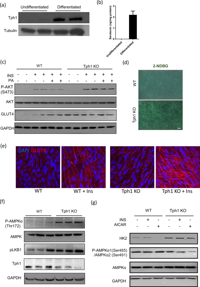
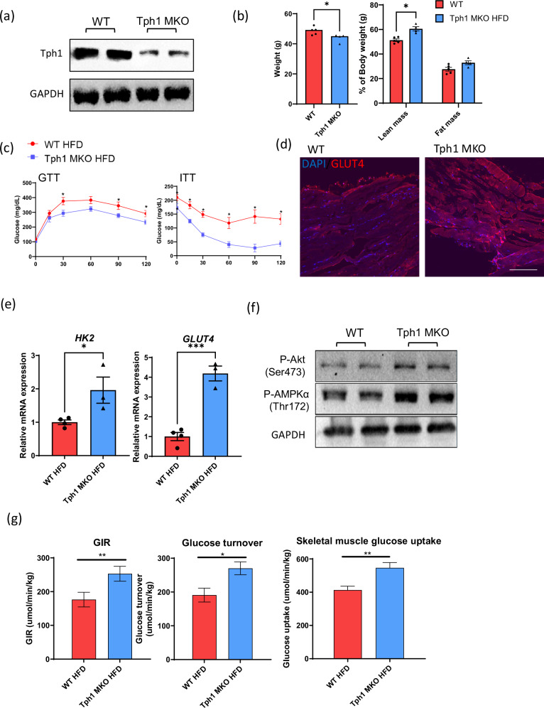
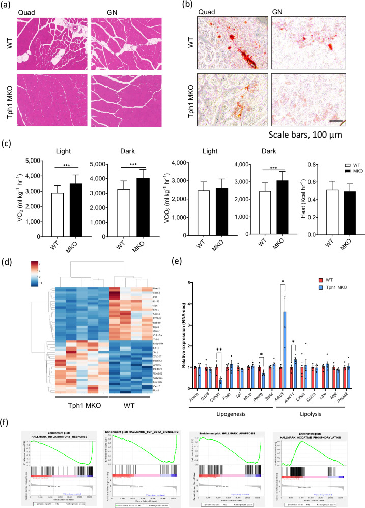
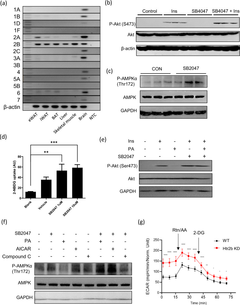
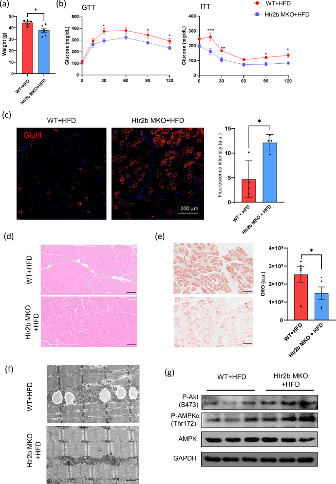
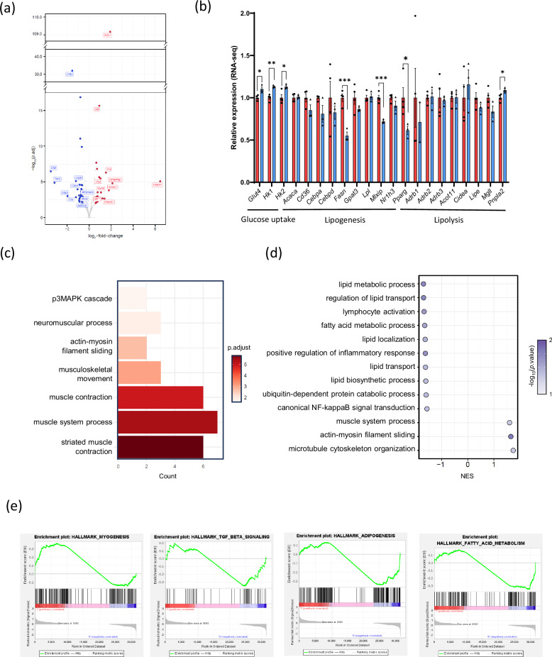
Obesity-induced insulin resistance is a major cause of metabolic disorders, including type 2 diabetes mellitus. Although peripheral serotonin (5-hydroxytryptamine, 5-HT) has been implicated in energy balance and metabolism, its effect on skeletal muscle insulin sensitivity remains unclear. Here we identified the 5-HT receptor 2b (Htr2b) as a critical regulator of insulin sensitivity and energy metabolism in the skeletal muscle. Using genetic and pharmacological approaches, we showed that muscle-specific Tph1-knockout (Tph1 MKO) mice fed a high-fat diet exhibited reduced body weight, increased lean mass and improved glucose tolerance compared with wild-type mice. The pharmacological inhibition of Htr2b in myotubes reversed palmitate-induced insulin resistance and increased glycolytic activity. Moreover, muscle-specific HTR2b-knockout (HTR2b MKO) mice exhibited improved glucose uptake, insulin sensitivity and overall metabolic health under high-fat-diet-induced obesity. Mechanistically, both Tph1 MKO and Htr2b MKO mice showed increased phosphorylation of AKT and AMPK, indicating improved insulin sensitivity and energy metabolism in the skeletal muscle. These findings demonstrate that 5-HT-Htr2b signaling negatively regulates insulin sensitivity and energy metabolism in skeletal muscles, providing new insights into the role of peripheral serotonin in muscle metabolism and potential therapeutic targets for metabolic disorders.


Conflict of interest statement

Competing interests: The authors declare no competing interests.

Figures

Fig. 1
Fig. 1. Effects of inhibiting serotonin on C2C12 myotubes.
a The western blot images of Tph1 expression in undifferentiated and differentiated C2C12 cells, with tubulin as the loading control. b The serotonin levels in undifferentiated and differentiated C2C12 cells. c Western blot images showing the levels of AKT, phosphorylated AKTS473, GLUT4 and GAPDH in WT and Tph1-KO C2C12 cells with or without insulin stimulation for 15 min after 24 h of 0.5 mM palmitate treatment. d The representative images of 2-NBDG glucose uptake in WT and Tph1-KO cells. Scale bar, 100 µm. e Representative immunofluorescence images of GLUT4 expression in C2C12 myotubes treated with or without 100 nM insulin for 30 min. f The western blot images of phosphorylated AMPKαThr172, AMPK, phosphorylated LKB1 and Tph1 in WT and Tph1-KO cells. g The western blot images of hexokinase (HK), phosphorylated AMPKα1Ser485/α2Ser491 and AMPKα in WT and Tph1-KO cells after treatment with either 100 nM insulin or 2 mM AICAR.
Fig. 2
Fig. 2. Tph1 KO in skeletal muscle protects against HFD-induced obesity and insulin resistance.
a The Tph1 expression in quadriceps muscle of WT and Tph1 muscle-specific-KO (Tph1 MKO) mice. b Body weight and body composition analysis (n = 4 or 5 per group) measured via dual energy X-ray absorptiometry after 12 weeks of HFD feeding. c The results of the GTT and ITT after 16 and 6 h of fasting, respectively (n = 4–6 per group). d Immunofluorescence staining of GLUT4 (red) in quadriceps muscle from WT and Tph1 MKO skeletal muscle. The nuclei were counterstained with DAPI (blue). Scale bar, 100 µm. e The relative mRNA expression of Hk2 and GLUT4 in quadriceps muscle from mice fed a HFD, assessed via qRT–PCR. f Western blot images showing phosphorylated AKT and phosphorylated AMPKαThr172 from WT and Tph1 MKO skeletal muscle after HFD feeding. g The measurement of insulin levels, glucose turnover and skeletal muscle glucose uptake in WT and Tph1 MKO mice during clamp studies. GIR, glucose infusion rate. *P < 0.05, **P < 0.01.
Fig. 3
Fig. 3. Effect of Tph1 muscle KO on energy metabolism.
a Hematoxylin and eosin (H&E) staining of quadriceps (Quad) and gastrocnemius (GN) muscles from WT and Tph1 MKO mice after 12 weeks of HFD feeding. b Oil Red O staining of skeletal muscle sections. Scale bar, 100 µm. c The metabolic rates of WT and Tph1 MKO mice under light and dark cycles after 12 weeks of HFD feeding. d A heat map showing the DEGs identified via bulk RNA-seq analysis of skeletal muscle from WT and Tph1-KO mice. e The relative expression of genes involved in lipogenesis and lipolysis from skeletal muscle transcriptome data of WT and Tph1 MKO mice. f The enrichment plots for the inflammatory response pathway, TGF-beta signaling pathway, apoptosis pathway and oxidative phosphorylation pathway. *P < 0.05, **P < 0.01, *** P < 0.001.
Fig. 4
Fig. 4. Effect of inhibiting Htr2b signaling on C2C12 myotubes.
a mRNA detection of 5-HT receptors (Htrs) in brain and peripheral tissues using RT–PCR. eWAT, epididymal white adipose tissue; iWAT, inguinal white adipose tissue; BAT, brown adipose tissue; NTC, no template control. b Western blot images showing the effect of SB204741 (an Htr2B inhibitor, SB2047, 50 nM) on phosphorylated AKTSer473 and total AKT levels in C2C12 myotubes with or without insulin treatment (100 nm for 30 min). c Western blot images showing the effect of SB2047 on phosphorylated AMPKαThr172 and total AMPK levels in C2C12 myotubes. d A quantification of 2-NBDG glucose uptake in C2C12 myotubes treated with SB2047. e Western blot images showing the effect of SB2047 on phosphorylated AKTSer473 and total AKT levels in C2C12 myotubes with palmitate (PA) (0.5 mM for 48 h). f Western blot images showing phosphorylated AMPKαThr172 in C2C12 myotubes with PA. CC, Compound C. g The ECAR was measured in WT and Htr2b knockdown C2C12 myotubes after sequential injections of compounds rotenone and antimycin cocktail (Rot/AA) and 2-deoxy-d-glucose (2-DG).
Fig. 5
Fig. 5. Htr2b KO in skeletal muscle protects against HFD-induced obesity and insulin resistance.
a The body weight of WT and HTR2b muscle-specific-KO (Htr2b MKO) mice after 12 weeks of HFD feeding. b GTT and ITT in WT and Htr2b MKO mice. c Immunofluorescence staining of Glut4 (red) in quadriceps muscle from WT and HTR2b MKO mice. The nuclei were counterstained with DAPI (blue). Scale bar, 100 µm. d Hematoxylin and eosin staining of quadriceps muscle from WT and HTR2b MKO mice after 12 weeks of HFD feeding. e Oil Red O staining of skeletal muscle sections. Scale bar, 100 µm. f Representative electron micrograph of skeletal muscle (quadriceps) from WT and Htr2b MKO mice. Scale bar, 10 µm. g Western blot images showing phosphorylated AKTSer473, phosphorylated AMPKαThr172 and total AMPK levels in skeletal muscle from WT and Htr2b MKO mice.
Fig. 6
Fig. 6. Transcriptome analysis of skeletal muscle of WT and Htr2b MKO mice.
a A heat map showing DEGs identified via bulk RNA-seq analysis of skeletal muscle from WT and HTR2b-KO mice. b The relative expression of genes involved in glucose uptake, lipogenesis and lipolysis from skeletal muscle transcriptome data of WT and Htr2b MKO mice. c d A pathway enrichment analysis of DEGs, GO (c) and GSEA (d). e Enrichment plots for the myogenesis pathway, TGF-beta signaling pathway, adipogenesis pathway and fatty acid metabolism pathway. *P < 0.05, **P < 0.01, ***P < 0.001.

References

    1. Kim, D.-S. & Scherer, P. E. Obesity, diabetes, and increased cancer progression. Diabetes Metab. J.45, 799–812 (2021). - PMC - PubMed
    1. Hepatology, T. L. G. The Lancet Gastroenterology. Obesity: another ongoing pandemic. Lancet Gastroenterol Hepatol.6, 411 (2021). - PMC - PubMed
    1. Boutari, C. & Mantzoros, C. S. A 2022 update on the epidemiology of obesity and a call to action: as its twin COVID-19 pandemic appears to be receding, the obesity and dysmetabolism pandemic continues to rage on. Metabolism133, 155217 (2022). - PMC - PubMed
    1. Jeong, S.-M. et al. 2023 Obesity fact sheet: prevalence of obesity and abdominal obesity in adults, adolescents, and children in Korea from 2012 to 2021. J. Obes. Metab. Syndr.33, 27–35 (2024). - PMC - PubMed
    1. Merz, K. E. & Thurmond, D. C. Role of skeletal muscle in insulin resistance and glucose uptake. Compr. Physiol.10, 785–809 (2011). - PMC - PubMed

LinkOut - more resources