. 2025 May 1;18(5):dmm052120.
doi: 10.1242/dmm.052120. Epub 2025 Jun 2.

Evaluating the feasibility of gene replacement strategies to treat MTRFR deficiency

Affiliations

Evaluating the feasibility of gene replacement strategies to treat MTRFR deficiency

Samia L Pratt et al. Dis Model Mech. .

Abstract

Mitochondrial translation release factor in rescue (MTRFR) catalyzes a termination step in protein synthesis, facilitating release of the nascent chain from mitoribosomes. Pathogenic variants in MTRFR cause MTRFR deficiency and are loss-of-function variants. Here, we tested gene replacement as a possible therapeutic strategy. A truncating mutation (K155*) was generated in mice; however, homozygotes die embryonically whereas mice heterozygous for this K155* allele are normal. We also generated transgenic strains expressing either wild-type human MTRFR or a partially functional MTRFR. Despite dose-dependent phenotypes from overexpression in vitro, neither transgene caused adverse effects in vivo. In K155* homozygous mice, the wild-type MTRFR transgene completely rescued the phenotype with only one copy present, whereas the mutant transgene rescued less efficiently. Detailed evaluation of mice rescued with the wild-type MTRFR transgene revealed no abnormalities. In human induced pluripotent stem cell (hiPSC)-derived knockdown neurons, mitochondrial phenotypes were corrected by AAV9-mediated delivery of MTRFR. Thus, we find no toxicity from truncated gene products or overexpression of MTRFR in vivo, and expression of MTRFR corrects phenotypes in both mouse and hiPSC models.

Keywords: C12ORF65; Behr's syndrome; CMT6; Leigh syndrome; MRPL58; Mitochondrial translation.


Conflict of interest statement

Competing interests The authors declare no competing or financial interests.

Figures
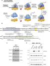
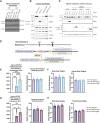
Fig. 1.
Fig. 1.
Structure and function of MTRFR. (A) The mechanism of MTRFR rescuing stalled mitochondrial translation is schematized. ‘GGQ catalytic domain’ is referring to the glycine-glycine-glutamine catalytic domain of MTRFR, and ‘PTC’ is referring to the peptidyl-transferase center of the mitoribosome. Created in BioRender by Pratt, S. L. (2025). https://BioRender.com/nzmjvti. This figure was sublicensed under CC-BY 4.0 terms. (B) Protein sequence alignment of human and mouse MTRFR. ‘|’ denotes an exact match between the sequences, ‘:’ denotes most highly conserved substitution, and ‘.’ denotes semiconserved substitution. The mitochondrial targeting sequence (MTS) is based on a prediction of the human protein using UniProt. Human MTRFR codes for a protein 166 amino acids long, and green highlight indicates homozygous or compound heterozygous pathogenic variants observed in patients. Yellow highlight indicates the highly conserved glycine-glycine-glutamine (GGQ) domain. Mouse Mtrfr encodes a protein 184 amino acids long; different colors depict variants used in our models. Red indicates premature truncation at amino acids 155 and 156 (K155,156*); blue indicates three lysines mutated to alanines at amino acids 146, 153 and 156; pink denotes mutations previously attempted to induce a premature truncation mouse model (amino acids 87 and 119). (C) Immunoblot of whole-cell lysates from mouse embryonic fibroblasts (MEFs) with the indicated Mtrfr genotypes. The steady-state abundance of MT-CO1 is a robust indicator for the defect in mitochondrial protein synthesis and is reduced in homozygous cells. The other three proteins in this blot [uL11m (also known as MRPL11), mS22 (also known as MRPS22) and TOM40 (also known as TOMM40)] are all nuclear-encoded genes that function within the mitochondria, showing that differences in Mtrfr do not affect cytosolic protein synthesis. (D) Immunoblot of whole-cell lysates from Mtrfr-knockout (KO) MEFs following retroviral transduction with an empty vector, and wild-type and mutated human MTRFR cDNAs with the indicated genotypes. The truncation at R141 results in a protein that is inactive, truncation at K151 results in a protein that retains a low level of activity, and substitution of lysines K146, K153 and K156 for alanines results in a protein that retains moderate activity.
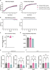
Fig. 2.
Fig. 2.
Overexpression of MTRFR and phenotyping. (A) Immunoblot of whole-cell lysates of Mtrfr-KO and wild-type MEFs following retroviral transduction with wild-type cDNA of mouse Mtrfr or human MTRFR shows that both rescue the deficit in mitochondrial translation (reduced MT-CO1 levels). (B) Immunoblots of wild-type human myoblasts treated with wild-type or mutated human MTRFR cDNAs. Expression of wild-type and MTRFR with a mutated catalytical site (GSQ) led to reduced steady-state abundance of MT-CO1. (C) Immunoblot of mouse embryonic cortical neuron cultures treated with an EGFP control vector or vectors expressing wild-type and mutated human MTRFR cDNAs at 2×10², 2×10³ and 2×10⁴ viral genomes/cell (vg/cell). High levels of MTRFR led to reduced steady-state abundance of MT-CO1. (D) A schematic of the transgene constructs inserted into the Rosa26 locus on mouse chromosome 6. Wild-type human gene (WTKI-Tg) or mutated human gene (3K>A-Tg) cDNAs were inserted into the genome driven by the CMV enhancer and beta-actin promoter. A LoxP-flanked stop cassette was deleted using a ubiquitous Cre transgenic strain to create constitutively expressing substrains used for these studies. ‘ex1’ and ‘ex2’ denote the first and second exon of the Rosa26 locus, respectively. WT, wild type. Created in BioRender by Pratt, S. L. (2025). https://BioRender.com/25hjouh. This figure was sublicensed under CC-BY 4.0 terms. (E) RT-qPCR was done using RNA extracted from spinal cord samples of wild-type (n=3), heterozygous (n=4) and homozygous (n=4) transgenic mice using primers specific to the mouse or human genes. The expression of human MTRFR was normalized to the endogenous expression of Mtrfr; endogenous Mtrfr expression was also measured using a different primer pair. (F) Body weights for males and females were measured at 9 months of age and did not differ with genotype. Mouse numbers were as follows: WTKI-Tg-negative, males n=11, females n=8; WTKI-Tg-heterozygote (het), males n=12, females n=9; WTKI-Tg-homozygote (hom), males n=12, females n=9. (G) RT-qPCR was done using RNA extracted from spinal cord samples of wild-type, heterozygous and homozygous transgenic 3K>A mice (n=3/genotype). The expression of transgenic MTRFR was normalized to the endogenous expression of mouse Mtrfr, and endogenous Mtrfr expression was also measured. (H) Body weights for both males and females were measured at 9 months of age and did not differ with genotype. Mouse cohorts were as follows: 3K>A-Tg-negative, males n=10, females n=9; 3K>A-Tg-het, males n=8, females n=8; 3K>A-Tg-hom, males n=10, females n=10. Error bars show s.d. Statistical analysis was performed using one-way ANOVA. ns, not significant; **P<0.01, ***P <0.001, ****P<0.0001.
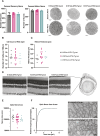
Fig. 3.
Fig. 3.
Rescue crosses of MtrfrK155* with wild-type or 3K>A MTRFR transgenes. (A) The breeding schematic for two generation crosses to test whether either transgene could rescue MtrfrK155* homozygous lethality (schematics of generation of K155*hom+WTKI-Tg-het and K155*hom+3K>A-Tg-het mice). Breeding schematic for the generation of K155*hom+3K>A-Tg-hom mice. (B) The number of WTKI-Tg-het mice (n=94) or 3K>A-Tg-het mice (n=84) that were K155 wild-type (wt)/het/hom were recorded, and a chi-squared analysis was performed to determine whether genotypes were being recovered at the expected rate. Although the WTKI-Tg rescued the homozygous K155* at a lower than predicted frequency, eight mice were recovered, whereas the 3K>A-Tg did not rescue. When the 3K>A-Tg was bred to produce homozygous transgenic mice (n=33), one K155* homozygote was rescued; however, that genotype was still under-represented. p.v, P-value. **P<0.01, ***P <0.001, ****P<0.0001.
Fig. 4.
Fig. 4.
Phenotyping of WTKI-Tg-het rescue in K155wt/*het/hom mice. (A) Male and female body weights of K155wt/het/hom mice, all heterozygous for WTKI-Tg, were taken weekly from birth to 18 weeks of age. No differences between genotypes were found. (B) Wire hang latency to fall was measured for male and female mice from 4 weeks to 18 weeks to test muscle and grip strength. No differences between genotypes were found. (A,B) Males: K155wt n=5, K155*het n=14, K155*hom n=4. Females: K155wt n=9, K155*het n=12, K155*hom n=4. Statistical analysis was performed using a one-way ANOVA. (C) Sciatic motor nerve conduction velocity of K155wt/*het/hom mice rescued with heterozygous WTKI-Tg was measured at 18 weeks of age. No differences with genotype were found. (D) Muscle weight (medial gastrocnemius) to body weight ratio of K155wt/*het/hom mice rescued with heterozygous WTKI-Tg was measured. No differences with genotype were found. (C,D) K155wt n=6, K155*het n=14, K155*hom n=4. Statistical analysis was performed using a one-way ANOVA. There were no sex differences, and data were pooled. (E) RT-qPCR analysis of mRNA levels of the integrated stress response (ISR) target genes Fgf21 and Gdf15 in spinal cord from K155wt/*het/hom mice rescued with heterozygous WTKI-Tg. (F) ISR target gene expression in liver from K155wt/*het/hom mice rescued with heterozygous WTKI-Tg. No elevated expression was found in either tissue. (E,F) K155wt n=3, K155*het n=3, K155*hom n=4. Statistical analysis was performed using a one-way ANOVA. ns, not significant. There were no sex differences, and data were pooled. Error bars±s.d. in all panels.
Fig. 5.
Fig. 5.
Rescue of K155* with heterozygous WTKI-Tg. (A) Axons from motor and sensory branches of femoral nerve were counted and did not differ by genotype. Representative images of each nerve in each genotype are shown. Femoral motor nerves: K155wt n=6, K155*het n=9, K155*hom n=3. Femoral sensory nerves: K155wt n=4, K155*het n=12, K155*hom n=4. There were no sex differences, and data were pooled. (B) Cells in the retinal ganglion cell (RGC) layer of the retina were counted in images from whole-eye histology. The data were normalized to number of cells per 100 µm of the RGC layer, and all counts were done adjacent to the optic nerve head. No differences with genotype were found. (C) The thickness of the retina from the base of the photoreceptor inner segments to the inner limiting membrane of the RGC layer was measured in the same retinal images, and no differences were found with genotype. (B,C) K155wt n=6, K155*het n=14, K155*hom n=4. (D) Representative images of retina histology for each genotype are shown. (E) The area of optic nerve cross-sections in each genotype was measured to assess possible optic nerve atrophy, but no differences were found. Mouse numbers were as follows: K155wt n=8, K155*het n=24, K155*hom n=8. (F) Using transmission electron microscopy, optic nerve axon diameters and myelin thicknesses for over 100 axons (103-136) were imaged per nerve and evaluated. Data are plotted as a cumulative histogram, showing that the distributions of axon sizes were not different with genotype. Optic nerves from K155wt (n=4) and K155*hom mice (n=4) were imaged. Statistical analysis was performed using one-way ANOVA (A-C,E) and Kolmogorov–Smirnov and Mann–Whitney tests (F). Graphs show mean±s.d. Scale bars: 50 µm (A), 100 µm (D) and 1 µm (F).
Fig. 6.
Fig. 6.
Rescue of mitochondrial translation in an hiPSC-derived neuronal model of MTRFR deficiency. (A) Representative brightfield images of NGN2-induced neuronal differentiation (left column), and immunofluorescence of the neuronal markers MAP2 and TUJ (also known as neuron-specific class III beta-tubulin) (right column). Scale bars: 25 μm. (B) Lentiviral transduction was used to express two single-guide RNAs targeting the promoter region of MTRFR to induce knockdown (KD) of MTRFR in human induced pluripotent stem cells (hiPSCs) constitutively expressing the dCas9-KRAB CRISPRi machinery. Successful KD of MTRFR expression was achieved with both guides based on RT-qPCR of MTRFR expression in neuronal progenitor cells (NPCs) using beta-actin as standard (n=3). A scrambled guide had no effect on MTRFR expression (n=3). For follow-up experiments, both KD lines were plotted together as ‘MTRFR KD’, and the scrambled line was plotted with the unedited cell line as ‘Control’. (C) KD of MTRFR in cortical neurons causes decreased mitochondrial translation efficiency 20 days after neuronal induction based on reduced MT-CO1 levels (n=4). Quantification was normalized to TOMM20. (D) Transduction of KD hiPSC-derived neurons with adeno-associated virus (AAV)9 expressing MTRFR at a multiplicity of infection of 500 restores expression levels by RT-qPCR 20 days after transduction (n=4). (E) AAV9 delivery of MTRFR restored mitochondrial translation based on western blotting and quantification of MT-CO1 levels in day 25 neurons before and after AAV9 treatment (n=4). Error bars are mean±s.e.m. One-way ANOVA (B,D,E) and unpaired two-tailed t-test (C) were used for statistical analysis. ns, not significant; *P<0.05, **P<0.01, ***P<0.001, ****P<0.0001.

References

    1. Amunts, A., Brown, A., Toots, J., Scheres, S. H. W. and Ramakrishnan, V. (2015). Ribosome. The structure of the human mitochondrial ribosome. Science 348, 95-98. 10.1126/science.aaa1193 - DOI - PMC - PubMed
    1. Anderson, N. S. and Haynes, C. M. (2020). Folding the Mitochondrial UPR into the Integrated Stress Response. Trends Cell Biol. 30, 428-439. 10.1016/j.tcb.2020.03.001 - DOI - PMC - PubMed
    1. Anderson, S., Bankier, A. T., Barrell, B. G., De Bruijn, M. H., Coulson, A. R., Drouin, J., Eperon, I. C., Nierlich, D. P., Roe, B. A., Sanger, F.et al. (1981). Sequence and organization of the human mitochondrial genome. Nature 290, 457-465. 10.1038/290457a0 - DOI - PubMed
    1. Antonicka, H., Ostergaard, E., Sasarman, F., Weraarpachai, W., Wibrand, F., Pedersen, A. M., Rodenburg, R. J., Van Der Knaap, M. S., Smeitink, J. A., Chrzanowska-Lightowlers, Z. M.et al. (2010). Mutations in C12orf65 in patients with encephalomyopathy and a mitochondrial translation defect. Am. J. Hum. Genet. 87, 115-122. 10.1016/j.ajhg.2010.06.004 - DOI - PMC - PubMed
    1. Armougom, F., Moretti, S., Poirot, O., Audic, S., Dumas, P., Schaeli, B., Keduas, V. and Notredame, C. (2006). Expresso: automatic incorporation of structural information in multiple sequence alignments using 3D-Coffee. Nucleic Acids Res. 34, W604-W608. 10.1093/nar/gkl092 - DOI - PMC - PubMed

Substances

LinkOut - more resources