. 2025 May 16:16:1571221.
doi: 10.3389/fimmu.2025.1571221. eCollection 2025.

Effector Tc17 cells resist shift from OXPHOS to aerobic glycolysis

Affiliations

Effector Tc17 cells resist shift from OXPHOS to aerobic glycolysis

Reni John et al. Front Immunol. .

Abstract

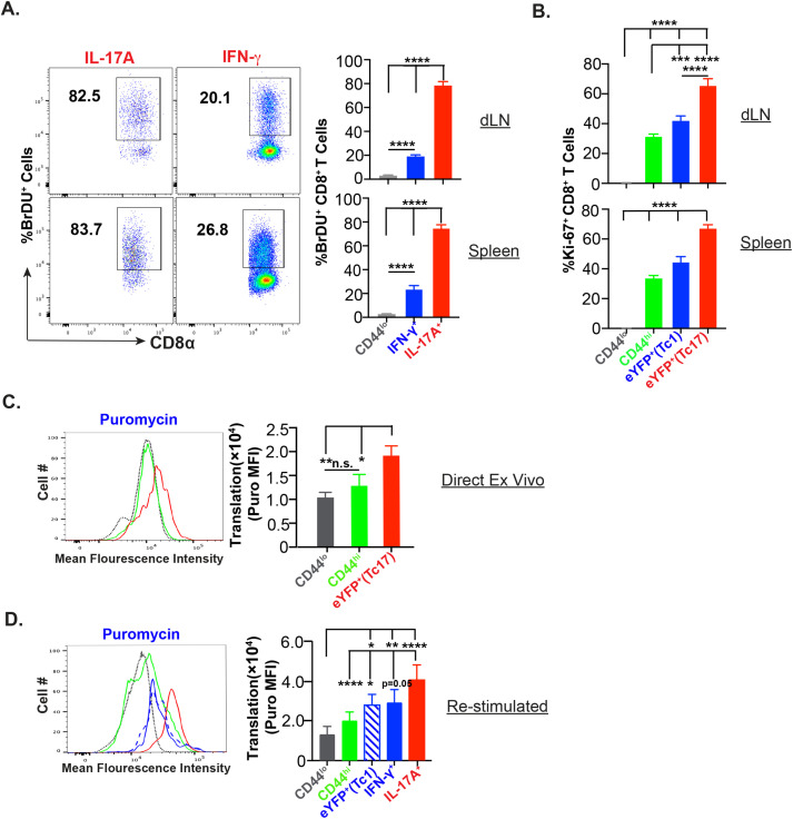
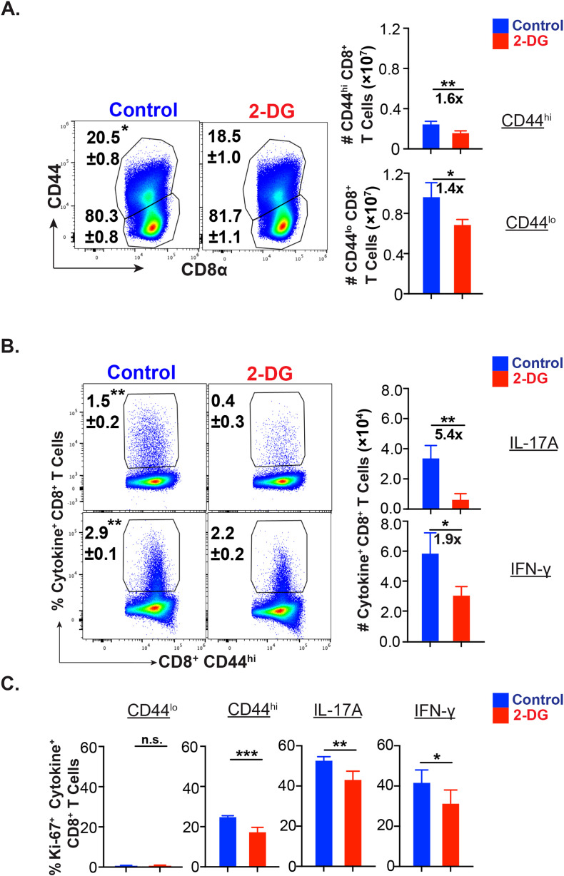
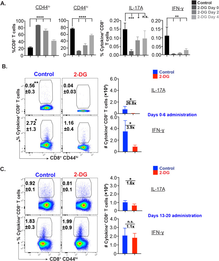
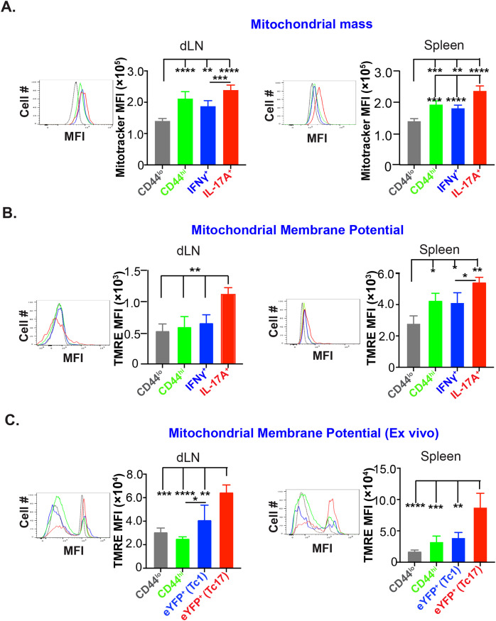
IL-17A-expressing lymphocytes, including Tc17 cells, are instrumental in immunity, immunopathology, and autoimmunity. We have previously shown that experimental attenuated live fungal vaccine-induced Tc17 cells are stable, long-lived without plasticity, and necessary to mediate sterilizing immunity during CD4+ T cell deficiency, which poses higher susceptibility to fungal infections. Cell metabolism is integral for T cell homeostasis but the metabolic adaptations of Tc17 cells are poorly defined. In this study, we hypothesized that effector Tc17 cells adopt high energy-yielding metabolic pathways to form stable, long-lived memory cells in vivo. Using a mouse model of attenuated fungal vaccination, we found that effector Tc17 cells were metabolically highly active with higher proliferation and protein synthesis than IFNγ+ CD8+ T (Tc1) cells. Glucose was necessary for effector Tc17 cell expansion but with less dependency during the late expansion despite the active metabolism. Contrary to established dogma, we found that the effector Tc17 cells preferentially channeled the glucose to OXPHOS than glycolysis, which was correlated with higher mitochondrial mass and membrane potential. Inhibition of OXPHOS shrunk the Tc17 responses while sparing Tc1 cell responses. Tc17 cells actively relied on OXPHOS throughout the expansion period, resisting adaptation to aerobic glycolysis. Our data showed that the effector Tc17 cells predominantly utilize glucose for metabolism through OXPHOS rather than aerobic glycolysis. Our study has implications in vaccine design to enhance the efficacy and immunotherapeutics to modulate the immunity and autoimmunity.

Keywords: CD8+ T cell; OXPHOS; T cell activation; Tc17 cell; antifungal; glycolysis; vaccine responses.


Conflict of interest statement

The authors declare that the research was conducted in the absence of any commercial or financial relationships that could be construed as a potential conflict of interest.

Figures

Figure 1
Figure 1
Effector Tc17 cells are metabolically active. (A) Proliferation of anti-fungal effector CD8+ T cells by BrdU: Naïve WT mice were vaccinated subcutaneously with ~105 CFU of attenuated #55 strain of B.dermatitidis (B.d. #55) and were pulsed with BrdU (0.8 mg/ml) in drinking water from day 5 to 15 post-vaccination (PV). On day 16PV, draining lymph nodes (dLNs) and spleens were harvested to prepared single-cell suspensions and were restimulated with αCD3/CD28 mAbs. Cells were surface- and intracellular cytokine stained before BrdU staining. Numbers represent the percent BrdU+ among IL-17A+/IFN-γ + CD8+ T cells. (B) Proliferation of effector CD8+ T cells by Ki67 staining: Single-cell suspensions from dLNs and spleens of vaccinated IL17aCreR26ReYFP and GREAT mice were stained with anti-Ki-67 monoclonal antibody (mAb) intracellularly following surface markers and cytokine staining, and the frequencies of Ki-67+ cells were analyzed by flow cytometry. The numbers represent the percent Ki-67+ cells among naïve (CD44lo), activated (CD44hi), and IL-17A or IFN-γ eYFP+ CD8+ T cells (C, D) Translational ability of CD8+ T cells: Single cell-suspensions from dLNs of vaccinated IL17aCreR26ReYFP and GREAT mice were stained direct ex vivo or following restimulation with surface markers and intracellular staining following incubation with puromycin. Data show the mean fluorescence intensity of incorporated puromycin in naïve (CD44lo), activated (CD44hi) and IL-17A/IFN-γ+ eYFP+ CD8+ T cells. Values are mean ± SD. N=4–5 mice/group. Data are representative of ≥2 individual experiments. *p≤ 0.05, **p≤ 0.01, ***p≤ 0.001 and ****p≤ 0.0001. ns-not significant. p≥0.05.
Figure 2
Figure 2
Glucose dependency of effector Tc17 cells. (A-C) Glucose utilization of antifungal CD8+ T cells for activation, proliferation, and cytokine expression: Naïve 6- to 8- week-old IL17aCreR26ReYFP mice were vaccinated with B.d. #55 (~ 105 CFU) and treated with PBS or glycolysis inhibitor 2-deoxy glucose (2-DG) intraperitoneally from day 5 to 15 PV. On day 16PV, single-cell suspensions from dLNs were restimulated with αCD3/CD28, surface and intracellular stained, and analyzed by flow cytometry. Data represent percent and total numbers of CD44lo and CD44hi cells gated on CD8+ T cells. (A) and IL-17A+/IFN-γ + cells gated on CD44hi CD8+ T cells (B) and percent of Ki-67+ cells among CD44lo, CD44hi, IL-17A+ and IFN-γ+ CD8+ T cells (C). Values are in mean ± SD. Data are representative of two independent experiments. n = 4–5 mice/group. *p≤ 0.05, and **p≤ 0.01.
Figure 3
Figure 3
Glucose dependency of effector CD8+ T cells during early and late expansion. (A) Phase specific glucose dependency of antifungal CD8+ T cells in vitro: © CD8+ T cells (WT mice) were enriched and cultured with bone marrow-derived dendritic cells pulsed with heat-killed yeast. On days 0-4, 2-4, and 4 post-culture, cells were treated with 250 µM 2-deoxy glucose. The cells were harvested on day 5 post-culture and stained for cytokine+ CD8+ T cells. Data show the percent naïve, activated, IL-17A+, and IFN-γ+ cells gated on (CD44hi) CD8+ T cells. (B, C) Glucose dependency of effector CD8+ T cells in vivo: Vaccinated were administered with vehicle or 2-deoxy glucose (2-DG) intraperitoneally on indicated days. On days 7 or 21 post vaccination (PV), single-cell suspensions from dLNs were restimulated with αCD3/CD28 mAbs, surface and intracellular cytokine-stained, and analyzed by flow cytometry. Data show percent and total numbers of IL-17A+ and IFN-γ+ cells gated on CD44hi CD8+ T cells on days 7 PV (days 0–6 PV administration; (B)) and 21PV (days 13–20 PV administration; (C)). Data are representative of two independent experiments, n= 5–6 replicates/group (A) and n= 4–5 mice/group (B, C). Values are mean ± SD. *p≤ 0.05, **p≤ 0.01, ***p≤ 0.001, and ****p≤ 0.0001. ns-not significant , p≥0.05.
Figure 4
Figure 4
High mitochondrial mass and membrane potential of effector Tc17 cells. (A) Mitochondrial mass: Naïve WT mice were vaccinated with B.d. #55. On day 16 post-vaccination (PV), mitochondrial mass of CD8+ T cells of draining lymph nodes (dLNs) and spleens was assessed by MitoTracker Deep Red staining and analyzed by flow cytometry. Data show the mean fluorescence intensities of MitoTracker Deep Red in CD44lo, CD44hi, IL-17A+, and IFN-γ+ CD8+ T cells. (B) Mitochondrial membrane potential: Single-cell suspensions from vaccinated mice (day 16PV) were restimulated (anti-CD3/CD28 mAbs), incubated with Tetramethyl Rhodamine, Ethyl Ester (TMRE), before staining for cell surface markers and intracellular cytokines for flow cytometry. Data show the mean fluorescence intensities of TMRE in CD44lo, CD44hi, IL-17A+, and IFN-γ+ CD8+ T cells. (C) Mitochondrial membrane potential of effector CD8+ T cells from reporter mice: Single-cell suspensions from dLNs and spleens of vaccinated IL17aCreR26ReYFP and GREAT mice (on day 21PV) were incubated with TMRE before staining for cell surface markers and analyzed by flow cytometry. Data show the mean fluorescence intensities of TMRE in naïve, activated, and eYFP+ CD8+ T cells. Values are mean ± SD. Data are representative of two independent experiments. n= 3–5 mice/group. *p≤ 0.05, **p≤ 0.01, ***p≤ 0.001, and ****p≤ 0.0001.
Figure 5
Figure 5
Mitochondrial metabolism by effector Tc17 cells. (A) OX-PHOS utilization by antifungal effector CD8+ T cells in vitro. Naive CD8+ T cells were enriched and cultured with bone marrow-derived dendritic cells (BMDCs) pulsed with heat-killed yeast. On days 2–4 culture, cells were treated with vehicle or Oligomycin. The cells were harvested on day 5 post-culture, stained for surface markers and intracellular cytokines, and analyzed by flow cytometry. Data show the percent and total numbers of IL-17A+ and IFN-γ+ cells gated on CD44hi CD8+ T cells. (B) Metabolic dependence of effector CD8+ T cells: Single-cell suspensions from dLNs of vaccinated GREAT mice (day 21 PV) were restimulated with αCD3/CD28 mAbs before treating with vehicle, 2-deoxyglucose (2-DG), oligomycin or both for 30 min followed by incubation with puromycin for 30 min. Cells were stained for cell surface markers and intracellular cytokine and puromycin and analyzed by flow cytometry. Data show the mean fluorescence intensities of puromycin incorporation in naïve, IL-17A+, and IFN-γ+ CD8+ T cells. (C) Metabolic dependence of IL-17A reporter effector CD8+ T cells: Single-cell suspensions from dLNs from vaccinated IL17aCreR26ReYFP mice (day 21PV) were treated with vehicle, 2-deoxyglucose (2-DG), oligomycin or 2-DG and oligomycin for 30 min followed by incubation with puromycin for 30 min. Cells were stained for cell surface markers and intracellular puromycin incorporation and analyzed by flow cytometry. Data show the mean fluorescence intensities of puromycin in naïve and eYFP+ CD8+ T cells. Values are mean ± SD. Data are representative of two independent experiments, n= 5–6 replicates/group (A) and n= 4–5 mice/group (B, C). *p≤ 0.05, **p≤ 0.01, ***p≤ 0.001 and ****p≤ 0.0001. ns-not significant , p≥0.05.
Figure 6
Figure 6
Glucose for OX-PHOS by Tc17 cells during early expansion. (A) Mitochondrial membrane potential of IL-17A reporter effector CD8+ T cells: Single-cell suspensions from dLNs of vaccinated IL17aCreR26ReYFP mice (on day 7 PV) were incubated with 50 nM tetramethyl rhodamine, ethyl ester (TMRE) for 30 min followed by staining for cell surface markers. Stained cells were analyzed by flow cytometry. Data show the mean fluorescence intensities of TMRE in naïve, activated, and eYFP+ CD8+ T cells. (B) Metabolic dependence of effector CD8+ T cells: Single-cell suspensions from dLNs of vaccinated IL17aCreR26ReYFP mice (on day 7 PV) were treated with vehicle, 2-DG, oligomycin or both 2-DG and oligomycin for 30 min followed by incubation with puromycin for 30 min. Cells were stained for cell surface markers and intracellular puromycin and analyzed by flow cytometry. Data show the mean fluorescence intensities of puromycin incorporation and percent metabolic dependence of naïve, activated, and eYFP+ CD8+ T cells. Values are mean ± SD Data are representative of two independent experiments. n= 4–5 mice/group. *p≤ 0.05, **p≤ 0.01, ***p≤ 0.001 and ****p≤ 0.0001.

Similar articles

Cited by

References

    1. Hamada H, Garcia-Hernandez Mde L, Reome JB, Misra SK, Strutt TM, McKinstry KK, et al. . Tc17, a unique subset of CD8 T cells that can protect against lethal influenza challenge. J Immunol. (2009) 182:3469–81. doi: 10.4049/jimmunol.0801814 - DOI - PMC - PubMed
    1. Harrison OJ, Linehan JL, Bouladoux N, Han S-J, Smelkinson M, Sen SK, et al. . Commensal-specific T cell plasticity promotes rapid tissue adaptation to injury. Science. (2019) 363:1–11. doi: 10.1126/science.aat6280 - DOI - PMC - PubMed
    1. Naik S, Bouladoux N, Linehan JL, Han S-J, Harrison OJ, Wilhelm C, et al. . Commensal-dendritic-cell interaction specifies a unique protective skin immune signature. Nature. (2015) 520:104–8. doi: 10.1038/nature14052 - DOI - PMC - PubMed
    1. Nanjappa SG, Heninger E, Wuthrich M, Sullivan T, Klein B. Protective antifungal memory CD8(+) T cells are maintained in the absence of CD4(+) T cell help and cognate antigen in mice. J Clin Invest. (2012) 122:987–99. doi: 10.1172/jci58762 - DOI - PMC - PubMed
    1. Cheuk S, Schlums H, Serezal IG, Martini E, Chiang SC, Marquardt N, et al. . CD49a expression defines tissue-resident CD8(+) T cells poised for cytotoxic function in human skin. Immunity. (2017) 46:287–300. doi: 10.1016/j.immuni.2017.01.009 - DOI - PMC - PubMed

LinkOut - more resources