Review
. 2025 May 16:16:1575917.
doi: 10.3389/fimmu.2025.1575917. eCollection 2025.

From sensation to regulation: the diverse functions of peripheral sensory nervous system

Affiliations
Review

From sensation to regulation: the diverse functions of peripheral sensory nervous system

Yixiao Mei et al. Front Immunol. .

Abstract

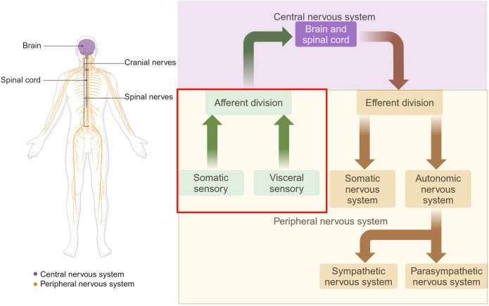
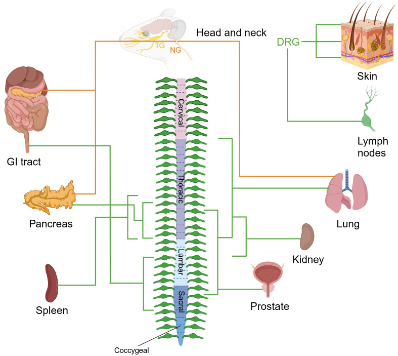
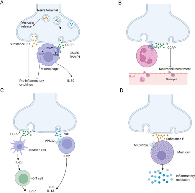
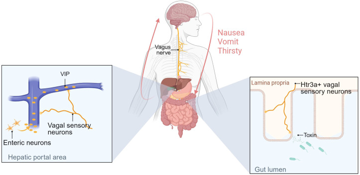
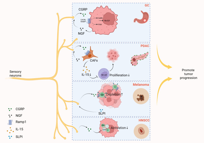
The peripheral sensory nervous system (PNS) has been widely recognized for its role in the collection, processing, and transmission of sensory information, including thermal, mechanical, chemical, and proprioceptive stimuli. In recent years, there has been a growing scholarly interest in the PNS attributable to its multiple physiological and pathophysiological non-sensory roles in the organs it innervates. The PNS exerts regulatory functions within the organs it innervates through direct interactions with local cells or through microbe-nerve-cell interactions that differ from the traditional feedback regulatory modes used by the hormonal and sensory brain-sympathetic/parasympathetic systems. The release of the neuropeptide calcitonin gene related peptide (CGRP) by nerves, through its action on CGRP receptors in peripheral cells, constitutes a primary molecular axis for PNS regulation of organ cells, maintaining tissue homeostasis, facilitating pathological processes, and modulating innate and adaptive immunity. This review highlights the non-sensory functions of the peripheral sensory nervous system in various tissues and organs, focusing on phenotypes, molecular mechanisms and their significance, while also exploring future research directions, methodologies and potential preclinical studies aimed at targeting these pathways for the development of novel therapies.

Keywords: CGRP signaling pathway; microbe-nerve-cell crosstalk; neuro-immune interaction; nonsensory regulatory functions; peripheral sensory nervous system (PNS); sensory signal transduction.


Conflict of interest statement

The authors declare that the research was conducted in the absence of any commercial or financial relationships that could be construed as a potential conflict of interest.

Figures

Figure 1
Figure 1
Organization of the nervous system. The nervous system is divided into two main parts: the CNS and the PNS. The CNS is composed of the brain and the spinal cord. The PNS consists of all the neural tissue outside the CNS, including nerves and ganglia. Functionally, the PNS is divided into the somatic and autonomic nervous systems. The sensory nervous system, also known as the afferent nervous system, is a crucial component of the PNS. Somatic sensory includes sensations of touch, pain, pressure, vibration and the special sensations of hearing, equilibrium and vision. Visceral sensory includes stretch, pain, temperature, chemical changes, irritation in viscera, nausea and hunger, as well as special sensations of taste and smell. Adapted from “The Major Components of the Nervous System”, by BioRender.com (2025) Retrieved from https://app.biorender.com/biorender-templates.
Figure 2
Figure 2
Sensory nerve supply to organs. The images depict the sensory innervation of various mouse organs from DRG, TG, and NG. The peripheral nervous system ganglia are organized symmetrically, and organs often receive innervation from the same ganglia from both sides of the body. Sensory nerve fibers from DRG and NG are marked in green and orange respectively. TG, trigeminal ganglion; NG, nodose ganglion; DRG, dorsal root ganglion; GI, gastrointestinal. Created with BioRender.com.
Figure 3
Figure 3
Neuro-immune cell units. (A) Substance P is released by nociceptors and acts on monocytes and macrophages through NF-κB activation mediated by ERK-p38 mitogen-activated protein kinase (MAPK), driving the expression of pro-inflammatory cytokines (left). CGRP acts on CALCRL-RAMP1, drives cAMP-protein kinase A (PKA) -dependent CREB regulation, upregulates IL-10 and induces cAMP early repressor (ICER)-dependent transcriptional inhibition of proinflammatory cytokines (right). (B) Nociceptors release CGRP, reduce neutrophil recruitment to the lungs and skin, and inhibit the ability of neutrophils to regulate phagocytosis and kill bacteria. (C) CGRP from nociceptive neurons drives dermal dendritic cells to produce IL-23, which in turn leads to the activation of γδT cells and the production of IL-17 (left). Neuropeptide vasoactive intestinal peptide (VIP) activates its receptor VPAC2 on ILC2s and drives the production of IL-5 and IL-13 (right). (D) Substance P activates MAS-related G protein-coupled receptor member B2 (MrgprB2 receptor) on mast cells, leading to mast cell degranulation and pro-inflammatory mediator release. Created with BioRender.com.
Figure 4
Figure 4
Sensory neurons are involved in gut-brain axis-mediated behavioral regulation and regulate gastrointestinal homeostasis. The figure illustrates the mechanisms by which two types of vagal sensory afferents regulate behavior. One mechanism is as follows: Changes in osmotic pressure in the gut mediate VIP secretion by intestinal neurons, and high concentrations of VIP in the portal vein activate vagal sensory afferents innervating the HPA region, thereby regulating thirst. The other is that Htr3a+ vagal sensory neurons can detect toxins secreted by intestinal bacteria and transmit signals to the brain, causing nausea and vomiting. Created with BioRender.com.
Figure 5
Figure 5
Sensory neurons promote tumor progression by releasing CGRP. Gastric cancer (GC) cells elevate the secretion of nerve growth factor (NGF), and then NGF induces CGRP release from sensory neurons. CGRP then engages the Calcrl/Ramp1 on gastric cancer cells, triggering dual activation of PI3K-Akt and CaMK-dependent signaling cascades. These pathways converge to amplify E2F-dependent transcriptional programs, fostering tumor progression through cell cycle deregulation and survival mechanisms. In the tumor microenvironment of pancreatic ductal adenocarcinoma (PDAC), cancer-associated fibroblasts (CAFs) secrete NGF that stimulates sensory neurons to release CGRP. This neuropeptide subsequently binds to receptor RAMP1 on CAFs, triggering downregulation of interleukin-15 (IL-15) expression. The reduced IL-15 levels impair NK cell infiltration and cytotoxic activity, ultimately fostering tumor progression in PDAC. In melanoma, tumor-derived secretory leukocyte protease inhibitor (SLPI) induces pain hypersensitivity and stimulates nociceptor neurite outgrowth and sustained release of CGRP, which binds to RAMP1 expressed on tumor-infiltrating CD8+ T cells, triggering upregulation of immune checkpoint receptors, driving functional exhaustion of cytotoxic CD8+ T cells, resulting in uncontrolled melanoma cell proliferation. In the microenvironment of head and neck squamous cell carcinoma (HNSCC), CGRP released by sensory neurons can directly suppress immune cells, namely T helper 1 (Th1) CD4+ T cells and activated CD8+ T cells, thereby promoting tumor growth. Created with BioRender.com.
Figure 6
Figure 6
Crosstalk between ligands secreted by cells in the mouse dorsal root ganglia and receptors in the organs. Heatmap of ligands that were identified by single nucleus RNA sequencing and bulk-seq analysis describes the expression levels of ligands within the cell clusters and dorsal root ganglia in both sham and SNI states. Sankey diagram reveals the ligands and the corresponding receptors predicted by Cellinker. Heatmap of receptors describing the expression levels of receptors within dorsal root ganglia in both sham and SNI states and different organs of an adult male C57BL/6 mouse (166).
Figure 7
Figure 7
Crosstalk between ligands secreted by cells in the mouse vagal ganglia and receptors in the organs. The left heatmap shows the expression levels of ligands across cell clusters within the vagal ganglia, based on single-nucleus RNA sequencing (snRNA-seq) data from Forster et al (167). The central Sankey diagram visualizes predicted ligand-receptor interactions, as computed using Cellinker, illustrating the diversity of VG-derived signals and their potential targets across tissues. The right heatmap displays the expression levels of corresponding receptors in various organs of an adult male C57BL/6 mouse, derived from reference transcriptomic datasets (166).

Similar articles

References

    1. Murtazina A, Adameyko I. The peripheral nervous system. Development. (2023) 150:9. doi: 10.1242/dev.201164 - DOI - PubMed
    1. Crawford LK, Caterina MJ. Functional anatomy of the sensory nervous system: updates from the neuroscience bench. Toxicol Pathol. (2020) 48:174–89. doi: 10.1177/0192623319869011 - DOI - PubMed
    1. Ran C, Boettcher JC, Kaye JA, Gallori CE, Liberles SD. A brainstem map for visceral sensations. Nature. (2022) 609:320–6. doi: 10.1038/s41586-022-05139-5 - DOI - PMC - PubMed
    1. Mehrpour V, Martinez-Trujillo JC, Treue S. Attention amplifies neural representations of changes in sensory input at the expense of perceptual accuracy. Nat Commun. (2020) 11:2128. doi: 10.1038/s41467-020-15989-0 - DOI - PMC - PubMed
    1. Donnelly CR, Chen O, Ji RR. How do sensory neurons sense danger signals? Trends Neurosci. (2020) 43:822–38. doi: 10.1016/j.tins.2020.07.008 - DOI - PMC - PubMed

MeSH terms

Substances

LinkOut - more resources