

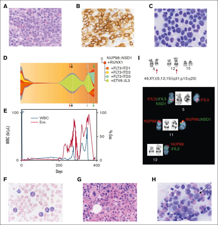
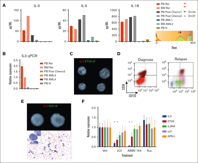
A novel 3-way translocation involving ETV6:: IL3 drives AML with eosinophilia
- PMID: 40453143
- PMCID: PMC12067892
- DOI: 10.1016/j.bneo.2025.100079
A novel 3-way translocation involving ETV6:: IL3 drives AML with eosinophilia
Conflict of interest statement
Conflict-of-interest disclosure: J.M. receives research funding paid to the institution from Incyte, Novartis, Celgene, Bristol Myers Squibb (BMS), Kartos, Karyopharm, PharmaEssentia, AbbVie, Geron, CTI BioPharma; and consulting fees from Incyte, Kartos, Karyopharm, Geron, Roche, AbbVie, CTIBiotech, GlaxoSmithKline (GSK), Pfizer, PharmaEssentia, Galecto, Celgene, BMS, and Novartis. D.T. receives contracted research funding paid to his institution from CTI BioPharma, Astellas Pharma, and Gilead; and consulting fees from CTI BioPharma, Novartis, AbbVie, Sierra Oncology, GSK, and Cogent Biosciences. J.F. receives contracted research funding paid to his institution from Syros Pharmaceuticals and ORYZON. M.K. receives research funding paid to the institution from Incyte, Celgene, BMS, MorphoSys, Protagonist, Ionis, Silence Therapeutics, Kura Oncology; and consulting fees from Incyte, AbbVie, MorphoSys, and Protagonist. The remaining authors declare no competing financial interests.
Figures
Similar articles
-
Novel ETV6::RAPGEF6 fusion gene in chronic eosinophilic leukemia: compiling evidence on the role of IL3 overexpression in tumorigenesis.Ann Hematol. 2025 Mar;104(3):2017-2021. doi: 10.1007/s00277-025-06217-0. Epub 2025 Feb 9. Ann Hematol. 2025. PMID: 39923209 Free PMC article.
-
A Novel IL3-ETV6 Fusion in Chronic Eosinophilic Leukemia Not Otherwise Specified With t(5; 12) (q31; p13): A Case Report and Literature Review.Front Oncol. 2022 Jun 7;12:887945. doi: 10.3389/fonc.2022.887945. eCollection 2022. Front Oncol. 2022. PMID: 35747804 Free PMC article.
-
A novel three-way rearrangement involving ETV6 (12p13) and ABL1 (9q34) with an unknown partner on 3p25 resulting in a possible ETV6-ABL1 fusion in a patient with acute myeloid leukemia: a case report and a review of the literature.Biomark Res. 2016 Aug 25;4(1):16. doi: 10.1186/s40364-016-0070-7. eCollection 2016. Biomark Res. 2016. PMID: 27570624 Free PMC article.
-
Eosinophilia in acute myeloid leukemia: Overlooked and underexamined.Blood Rev. 2019 Jul;36:23-31. doi: 10.1016/j.blre.2019.03.007. Epub 2019 Mar 30. Blood Rev. 2019. PMID: 30948162 Review.
-
T cell phenotype and lack of eosinophilia are not uncommon in extramedullary myeloid/lymphoid neoplasms with ETV6::FLT3 fusion: a case report and review of the literature.Virchows Arch. 2024 May;484(5):853-857. doi: 10.1007/s00428-023-03693-5. Epub 2023 Nov 20. Virchows Arch. 2024. PMID: 37985498 Review.
References
-
- Naymagon L, Marcellino B, Mascarenhas J. Eosinophilia in acute myeloid leukemia: overlooked and underexamined. Blood Rev. 2019;36:23–31. - PubMed
-
- Pulsoni A, Iacobelli S, Bernardi M, et al. M4 acute myeloid leukemia: the role of eosinophilia and cytogenetics in treatment response and survival. The GIMEMA experience. Haematologica. 2008;93(7):1025–1032. - PubMed
-
- Gotlib J, Cools J. Five years since the discovery of FIP1L1-PDGFRA: what we have learned about the fusion and other molecularly defined eosinophilias. Leukemia. 2008;22(11):1999–2010. - PubMed
-
- De Braekeleer E, Douet-Guilbert N, Morel F, Le Bris MJ, Basinko A, De Braekeleer M. ETV6 fusion genes in hematological malignancies: a review. Leuk Res. 2012;36(8):945–961. - PubMed
LinkOut - more resources
Full Text Sources
