Review
. 2025 May 2:50:603-639.
doi: 10.1016/j.bioactmat.2025.04.026. eCollection 2025 Aug.

Angiogenesis in rheumatoid Arthritis: Pathological characterization, pathogenic mechanisms, and nano-targeted therapeutic strategies

Affiliations
Review

Angiogenesis in rheumatoid Arthritis: Pathological characterization, pathogenic mechanisms, and nano-targeted therapeutic strategies

Fang Zhao et al. Bioact Mater. .

Abstract

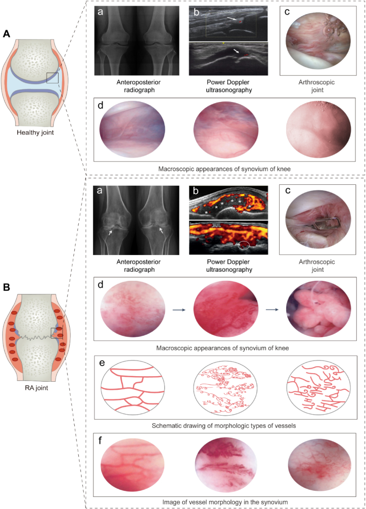
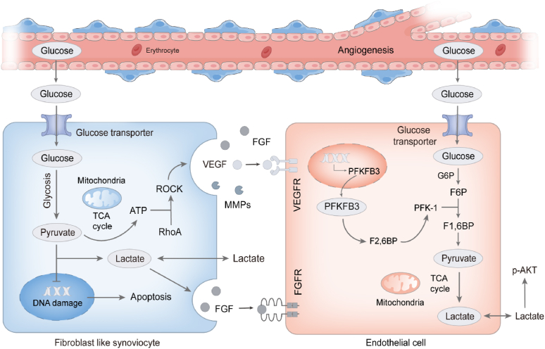
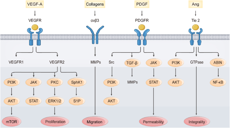
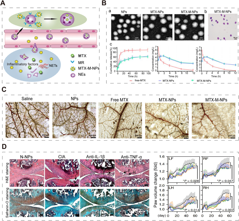
Angiogenesis is critical from early development through the progression of life-threatening diseases. In rheumatoid arthritis (RA), angiogenesis is markedly heightened and undergoes aberrant changes that exacerbate the progression of synovitis. However, the intricate mechanisms underlying these changes remain poorly understood. Despite the development of numerous anti-angiogenic agents, their clinical efficacy is often compromised by adverse effects and the emergence of adaptive resistance, leading to disease relapse or progression. Nanomedicine has gained significant attention owing to its excellent biocompatibility, precise biological targeting, and enhanced therapeutic outcomes. Anti-angiogenic nanoagents have shown transformative potential in treating cancer and retinal diseases. Nevertheless, a comprehensive review addressing the fundamental mechanisms of anti-angiogenic nanoagents in RA has yet to be undertaken. Herein, this review provides an in-depth description of the unique structural and functional aspects of pathological angiogenesis in RA and its negative consequences. The mechanisms of pro-angiogenic mediators contributing to RA angiogenesis are further explored. Subsequently, biological activities of nanomedicines for the treatment of RA are summarized. Finally, the cutting-edge developments in the anti-angiogenic nanoagents of RA engineered based on these mechanisms and bioactivities are outlined. A helpful introduction to anti-angiogenic strategies for treatment of RA, which may offer novel perspectives for the development of nanoagents, is opening a new horizon in the fight against RA.

Keywords: Angiogenesis; Nanoagents; Pro-angiogenic factor; Rheumatoid arthritis.


Conflict of interest statement

The authors declare no conflict of interest, financial or otherwise.

Figures

Image 1
Graphical abstract
Scheme 1
Scheme 1
Schematic diagram of anti-angiogenic nanoagents constructed by targeting activated stromal cells (fibroblast-like synoviocytes, neutrophils, macrophages, and endothelial cells) and specific removal pro-angiogenic mediators (vascular endothelial growth factor, platelet-derived growth factor, and selectin) for treatment of RA.
Fig. 1
Fig. 1
Steps of Angiogenesis under Physiological Conditions. (A) The steps of angiogenesis under physiological conditions include the following: (Step.1) ECs receive pro-angiogenic signals, accelerating activation. (Step.2) Once activated, ECs dissociate from their parent vessels and secrete proteolytic enzymes, such as MMPs, that degrade the basement membrane, detach pericytes, and increase vascular permeability. (Step.3) Tip cells navigate the environment and invade the target tissue, guiding the sprouting process and transforming into more proliferative phenotypes, called stem cell. (Step.4) During sprouting angiogenesis, ECs proliferate and migrate, forming lumens through intercellular junctions. (Step.5) The DLL4/Notch-1 pathway is activated, increasing the expression of VEGFR1 while inhibiting VEGFR2 expression, facilitating vessel fusion and pruning. (Step.6) Tip cells also secrete PDGF-β, which binds to PDGFR-β on pericytes, leading to pericyte recruitment and BM deposition, thus forming stable and mature vessels. (B) Stromal cells in the surrounding tissue release various pro-angiogenic growth factors such as VEGF, FGF, HGF, and Ang, which bind to receptors on ECs like VEGFR, FGFR, c-Met, and Tie, initiating pro-angiogenic signaling and activating ECs. (C) Under the stimulation of VEGF, tip cells secrete DLL4, which interacts with Notch receptors in stem cells. Notch is cleaved by γ-secretase, releasing the Notch intracellular domain (NICD), which inhibits VEGFR2 expression while upregulating VEGFR1 expression, promoting vessel fusion and pruning. Endothelial cell: EC, VEGF: Vascular endothelial growth factor, FGF: Fibroblast growth factor, HGF: Hepatocyte growth factor, PDGF: Platelet-derived growth factor, VEGFR: Vascular endothelial growth factor receptor, FGFR: Fibroblast growth factor receptor, PDGFR: Platelet-derived growth factor receptor, c-Met: Cellular-mesenchymal to epithelial transition factor, Tie: Tyrosine kinase receptors with immunoglobulin and EGF homology domains, MMP: Matrix metalloproteinase, Ang: Angiopoietin.
Fig. 2
Fig. 2
The Critical Role of Pathological Angiogenesis in the Progression of RA. (A) Susceptible individuals exposed non-genetic risk factors exhibited asymptomatic synovitis. As the disease advances, the aggressive pannus invades and erodes cartilage and joints, resulting in clinical symptoms such as joint tenderness, stiffness, swelling, and diminished range of motion. (B) The vascular layer of normal synovium contains only a few macrophages and collagen fibers (left). In RA synovial vascular layer, angiogenesis can be divided into an early phase characterized by intense inflammation (middle) and a later phase marked by significant angiogenesis (right). (C) Mechanism of invasion of pannus into adjacent bone and cartilage. T cells and macrophages migrate from the pannus and produce inflammatory cytokines such as TNF-α, IL-1, IL-6, and IL-17. Inflammatory cytokines-stimulated fibroblast-like synoviocytes not only activates osteoclasts by inducing RANKL, but also inhibits osteoblast differentiation by producing DKK1 and sclerostin. RANKL: receptor activator of nuclear factor κB ligand. DKK1: Dickkopf-related protein 1.
Fig. 3
Fig. 3
Synovial Vascular Morphology in Healthy and RA patients under Macroscopic Examination. (A) In the healthy joint (a), PDUS images show a physiological vessel (arrow) in the quadriceps fat pad (b). Adapted with permission from Refs. [27,30,47], copyright 2022. Adapted with permission from Ref. [30], copyright 2018. Adapted with permission from Ref. [27], copyright 2014. Under arthroscopy, sparse blood vessels and avascular joint cartilage can be macroscopically observed (c). Thin veins, arterioles, and capillaries in the normal synovial vascular layer form a closed quadrangular array (d). Adapted with permission from Ref. [48], copyright 2019. (B) In RA joint, anteroposterior radiograph results show a narrow symmetric joint space with subchondral cysts (arrow) in bilateral knee joints (a). Adapted with permission from Ref. [49], copyright 2017. Intra-articular synovial hypertrophy (asterisks), areas of active inflammation (red), and erosion of bone by pannus (circled) could be seen on PDUS (b). Adapted with permission from Ref. [31], copyright 2023. The main feature of RA synovium is increased number of blood vessels, eroding articular cartilage (c). Congested and increased blood vessels are observed in mild synovitis (d, left). As the condition advances to severe synovitis, there is significant angiogenesis (d, middle). Blood vessels become congested, hypertrophic, rod-shaped, pale, swollen even forming villi (d, right). Adapted with permission from Ref. [33], copyright 2016. The vascular morphology patterns in RA (e) can be categorized into three types: linear (f, left), tortuous (f, middle), and mixed (f, right). Adapted with permission from Ref. [35], copyright 2003.
Fig. 4
Fig. 4
Abnormalities in EPC Phenotype and Vascular Barriers in RA Synovium. (A) In RA, EPCs home to inflamed synovium, primarily through the CXCL13/CXCR5 axis. Then, this axis activates the downstream PLC/MEK signaling pathway, promoting VEGF release. Additionally, the VCAM-1/VLA4 interaction enhances the adhesion of EPCs to ECs, mediating the selective recruitment of EPCs to inflamed joint tissues. (B) In healthy, normal vessels are characterized by pericyte coverage, mature and homogenized BM barriers, and ECs arranged in a squared pattern (left). In the hypoxic and highly inflammatory RA microenvironment, in certain newly formed vessels display activated ECs, detachment of pericytes, and disrupted vascular barriers, resulting in immature vessels (right). EPC: Endothelial progenitor cell. EC: Endothelial cell. BM: Basilar membrane. RAM: Rheumatoid arthritis microenvironment.
Fig. 5
Fig. 5
Mechanisms Underlying Loss of Lymphatic Contractility and Insufficient Vascular Perfusion in RA. (A) In healthy conditions, lymphatic endothelial cells (LEC) and lymphatic muscle cells (LMC) form cohesive units that facilitate efficient lymph transport (left). RA joint exhibits no lymphatic contraction. Mechanically, the accumulation of macrophages and B cells promotes NO secretion by damaged LECs. This secretion keeps the LMC in a diastolic state, further culminating in decreased lymph flow (right). (B) Causes of reduced blood in RA synovium: Chaotic blood flow (due to the increased microvascular density and depth, and altered vascular morphology), and vascular compression (due to the thickening of FLS layer, increased synovial effusion, and the rigidity of joint movement). Causes of hypoxic microenvironment in RA synovium: Increased oxygen consumption (due to FLS proliferation and enhanced metabolism), inadequate oxygen supply (due to the increased distance between FLS and blood vessels, as well as compression by synovial effusion), and increased expression of vasoconstrictive substances. FLS: Fibroblast-like synoviocytes. Ang-1: Angiopoietin-1. NO: Nitric oxide. iNOS: Inducible nitric oxide synthase. eNOS: Endothelial nitric oxide synthase.
Fig. 6
Fig. 6
Impact of Glycolysis in RA Angiogenesis. FLS in RA efficiently produce ATP via glycolysis, providing essential energy that facilitates the activation of the Rho/ROCK signaling pathway. This pathway plays a pivotal role in upregulating the secretion of various pro-angiogenic factors, including VEGF, FGF, and MMPs. Moreover, lactate, a metabolic byproduct of glycolysis, further stimulates the expression of endogenous FGF, thus expediting the process of angiogenesis (left). In ECs located in RA synovium, the glycolytic process is likely influenced by the enzyme phosphofructokinase-2/fructose-2,6-bisphosphatase isozyme 3 (PFKFB3). PFKFB3 enhances the conversion of fructose-6-phosphate to fructose-2,6-bisphosphate, leading to increased lactate production. This, in turn, promotes the phosphorylation of Akt, facilitating pathological angiogenesis (right).
Fig. 7
Fig. 7
The Relationship between Oxidative Stress and Angiogenesis in RA. (A) In the hypoxic microenvironment of RA synovium, NOX and XDH catalyze the conversion of O2 to ROS. ROS further promotes the formation of lipid peroxidation products such as MDA and 4-HNE, which in turn enhance the expression of VEGF. (B) Sites of ROS production within mitochondria: ROS is generated in the mitochondrial matrix, including complexes I and III and various metabolic enzymes. ROS generation also occurs at complex III in the mitochondrial intermembrane space. (C) Excessive ROS can induce cell death, triggering the cGAS/STING signaling pathway and producing a cascade of inflammatory cytokines. (D) Highly infiltrated inflammatory cells in RA joints, such as neutrophils and macrophages, secrete various pro-inflammatory cytokines. These cells can produce excessive ROS via various oxidases (NOX, MPO, XO, and iNOS) in the synovium, thereby triggering angiogenesis. NOX: NADPH oxidase; XDH: xanthine dehydrogenase; SOD: superoxide dismutase; MDA: malondialdehyde; 4-HNE: 4-hydroxynonenal; HRE: hypoxia response element; OMM: Outer mitochondrial membrane; IMS: intermembrane space; IMM: inner mitochondrial membrane; OGDH: 2-oxoglutarate dehydrogenase; PDH: pyruvate dehydrogenase; GPDH: glycerol-3-phosphate dehydrogenase; FQR: flavin-containing quinone oxidoreductase; mtDNA: mitochondrial DNA; PTPC: permeability transition pore complex; MPO: myeloperoxidase; XO: xanthine oxidase.
Fig. 8
Fig. 8
Schematic diagram showing crosstalk of signaling pathways mediated by pro-angiogenesis factors during RA angiogenesis.
Fig. 9
Fig. 9
Anti-Angiogenic Nanoagents for treating RA.
Fig. 10
Fig. 10
(A) Schematic illustration of the mechanism of SMSCs-Exos in RA. (B) Representative micro-CT photograph, clinical scores and histological assessment scores. Adapted with permission from Ref. [240], copyright 2021. (C) MMP14 and VEGF mRNA (a) and protein (b) expression, and image of tube formation assay, MMP14 (c), VEGF, and CD31 (d) immunohistochemistry in RA FLS and CIA mice with Exo-150 treatment. Adapted with permission from Ref. [233], copyright 2018. (D) Fluorescent distribution of HA@RFM@GP@SIN NPs at various time points in the arthritis site. Adapted with permission from Ref. [241], copyright 2024.
Fig. 11
Fig. 11
(A) Schematic illustration of treatment of RA by MTX-M-NPs to target neutrophils. (B) TEM microscopy results (a) and characterization (b) of NPs, MTX-NPs and MTX-M-NPs, and pharmacokinetics (c) of free MTX, MTX-NPs and MTX-M-NPs. (C) Morphology of neutrophils after incubation with MTX-M-NPs and effect of different formulations on neovascularization in the CAM assays. Adapted with permission from Ref. [242], copyright 2021. (D) Representative images of H&E staining and safranin-O staining on knee sections from CIA mice treated with N-NPs, CIA, anti-IL-1β antibody or anti-TNF-α antibody. Values of paw volume recorded every other day for a total of 60 days. Adapted with permission from Ref. [244], copyright 2018.
Fig. 12
Fig. 12
(A) Confocal microscopy images showing ML-Morin uptake by synovial macrophages (a) and VEGF expression in synovial macrophages incubated with ML-Morin (b–c). (B) Effects of ML-Morin on the ankle joint of AIA model rats. Adapted with permission from Ref. [245], copyright 2017. (C) Particle size distribution and TEM images of DL-AA NPs. (D) Characterization of NPs, MTX-NPs, and MTX-M-NPs and drug concentrations in blood. Adapted with permission from Ref. [246], copyright 2024. (E) Super-resolution microscopy images and fluorescence intensities of co-localization of DiI-labelled MSCNVs and Cy5-labelled Ce. (F) Histological and immunohistochemical evaluation of the knee joints 45 days after immunization. Adapted with permission from Ref. [204], copyright 2018.
Fig. 13
Fig. 13
(A) Schematic diagram of the mechanism by which c(RGDyk) suppressed integrin αVβ3 to inhibit angiogenesis. Adapted with permission from Ref. [252], copyright 2024. (B) Representative photographs of hind legs were taken on the 27th day. Adapted with permission from Ref. [142], copyright 2019. (C) The exact mechanism of Se@RuNPs inducing NO to recruit immune cells and regulate inflammatory response. Adapted with permission from Ref. [248], copyright 2021. (D) Schematic representation of Fum-PD NP delivery mechanism. Adapted with permission from Ref. [207], copyright 2014. (E) Schematic representation showing that tBNPs-MTX target endothelial progenitor CD34+ cells. Adapted with permission from Ref. [249], copyright 2019.
Fig. 14
Fig. 14
(A) Schematic illustration of a single IA injection of Nano-PAZII for long-term OA treatment. (B) The amount of pazopanib was quantified using high-performance liquid chromatography and in vitro release of pazopanib from the PEG-b-PCL nanoparticles was fitted with Higuchi model with R2 = 0.9672, first-order model with R2 = 0.9731, and zero-order kinetics model with R2 = 0.9956. (C) HE staining and quantitative results for the presence of CD31+ vessels in the synovium. Adapted with permission from Ref. [153], copyright 2024.
Fig. 15
Fig. 15
(A) Distribution of NPs in paws and major organs. (B) Flow cytometry analysis of neutrophils in the paws of CIA mice treated with different formulations and quantitative analysis of fluorescent-labelled neutrophils attached to HUVECs after separate incubation with different formulations. (C) Representative CLSM images of the joints of CIA mice. Adapted with permission from Ref. [181], copyright 2021. (D) Schematic illustration of MNP targeting sites of RA. MNP could target sites of RA through ICAM-1 or P-selectin adhesion. (E) Western blot of CD44 and Mac-1 in MMV or MNP. Adapted with permission from Ref. [258], copyright 2019. (F) Molecular docking simulations of sLex and STMF in complex with E-selectin. (G) IF analysis of E-selectin and Cy7 at joints of RA mice. Adapted with permission from Ref. [259], copyright 2024.

References

    1. Baig M., Wong L.K., Zia A.W., et al. Development of biomedical hydrogels for rheumatoid arthritis treatment. Asian J. Pharm. Sci. 2024;19 doi: 10.1016/j.ajps.2024.100887. - DOI - PMC - PubMed
    1. Radu A.F., Bungau S.G. Management of rheumatoid arthritis: an overview. Cells. 2021;10 doi: 10.3390/cells10112857. - DOI - PMC - PubMed
    1. Figus F.A., Piga M., Azzolin I., et al. Rheumatoid arthritis: extra-articular manifestations and comorbidities. Autoimmun. Rev. 2021;20 doi: 10.1016/j.autrev.2021.102776. - DOI - PubMed
    1. Zahid S., Mohamed M.S., Rajendran A., et al. Rheumatoid arthritis and cardiovascular complications during delivery: a United States inpatient analysis. Eur. Heart J. 2024;45:1524–1536. doi: 10.1093/eurheartj/ehae108. - DOI - PMC - PubMed
    1. Zhao F., He Y., Zhao Z., et al. The notch signaling-regulated angiogenesis in rheumatoid arthritis: pathogenic mechanisms and therapeutic potentials. Front. Immunol. 2023;14 doi: 10.3389/fimmu.2023.1272133. - DOI - PMC - PubMed

LinkOut - more resources