Review
. 2025 Apr;20(2):101040.
doi: 10.1016/j.ajps.2025.101040. Epub 2025 Feb 26.

Navigating the brain: Harnessing endogenous cellular hitchhiking for targeting neoplastic and neuroinflammatory diseases

Affiliations
Review

Navigating the brain: Harnessing endogenous cellular hitchhiking for targeting neoplastic and neuroinflammatory diseases

Suraj S Wagh et al. Asian J Pharm Sci. 2025 Apr.

Abstract

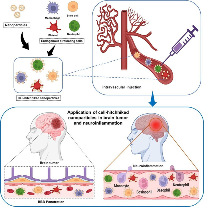
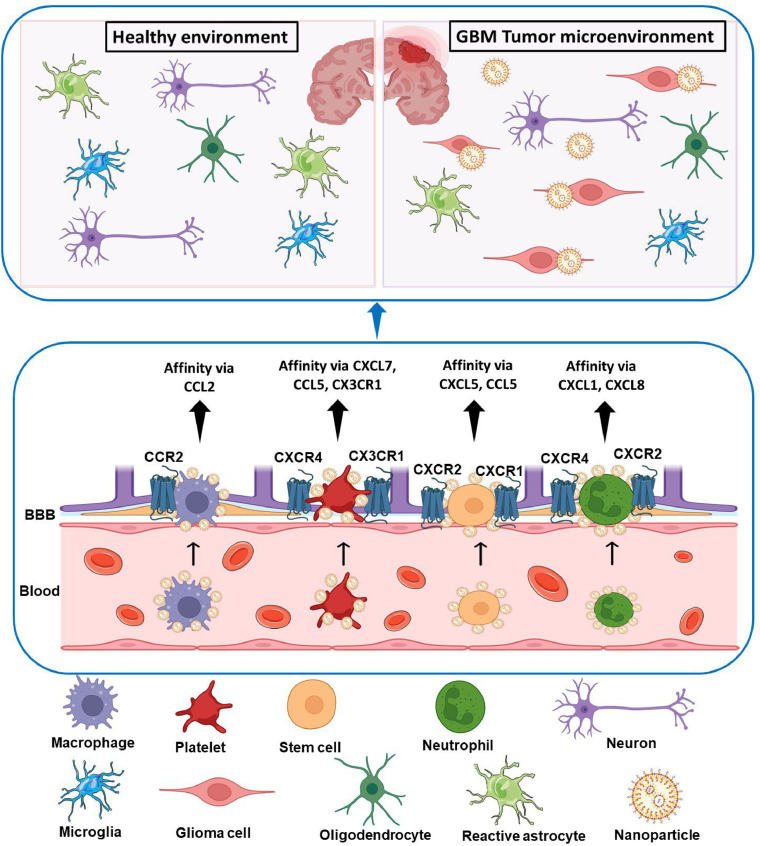
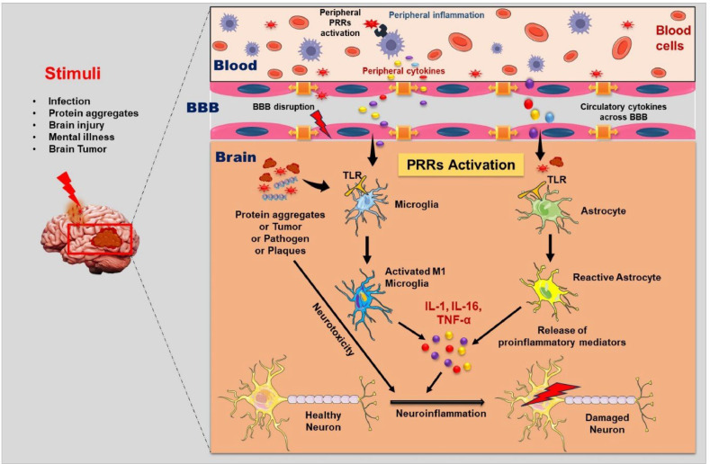
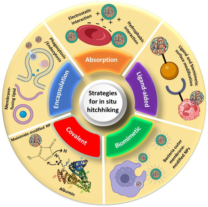
Cellular hitchhiking is an emerging therapeutic strategy that uses an endogenous cell migration mechanism to deliver therapeutics to specific sites in the body. Owing to the low permeability and presence of the blood-brain barrier (BBB), the targeted delivery of therapeutics is limited, leading to inadequate localization in the brain. NCs fail to extravasate significantly into the tumor microenvironment (TME), demonstrating poor accumulation and tumor penetration. The novel cellular hitchhiking concept has been utilized to promote systemic half-life and therapeutic targeting. Neoplastic and neuroinflammatory diseases of the brain, including glioblastoma and neuroinflammation, face critical hurdles for efficiently delivering therapeutic entities owing to the BBB. Cellular hitchhiking can surmount these hurdles by utilizing various cell populations, such as stem cells, monocytes/macrophages, neutrophils, and platelets, as potential functional carriers to deliver the therapeutic cargo through the BBB. These carrier cells have the innate capability to traverse the BBB, transit through the brain parenchyma, and specifically reach disease sites such as inflammatory and neoplastic lesions owing to chemotactic navigation, i.e., movement attributed to chemical stimuli. Chemotherapeutic drugs delivered by cellular hitchhiking to achieve tumor-specific targeting have been discussed. This article explores various cell types for hitchhiking NCs to the TME with in-depth mechanisms and characterization techniques to decipher the backpack dissociation dynamics (nanoparticle payload detachment characteristics from hitchhiked cells) and challenges toward prospective clinical translation.

Keywords: Brain-targeting; Cellular hitchhiking; Glioblastoma.


Conflict of interest statement

The authors report no conflicts of interest. The authors alone are responsible for the content and writing of this article.

Figures

Image, graphical abstract
Graphical abstract
Fig 1
Fig. 1
Mechanism of nanocarrier delivery to the brain mediated by cellular hitchhiking.
Fig 2
Fig. 2
Crosstalk between peripheral and central immune cells in neuroinflammation. Peripheral and central immune cells play pivotal roles in neuroinflammation across various CNS complications. External and internal stimuli, such as infections, brain injury, mental illness, and brain tumors, can trigger this process. Activation of PRRs on microglia and astrocytes promotes neuroinflammation, releasing pro-inflammatory cytokines (e.g., IL-1, IL-6, TNF-α), ultimately resulting in neuronal death. Furthermore, protein aggregates, demyelinated plaques, and brain tumors can directly induce neurotoxicity, contributing to neuronal demise. Tumor-associated inflammation, characterized by infiltrating immune cells and cytokine release within the brain microenvironment, exacerbates neuroinflammatory responses and neuronal injury. Abbreviations: PRRs: Pattern recognition receptors; TLR, Toll-like receptors.
Fig 3
Fig. 3
Schematic diagram illustrating the primary strategies for in situ cellular hitchhiking of NPs. Created with Biorender.
Fig 4
Fig. 4
Adsorption of PLGA NPs to human, mouse and rabbit RBCs. SEM images of (A) DEX-PLGA NPs, (B) Adsorption of PLGA-NP onto human RBCs and (C) Adsorption of PLGA-NP onto mouse RBCs. (D) Coupling efficiency of PLGA NPs onto human, mouse, and rabbit RBCs. (E) Percentages of RBC populations carrying NPs. Reproduced with permission from [114] Copyright© 2022 Springer Nature. Effect of protein coronas on layer-by-layer assembled polymer particles functionalized with monoclonal antibodies. (F) Schematic illustration of the assembly of monoclonal antibody-functionalized capsules and particles. (G) Fluorescence and transmission electron microscopy images of Alexa Fluor 633-labeled (red) capsules (1, 2) and particles (3, 4) functionalized with Alexa Fluor 488-labeled huA33 mAbs (green). (H) Deconvolution microscopy of LIM2405+ and LIM2405− cells incubated with the huA33 mAb-functionalized capsules (1, 2) or particles (3, 4) in the absence (1, 3) or presence (2, 4) of a protein corona. Reproduced with permission from [116] Copyright © 2015 American Chemical Society.
Fig 5
Fig. 5
Hitchhiking of D@MLL NPs on monocytes after low-dose RT for treatment of GBM. (A) DiR fluorescence intensity in the brain of GL261-bearing mice with varying radiation dosages and normal mice in the absence of radiation by tail vein injection using an IVIS system. (B) Bioluminescence and DiR fluorescence imaging in GL261-luc bearing C57/BL6 mice. (C) Quantified intensity of PpIX in D@ML, DP@ML, and DP@MLL. (D) Representative fluorescence images of PpIX were obtained at various time points. (E) Survival curves and body weight variation in mice with various treatments. (F) In vivo bioluminescence imaging of orthotopic GL261-luc GBM bearing mice subjected to several treatments. Reproduced with permission from Kuang et al. [42] Copyright© 2023 ACS Publications.
Fig 6
Fig. 6
Effect of DOX@M1-NPs in U87 glioma-bearing mouse model. (A) Cytotoxic effect of DOX@NPs on M1 macrophages post 8 h. (B) Release of DOX from M1 macrophages and its retention in them. (C) Distribution of DOX@NPs at varying time points. (D) Whole-body imaging of DiR loading NPs and M1-NPs after tail vein injection. (E) ROI examination of mean fluorescence intensity (MFI) in the brain. (F) Ex vivo imaging of brains after 24 h injection. (G) In vivo anti-cancer effect on orthotopic U87 glioma-bearing mouse. (H) Caspase-3 expression in brain tumor. Reproduced with permission from [109] Copyright© 2018 Taylor and Francis.
Fig 7
Fig. 7
M1 macrophage incubated in situ fibrin gel for the treatment of post-operative GBM. (A) Pictographic representation of the biological characteristics of M, MD, MD-NVs, and MPDA-DOX-NVs. NIR-triggered release of DOX from internalized MD-NVs is depicted. Further cellular viability of GL261 and BV-2 cells at different concentrations is depicted; (B) H&E staining of major organs of GBM mice model after treatment for 24 d; (C)In vivo bioluminescence images and MRI images of treated GBM mice after surgical resection; (D) Flow cytometry plots of MDSCs, M2 and M1-like macrophages, and T cells in postresection- glioma brain displaying the immune modulation capability of the hydrogel. Reproduced with permission from [146] Copyright© 2023 American Chemical Society.
Fig 8
Fig. 8
Administration of ND-MMSNs in postsurgical glioma-bearing mice. (A) Flow cytometry, CLSM, and T2-weighted MRI imaging of NEs after coincubation with D-MMSNs and D-FMMSNs. (B) CLSM images of NETs following ND-FMMSNs incubation with U87 cells for 2 h; (C)In vivo T2-weighted MR imaging (D)In vivo biodistribution and fluorescence images (E) Pictographic representation of in vivo bioluminescent images for determining tumor volume. Reproduced with permission from [156] Copyright© 2018 Springer Nature.
Fig 9
Fig. 9
Evaluation of targeting ability of Cur-HPPS to inflammatory monocytes in the brain. (A) Typical images of DiR-BOA signal. (B) Percentage of DiR-BOA+ and MFI of DiR-BOA cells in the neutrophil and monocyte populations after 6 and 24 h i.v. injection of HPPS(DiR-BOA). (C) Spinal cord sections of EAE and normal mice after 24 h. (D) Intravital imaging of brain surface after 3 and 24 h. (E) Confocal microscopy of NF-κB activated monocytes. Reproduced with permission from [161] Copyright© 2020 Elsevier.
Fig 10
Fig. 10
Evaluation of the ability of CPSAT and ROS-responsive liposomes to target thrombus in stroke mice. (A) Fluorescence intensity of various organs post administration in stroke mice for 24 h. (B) Representation of relative fluorescence intensity in stroke sites. (C) Representative thrombolytic images in mice treated with different groups after visible thrombus formation. (D) Relative fluorescence intensity of thrombus site at different time intervals. (E) Representative MRI, laser speckle and OCT images after 48 h of administration. (F-H) Quantitative evaluation of MRI, laser speckle, and OCT images, respectively. (I) Immunofluorescence (IF) staining site in stroke brain, IF images of NeuN (red, neurons) and Brdu (green, apoptosis cells) and IF images of glial fibrillary acidic protein (GFAP) staining, ionized calcium binding adapter molecule 1 (Iba-1) staining, and Gr-1 respectively. Reproduced with permission from [172] Copyright © 2023, American Chemical Society.

Similar articles

References

    1. Liu Q., Li X., Wang Y., Liu X., Cao Y., Zhang H., et al. Neutrophil hitchhiking for nanoparticle delivery to the central nervous system. Appl Mater Today. 2024;38
    1. Liu X., Yin L., Shen S., Hou Y. Inflammation and cancer: paradoxical roles in tumorigenesis and implications in immunotherapies. Genes Dis. 2023;10:151–164. - PMC - PubMed
    1. Patterson C.M., Shaw T.D., Gerovasili V., Khatana U., Jose RJ. Emerging therapies and respiratory infections: focus on the impact of immunosuppressants and immunotherapies. Clinical Medicine. 2024;24 - PMC - PubMed
    1. Kaur N., Chugh H., Sakharkar M.K., Dhawan U., Chidambaram S.B., Chandra R. Neuroinflammation mechanisms and phytotherapeutic intervention: a systematic review. ACS Chem Neurosci. 2020;11:3707–3731. - PubMed
    1. Euteneuer F., Dannehl K., del Rey A., Engler H., Schedlowski M., Rief W. Immunological effects of behavioral activation with exercise in major depression: an exploratory randomized controlled trial. Transl Psychiatry. 2017;7:e1132. - PMC - PubMed

LinkOut - more resources