

Ionized calcium and ionized magnesium disturbances in dogs and cats with septic peritonitis
- PMID: 40454174
- PMCID: PMC12123877
- DOI: 10.3389/fvets.2025.1550701
Ionized calcium and ionized magnesium disturbances in dogs and cats with septic peritonitis
Abstract
Introduction: Hypocalcemia and magnesium disturbances are linked to vasoplegia, cardiac arrhythmias, gastrointestinal ileus, and coagulopathies. In human critical care patients, these imbalances are associated with higher mortality and longer hospital stay. Little is known about such associations in companion animals. Our study assessed the prevalence of ionized calcium (iCa) and ionized magnesium (iMg) disturbances in dogs and cats with septic peritonitis at presentation, and their association with administration of antiarrhythmics, vasopressors, prokinetics, and plasma, hospitalization duration and mortality.
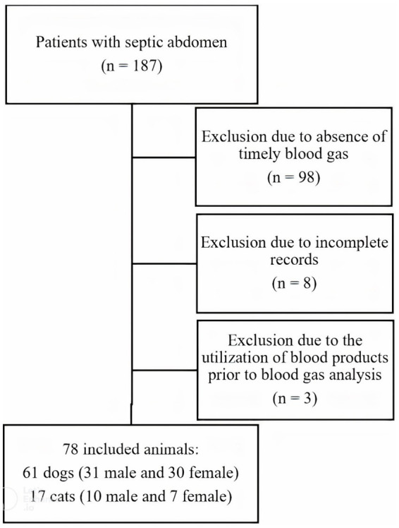
Methods: Medical records of patients with septic peritonitis from January 2018 to December 2023 were reviewed. Inclusion criteria were confirmed septic peritonitis and blood gas analysis with ionized calcium and magnesium values at admission. Data collected included signalment, diagnosis and cause, calcium and magnesium levels, administration of vasopressors, antiarrhythmics, prokinetics, and plasma, length of hospitalization, and survival. Comparisons were made using Chi-square, Fisher exact test, and ANOVA. Correlations were assessed with the Spearman coefficient.
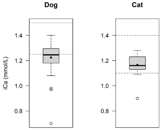
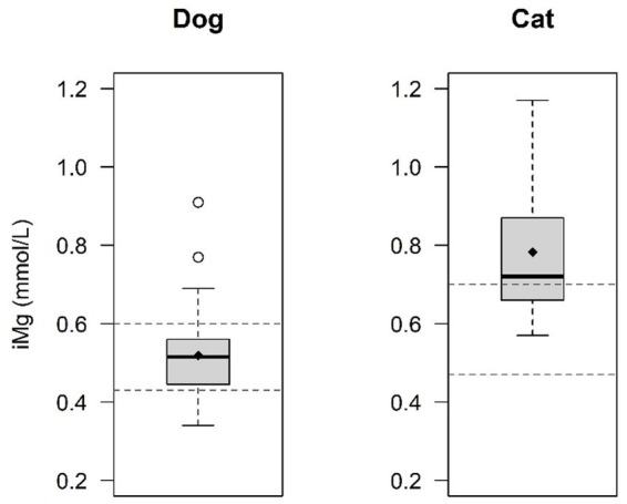
Results: Sixty-one dogs and 17 cats were included. Hypocalcemia (iCa < 1.25 mmol/L in dogs; <1.10 mmol/L in cats) was diagnosed in 51.7% of dogs and 23.5% of cats. Hypomagnesemia (iMg < 0.43 mmol/L in dogs; <0.47 mmol/L in cats) was found in 13.5% of dogs and 0% of cats, and hypermagnesemia (iMg > 0.6 mmol/L in dogs; >0.7 mmol/L in cats) in 15.4% of dogs and 60% of cats. Hypocalcemia and hypermagnesemia were significantly more common in dogs and cats, respectively. Ionized hypocalcemia was associated with plasma administration in dogs (p = 0.038). No significant correlation was found between iCa and iMg disturbances and length of hospitalization in dogs (respectively p = 0.62 and p = 0.62) or cats (respectively p = 0.62 and p = 0.27). The survival rate was 50%, with no association between iCa and iMg derangements and survival in dogs (p = 0.30 and p = 0.38 respectively), nor in cats (p = 0.29 and p = 0.29 respectively). A significant difference was observed in the mean iMg values between survivors (0.49 ± 0.09 mmol/L) and non-survivors (0.55 ± 0.11 mmol/L) in dogs (p = 0.042).
Discussion: In this population with septic peritonitis, about half of the dogs and a quarter of the cats had ionized hypocalcemia. Hypo- and hypermagnesemia were rare in dogs, while hypermagnesemia was common in cats and hypomagnesemia was not seen in cats. Dogs and cats with hypocalcemia were more likely to receive plasma. Mean iMg values were lower in survived dogs than dogs that did not survive.
Keywords: calcium metabolism; critical illness; ionized hypermagnesemia; ionized hypocalcemia; ionized hypomagnesemia; septic abdomen.
Copyright © 2025 Debie, Giberto, Noel, Paepe and Gommeren.
Conflict of interest statement
The authors declare that the research was conducted in the absence of any commercial or financial relationships that could be construed as a potential conflict of interest. The author(s) declared that they were an editorial board member of Frontiers, at the time of submission. This had no impact on the peer review process and the final decision.
Figures


References
-
- Schenck PA, Chew DJ, Nagode LA, Rosol TJ. Disorders of Calcium: Hypercalcemia and Hypocalcemia. In: DiBartola SP, editor. Fluid, electrolyte, and acid-base disorders in small animal practice, Fourth Edition. St. Louis, Missouri: Elsevier, (2012), pp. 120–194.
LinkOut - more resources
Full Text Sources
Miscellaneous