. 2025 Jun 2;135(11):e177533.
doi: 10.1172/JCI177533.

Polybromo 1/vimentin axis dictates tumor grade, epithelial-mesenchymal transition, and metastasis in pancreatic cancer

Affiliations

Polybromo 1/vimentin axis dictates tumor grade, epithelial-mesenchymal transition, and metastasis in pancreatic cancer

Munenori Kawai et al. J Clin Invest. .

Abstract

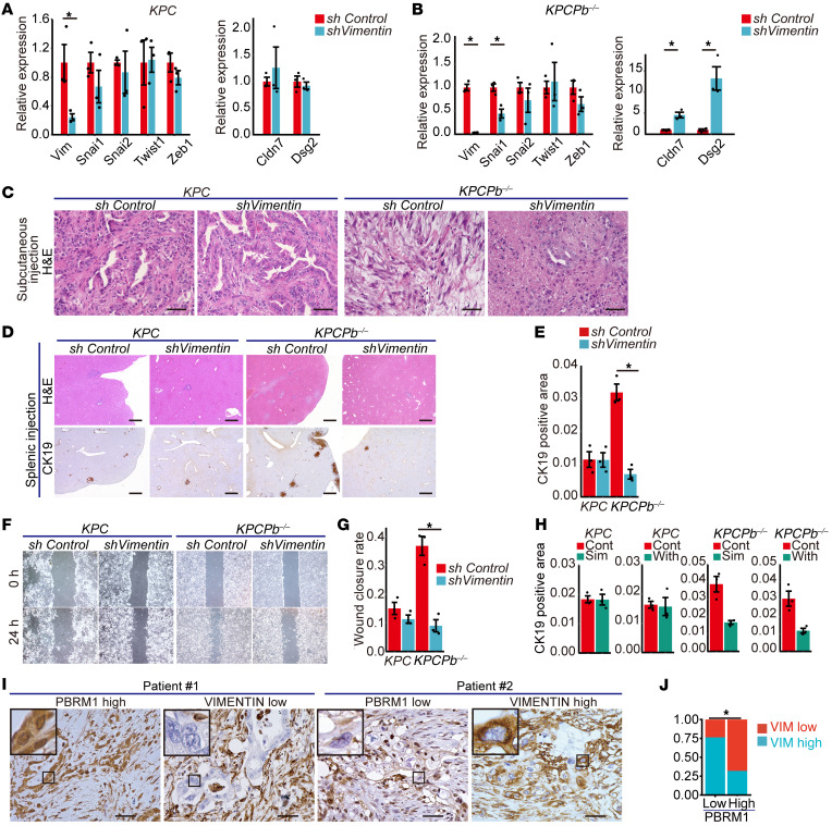
Mutations in Polybromo 1 (PBRM1), a subunit of the switch/sucrose nonfermentable (SWI/SNF) chromatin remodeling complex, are frequently observed in several cancers, including pancreatic ductal adenocarcinoma (PDAC). In this study, we demonstrated that pancreas-specific loss of Pbrm1 in mice harboring Kras mutations and Trp53 deletions accelerated the development of poorly differentiated PDAC, epithelial-mesenchymal transition (EMT), and metastasis, resulting in worsened prognosis. Pbrm1 loss in preexisting PDAC shifted the tumor grade from a well- to a poorly differentiated state and elevated vimentin expression. Pbrm1-null PDAC exhibited downregulation of apical junction genes and upregulation of EMT pathway genes, including the vimentin and squamous molecular subtype signature genes. Mechanistically, PBRM1 bound to the vimentin gene promoter and directly downregulated its expression. Furthermore, suppression of vimentin in Pbrm1-null PDAC cells reversed the dedifferentiation phenotype and reduced EMT and metastasis. Consistently, reduced PBRM1 expression correlated with high vimentin expression, poorly differentiated histology, a high recurrence rate, and reduced overall survival in human PDACs. Additionally, PDAC with PBRM1 deletion was associated with the aggressive squamous molecular subtype. Our data established PBRM1 as a tumor suppressor that controls tumor grade and metastasis of PDAC by regulating vimentin expression.

Keywords: Cancer; Epigenetics; Gastroenterology; Mouse models; Oncology.


Conflict of interest statement

Conflict of interest: YF is employed by Sumitomo Pharma Co., Ltd.

Figures

Figure 1
Figure 1. Low PBRM1 expression is associated with high tumor grade, high recurrence rate, poor prognosis, and the squamous molecular subtype.
(A) Representative immunohistochemical analysis (IHC) of PBRM1 in human pancreatic samples. Scale bar: 50 μm. Data are representative of 3 independent experiments. (B) Recurrence rates at high (n = 50) and low (n = 52) PBRM1 expression levels in human PDACs. (C) Rates of high and low PBRM1 IHC levels in human well- and moderately differentiated pancreatic ductal adenocarcinoma (well+mod) (n = 85) and poorly differentiated pancreatic ductal adenocarcinoma (por) (n = 20). (D and E) Kaplan-Meier plots showing overall survival or disease-free survival in a cohort of pancreatic cancer patients with high (n = 50) and low (n = 55 (D), 54 (E)) PBRM1 protein expression levels. (F) Multivariate Cox proportional hazard analysis for overall survival in a cohort of patients with pancreatic cancer. HR, hazard ratio. CI, confidence interval. (G) PBRM1 mRNA expression with each putative copy number alteration status from a cohort of 147 patients in TCGA dataset. (H) GSEA enrichment plots of BAILEY GP4 SQUAMOUS and BAILEY GP5 SQUAMOUS in PDAC tumors from PBRM1 diploid PDAC (n = 100) and PDAC with PBRM1 deletion (n = 42) in a cohort of 142 patients in TCGA dataset. NES, normalized enrichment score; FDR, false discovery rate. (I) Representative IHC results of ΔNp63 and PBRM1 in human adenosquamous carcinoma samples (n = 11). Scale bar: 50 μm. (J) Rates of high and low PBRM1 IHC levels in human well- and moderately differentiated PDAC (well+mod) (n = 85) and adenosquamous carcinoma (ASC) (n = 11) and squamous cell carcinoma of the pancreas (SCC) (n = 1). *P < 0.05. G, Data shown as mean ± SE. B, Pearson’s χ2 test. C and J, Fisher’s exact test. D and E, Log-rank (Mantel-Cox) test. G, 1-way ANOVA, followed by the Tukey’s multiple comparison test.
Figure 2
Figure 2. Pancreatic PBRM1 deletion accelerates the formation of precancerous lesions and leads to poorly differentiated pancreatic ductal adenocarcinoma with a poor prognosis.
(A) The genetic strategy employed to activate oncogenic Kras and delete Pbrm1, specifically in the pancreas, obtained from the embryonic stage. (B) Representative hematoxylin and eosin (H&E), Alcian blue, and PBRM1 staining in Ptf1aCre; LSL-KrasG12D(KC), Ptf1aCre; LSL-KrasG12D; Pbrm1f/wt (KCPb+/–), and Ptf1aCre; LSL-KrasG12D; Pbrm1f/f (KCPb–/–) mice at 20 weeks of age. Scale bar: 200 μm (H&E and alcian blue); 50 μm (Pbrm1). Data are representative of 3 independent experiments. (C) Quantification of Alcian blue–positive late acinar-to-ductal metaplasia and PanINs determination by combining 3 independent sections from KC (n = 3), KCPb+/– (n = 3), and KCPb–/– (n = 3) mice at 8 weeks of age. *P < 0.05, 1-way ANOVA, followed by Tukey’s multiple comparison test. Data are shown as mean ± SE. (D) Quantification of Alcian blue–positive late acinar-to-ductal metaplasia and PanINs determination by combining 3 independent sections from KC (n = 4), KCPb+/– (n = 3), and KCPb–/– (n = 3) mice at 20 weeks of age. *P < 0.05, 1-way ANOVA, followed by Tukey’s multiple comparison test. Data are shown as mean ± SE. (E) Representative H&E, CK19, and PBRM1 staining in PDACs from KC, KCPb+/–, and KCPb–/– mice. Scale bar: 50 μm. Data are representative of 3 independent experiments. (F) Kaplan-Meier plots showing the overall survival in the cohorts of KC (n = 86) and KCPb–/– (n = 54) mice. The log-rank (Mantel-Cox) test was used to assess statistical significance. (G) Rate of PDAC incidence in KC (n = 25), KCPb+/– (n = 14), and KCPb–/– (n = 12) mice aged 20–30 weeks. *P < 0.05, Fisher’s exact test.
Figure 3
Figure 3. Pancreatic PBRM1 loss synergizes with oncogenic KRAS and heterozygous Trp53 deletion to yield poorly differentiated PDAC and induce liver metastasis with a poor prognosis.
(A) Genetic strategy used to activate oncogenic Kras and delete Pbrm1 and Trp53 specifically in the pancreas from the embryonic stage. (B) Representative H&E staining in PDAC from Ptf1aCre; LSL-KrasG12D; Trp53f/wt (KPC) (n = 16), Ptf1aCre; LSL-KrasG12D; Trp53f/wt; Pbrm1f/wt (KPCPb+/–) (n = 18) and Ptf1aCre; LSL-KrasG12D; Trp53f/wt; Pbrm1f/f (KPCPb–/–) (n = 20) mice at the primary site. Scale bar: 50 μm. (C) Rate of tumor grade 3 or 4 of PDACs in KPC (n = 16), KPCPb+/– (n = 19), and KPCPb–/– (n = 20) mice. (D) Representative H&E staining in the livers of KPC (n = 16) mice and metastatic PDAC in the livers of KPCPb+/– (n = 7) and KPCPb–/– (n = 11) mice. Scale bar: 50 μm. (E) Quantification of liver metastasis incidence in KPC (n = 16), KPCPb+/– (n = 15), and KPCPb–/– (n = 18) mice during moribund state. (F) Kaplan-Meier plots showing overall survival of KPC (n = 37) and KPCPb–/– (n = 52) mice. (G) Representative H&E staining in PDAC allografted subcutaneously or orthotopically with PDAC cells from KPC (n = 3) and KPCPb–/– (n = 3) mice. Scale bar: 50 μm. (H) Representative H&E and CK19 staining of liver metastases after injection of PDAC cells into the spleen of KPC (n = 3) and KPCPb–/– (n = 3) mice. Metastatic lesions were circled by blue lines in H&E staining. Scale bar: 500 μm. (I) Rate of CK19-positive areas determined by combining 3 independent sections of liver metastases after injection of PDAC cells into the spleen of KPC (n = 3) and KPCPb–/– (n = 3) mice. (J) Representative image of the scratch assay with PDAC cells from KPC (n = 3) and KPCPb–/– (n = 3) mice. (K) Quantification of the scratch assay using PDAC cells from KPC (n = 3) and KPCPb–/– (n = 3) mice. (L) Representative coimmunostaining of vimentin, CK19, and Hoechst in primary and metastatic lesions in KPCPb–/– mice (n = 3). Scale bar: 50 μm. (M) Quantification of the rate of the vimentin-positive cancer cells in primary and metastatic lesions in KPCPb–/– mice (n = 3). *P < 0.05. C, E, Fisher’s exact test. F, Log-rank (Mantel-Cox) test. I, K Student t test. M, paired t test. Data shown as mean ± SE.
Figure 4
Figure 4. Pancreatic PBRM1 loss synergizes with oncogenic KRAS and homozygous Trp53 deletion to accelerate the development of poorly differentiated PDAC and to facilitate the EMT of PDAC cells, resulting in a poor prognosis.
(A) Representative H&E staining of the pancreas from Ptf1aCre; LSL-KrasG12D; Trp53f/f (KP–/–C), Ptf1aCre; LSL-KrasG12D; Trp53f/f; Pbrm1f/wt (KP–/–CPb+/–), and Ptf1aCre; LSL-KrasG12D; Trp53f/f; Pbrm1f/f (KP–/–CPb–/–) mice at 3 weeks of age and 6-to-9 weeks of age in the moribund state. Scale bar: 50 μm. Data are representative of 3 independent experiments. (B) Rate of tumor grade 3 or 4 of PDACs in KP–/–C (n = 10), KP–/–CPb+/– (n = 14), and KP–/–CPb–/– (n = 23) mice. *P < 0.05, Fisher’s exact test. (C) Kaplan-Meier plots showing overall survival of KP–/–C (n = 31) and KP–/–CPb–/– (n = 51) mice. The log-rank (Mantel-Cox) test has been used to assess the statistical significance. (D) Representative coimmunostaining of vimentin and tdTomato in PDAC from Ptf1aCre; LSL-KrasG12D Trp53f/f; LSL-Rosatd–tomato (KP–/–CTomato) (n = 3) and Ptf1aCre; LSL-KrasG12D; Trp53f/f; Pbrm1f/f; LSL-Rosatd–tomato (KP–/–CPb–/–Tomato) (n = 3) mice. Scale bar: 50 μm. (E) Kaplan-Meier plots showing overall survival of KP–/–C mice treated with gemcitabine (n = 4) and KP–/–C mice without treatment with gemcitabine (n = 31). The log-rank (Mantel-Cox) test has been used to assess the statistical significance. (F) Kaplan-Meier plots showing overall survival of KP–/–CPb–/– mice treated with gemcitabine (n = 6) and KP–/–CPb–/– mice without treatment with gemcitabine (n = 51). The log-rank (Mantel-Cox) test has been used to assess the statistical significance. (G) Representative coimmunostaining of CK19 and ΔNp63 in PDAC from KP–/–C (n = 3) and KP–/–CPb–/– (n = 3) mice. Scale bar: 50 μm. (H) Quantification of the rate of the ΔNp63-positive cancer cells in PDAC from KP–/–C (n = 3) and KP–/–CPb–/– (n = 3) mice. *P < 0.05, Student t test. Data shown as mean ± SE.
Figure 5
Figure 5. PBRM1 ablation in established PDAC results in the conversion of tumor grade into poorly differentiated PDAC in mice.
(A) Genetic strategy used to activate oncogenic Kras and delete Trp53 heterozygously at the embryonic stage and delete Pbrm1 at the tumor-bearing adult stage. (B) Experimental design for tamoxifen administration and analysis. (C) Representative H&E, CK19, and PBRM1 staining in PDAC from Pdx1-Flp; FSF-KrasG12D; Trp53fr/wt; FSF-Rosa26CAG–CreERT2 (KPF), Pdx1-Flp; FSF-KrasG12D; Trp53fr/wt; FSF-Rosa26CAG–CreERT2; Pbrm1f/wt (KPFPb+/–) and Pdx1-Flp; FSF-KrasG12D; Trp53fr/wt; FSF-Rosa26CAG–CreERT2; Pbrm1f/f (KPFPb–/–) mice 2 weeks after tamoxifen administration. Scale bar: 50 μm. Data are representative of 3 independent experiments. (D) Representative H&E, vimentin, PBRM1, and CK19 staining of PDAC from KPFPb–/– mice 2 weeks after tamoxifen administration, which exhibited a transient state of degradation of the tubular component to undifferentiated carcinoma. Scale bar: 50 μm. Data are representative of 3 independent experiments. (E) Rate of tumor grade 3 or 4 of PDACs in KPF (n = 15), KPFPb+/– (n = 10), and KPFPb–/– (n = 15) mice 2 weeks after tamoxifen administration and PDACs in KPF (n = 5), KPFPb+/– (n = 7), and KPFPb–/– (n = 16) mice without tamoxifen administration. *P < 0.05, Fisher’s exact test. (F) Rate of liver metastasis in KPF (n = 15), KPFPb+/– (n = 10), and KPFPb–/– (n = 15) mice 2 weeks after tamoxifen administration. *P < 0.05, Fisher’s exact test.
Figure 6
Figure 6. PBRM1 binds to the vimentin gene promoter to directly regulate its expression.
(A) GSEA of PDAC cells from KPC and KPCPb–/– mice using “Hallmark gene sets.” NES, normalized enrichment score. *P < 0.05. (B) GSEA enrichment plots of the HALLMARK apical junction. FDR, false discovery rate. (C) GSEA of PDAC cells from KPC and KPCPb–/– mice using the vimentin gene set. (D) Quantitative real-time PCR analysis of the relative mRNA expression of Cdh1, Cdh2, Cldn4, Cldn7, Dsc2, and Dsg2 in KPCPb–/– (n = 3) PDAC cells compared with KPC (n = 3) PDAC cells. *P < 0.05, Student t test. Data shown as mean ± SE. (E) Quantitative real-time PCR analysis of the relative mRNA expression of Vim, Snai1, Snai2, and Twist1 in KPCPb–/– (n = 3) PDAC cells compared with KPC (n = 3) PDAC cells. *P < 0.05, Student t test. Data shown as mean ± SE. (F) Venn diagram of the analysis of genes bound by PBRM1 and differentially expressed genes identified by RNA-seq. Sixty-eight genes are bound by H3K27Ac out of the 164 genes that are bound by PBRM1 and upregulated in KPC PDAC cells and 25 genes are bound by H3K27ac out of 60 genes that are bound by PBRM1 and upregulated in KPCPb–/– PDAC cells. (G) ChIP data of the PBRM1 and H3K27ac binding region in the vimentin gene promoter and coding regions. TSS, transcription start site. (H) Representative vimentin staining in PDAC of KPC and KPCPb–/– mice. Scale bar: 50 μm. Data are representative of 3 independent experiments.
Figure 7
Figure 7. Vimentin inhibition reverses the dedifferentiation phenotype and reduces metastasis of Pbrm1-null PDAC in mice.
(A) Quantitative real-time PCR analysis of the relative mRNA expression of Zeb1, Vim, Snai1, Snai2, Twist1, Cldn7, and Dsg2 in KPC PDAC cells with shRNA knockdown of the vimentin gene (shVimentin) (n = 3) compared with KPC PDAC cells with shRNA control (shControl) (n = 3). *P < 0.05, Student t test. Data shown as mean ± SE. (B) Quantitative real-time PCR analysis of the relative mRNA expression of Zeb1, Vim, Snai1, Snai2, Twist1, Cldn7, and Dsg2 in KPCPb–/– PDAC cells with shRNA knockdown of the vimentin gene (shVimentin) (n = 3) compared with KPCPb–/– PDAC cells with shRNA control (shControl) (n = 3). *P < 0.05, paired t test. Data shown as mean ± SE. (C) Representative H&E staining in PDACs allografted subcutaneously with KPCshControl, KPCshVimentin, KPCPb–/–shControl, and KPCPb–/–shVimentin PDAC cells. Scale bar: 50 μm. Data are representative of 3 independent experiments. (D) Representative H&E and CK19 staining in metastatic PDAC after injection into the spleen with KPCshControl, KPCshVimentin, KPCPb–/–shControl, and KPCPb–/–shVimentin PDAC cells. Scale bar: 500 μm. Data are representative of 3 independent experiments. (E) Quantification of CK19-positive liver metastasis with splenic injection of KPC shControl (n = 3), KPCshVimentin (n = 3), KPCPb–/–shControl (n = 3), and KPCPb–/–shVimentin (n = 3) PDAC cells, determined by combining 3 independent sections. *P < 0.05, Student t test. Data are represented as mean ± SE. (F) Representative images of the scratch assay with KPC shControl, KPCshVimentin, KPCPb–/–shControl, and KPCPb–/–shVimentin PDAC cells. Data are representative of 3 independent experiments. (G) Quantification of the scratch assay with KPC shControl (n = 3), KPCshVimentin (n = 3), KPCPb–/–shControl (n = 3), and KPCPb–/–shVimentin (n = 3) PDAC cells. *P < 0.05, Student t test. Data shown as mean ± SE. (H) Quantification of CK19-positive liver metastasis in mice treated with simvastatin (n = 3), Withaferin A (n = 3), and each vehicle control (n = 3) with splenic injection of KPC and KPCPb–/– PDAC cells, determined by combining 3 independent sections. *P < 0.05, Student t test. Data are represented as mean ± SE. (I) Representative IHC analysis of PBRM1 and vimentin in human PDACs. Patient #1 shows high PBRM1 expression and low expression of vimentin. Patient #2 shows the low expression of PBRM1 and the high expression of vimentin. Scale bar: 50 μm. Data are representative of 3 independent experiments. (J) Analysis of high vimentin expression in human PDACs (n = 105) surgically resected with high (n = 50) or low (n = 55) PBRM1 expression as determined by IHC. *P < 0.05, Pearson’s χ2 test.

Comment in

  • Switching on the evolutionary potential of pancreatic cancer: the tumor suppressor functions of PBRM1

References

    1. Siegel RL, et al. Cancer statistics, 2023. CA Cancer J Clin. 2023;73(1):17–48. doi: 10.3322/caac.21763. - DOI - PubMed
    1. Bednar F, Di Magliano MP. Chemotherapy and tumor evolution shape pancreatic cancer recurrence after resection. Cancer Discov. 2020;10(6):762–764. doi: 10.1158/2159-8290.CD-20-0359. - DOI - PubMed
    1. Hidalgo M. Pancreatic cancer. N Engl J Med. 2010;362(17):1605–1617. doi: 10.1056/NEJMra0901557. - DOI - PubMed
    1. Park W, et al. Pancreatic cancer: a review. JAMA. 2021;326(9):851. doi: 10.1001/jama.2021.13027. - DOI - PMC - PubMed
    1. Herman JM, et al. Analysis of fluorouracil-based adjuvant chemotherapy and radiation after pancreaticoduodenectomy for ductal adenocarcinoma of the pancreas: results of a large, prospectively collected database at the Johns Hopkins Hospital. J Clin Oncol. 2008;26(21):3503–3510. doi: 10.1200/JCO.2007.15.8469. - DOI - PMC - PubMed

MeSH terms