. 2025 Jun 18;139(12):667-681.
doi: 10.1042/CS20255840.

DAPK1 acts as a positive regulator of hypertension via induction of vasoconstriction

Affiliations

DAPK1 acts as a positive regulator of hypertension via induction of vasoconstriction

Xiuli Zhang et al. Clin Sci (Lond). .

Abstract

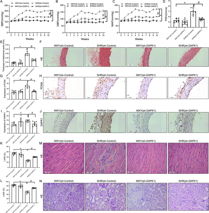
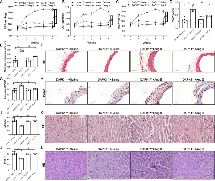
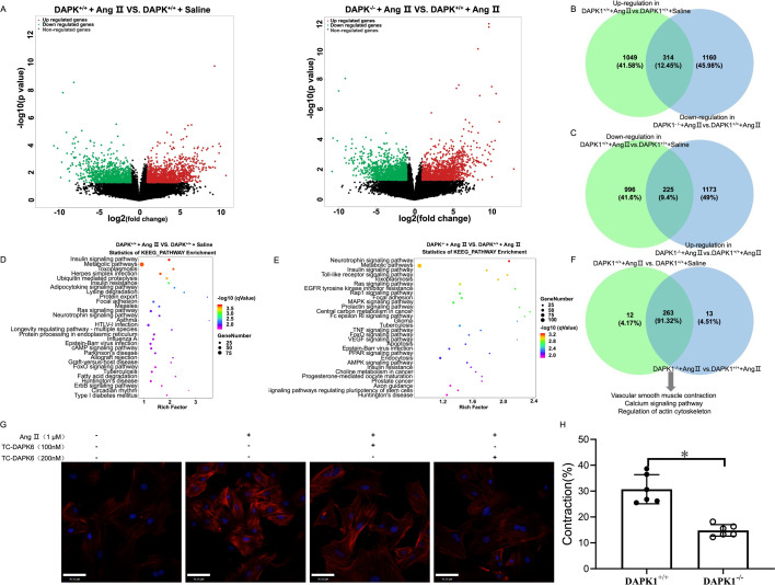
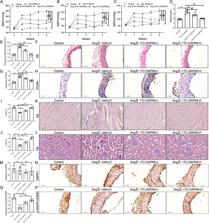
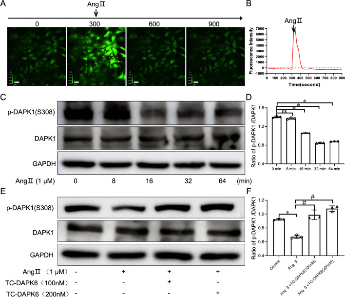
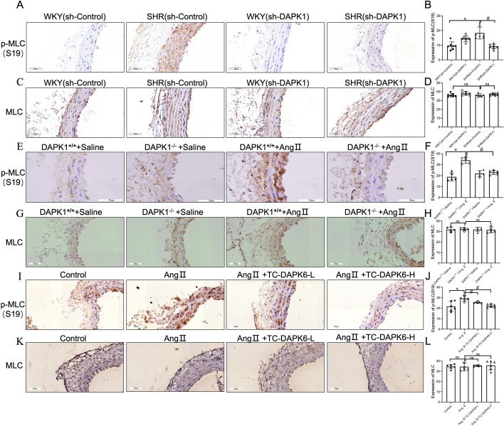
Death-associated protein kinase 1 (DAPK1) is a tumor suppressor gene involved in apoptosis, autophagy, and tumor progression. However, its role in hypertension (HTN) remains largely unexplored and lacks systematic evaluation. We administered adeno-associated virus (AAV) harboring short hairpin RNA targeting DAPK1 or control short hairpin RNA to male spontaneously hypertensive rats (SHRs) and Wistar-Kyoto rats. Additionally, wildtype and DAPK1 knockout mice were infused with angiotensin II (Ang II) or saline for four weeks. Male C57BL/6 mice underwent a four-week Ang II infusion and were treated with TC-DAPK6, a selective DAPK1 inhibitor. We examined the abdominal aortas (AAs) of mice and rats for pathological changes, measured blood pressure (BP) and pulse wave velocity using noninvasive BP methods, ultrasound, and hematoxylin and eosin staining. The role of DAPK1 in early HTN was further assessed through immunofluorescence, ex vivo isometric constriction of the AA, RNA sequencing, Western blot, and immunohistochemistry. Our study demonstrated that the targeted inhibition of DAPK1 with AAV significantly ameliorated HTN in SHRs and reduced damage to the AAs and target organs, including the heart and kidneys. Meanwhile, DAPK1 knockout or inhibition in mice significantly ameliorates Ang II-induced HTN in mice, as well as reducing damage to the AAs and target organs, including the heart and kidneys. Mechanistically, DAPK1 inhibition prevents myosin light chain (MLC) phosphorylation at serine 19, reducing vasoconstriction and protecting against HTN. In conclusion, DAPK1 is involved in HTN pathogenesis by regulating the MLC pathway to mediate vascular constriction, highlighting potential as a therapeutic target for HTN.

Keywords: DAPK1; MLC; hypertension; kinase activity; vasoconstriction.


Conflict of interest statement

The authors declare that they have no known competing financial interests or personal relationships that could have appeared to influence the work reported in this paper.

Figures

Figure 1:
Figure 1:. Death-associated protein kinase 1 (DAPK1) suppression lowers blood pressure and reduces vascular and organ damage in spontaneously hypertensive rats (SHRs).
(A–C) Tail-cuff plethysmography: systolic blood pressure (SBP), diastolic blood pressure (DBP), and mean arterial pressure (MAP). (D,E) Ultrasound: pulse wave velocity (PWV) and aortic wall thickness. (F) Hematoxylin-eosin (H&E)-stained abdominal aorta sections (400× magnification). (G–J) immunohistochemical (IHC) analysis: proliferating cell nuclear antigen (PCNA) (G,H) and DAPK1 (I,J) expression in aortic tissues (scale bar = 50 µm). (K,L) Echocardiography: left ventricular fractional shortening (LVFS) and left ventricular ejection fraction (LVEF). (M,N) H&E-stained cardiac and renal tissues (scale bar = 50 µm). n=7, *,#p<0.05 vs. sh-Control and sh-DAPK1, respectively.
Figure 2:
Figure 2:. DAPK1 deficiency alleviates angiotensin II (Ang II)-induced hypertension and vascular pathology.
(A–C) SBP, DBP, and MAP in each group. (D,E) Ultrasonography: PWV and aortic wall thickness. (F) H&E-stained cross-sections of abdominal aorta tissues (scale bar = 50 µm). (G,H) IHC analysis: PCNA expression in aortic tissues (scale bar = 50 µm). (I,J) LVFS and LVEF. (K,L) H&E-stained cardiac and renal tissues (scale bar = 50 µm). n=5, *,#p<0.05 vs. DAPK1+/+ + Ang II and DAPK1−/− +Ang II, respectiely.
Figure 3:
Figure 3:. DAPK1 deficiency mitigates Ang II-provoked aortic constriction in hypertensive mice.
(A) Volcano plots: differentially expressed transcripts (DETs) in abdominal aorta tissues (p<0.05, |fold change| ≥ 2). (B,C) Comparative transcript analysis: increased and decreased DETs in DAPK1+/++ Ang II vs. DAPK1+/+ + saline groups and DAPK1−/− + Ang II vs. DAPK1+/+ + Ang II. (D–F) KEGG analysis: Top 30 enriched pathways in DAPK1+/+ + Ang II vs. DAPK1+/+ + saline (D) and DAPK1−/− + Ang II vs. DAPK1+/+ + Ang II (E), with integrated pathway overlaps (F). (G) Immunofluorescence staining: F-actin (red) with DAPI-labeled nuclei (blue); scale bar = 20 µm. (H) Effect of DAPK1 knockdown on tension in Ang II pre-constricted abdominal aorta rings.
Figure 4:
Figure 4:. DAPK1 inhibition reverses Ang II-induced hypertension and vascular damage.
(A–C) TC-DAPK6 reduces SBP, DBP, and MAP in Ang II-triggered hypertensive mice. (D,E) Ultrasonography: PWV and aortic wall thickening. (F) H&E-stained cross-sections of the abdominal aorta. (G,H) IHC analysis: PCNA expression in aortic tissues (scale bar = 50 µm). (I,J) LVFS and LVEF measurements. (K,L) H&E-stained cardiac and renal tissues. (M–P) IHC analysis: DAPK1 (M) and p-DAPK1(S308) (O) expression in aortic tissues, with statistical representation. n=6,*,#p<0.05 vs. Control and Ang II + ddH₂O, respectively.
Figure 5:
Figure 5:. Ang II increases intracellular calcium and activates DAPK1 in vascular smooth muscle cells (VSMCs).
(A,B) Real-time confocal microscopy: VSMCs loaded with Fluo-4 showing intracellular Ca²+ increase after Ang II (1 μM) stimulation (200× magnification). Ca²+ levels were normalized to baseline. (C,D) Western blotting: p-DAPK1(S308)/DAPK1 ratio over time (*P<0.05 vs. 0 min). (E,F) Western blot analysis was performed to determine the ratio protein expression of p-DAPK1(S308)/DAPK1 protein expression, *p<0.05 vs. the Control group, #p<0.05 vs. Ang II + group.
Figure 6:
Figure 6:. DAPK1 lowers blood pressure by inhibiting myosin light chain (MLC)-mediated vasoconstriction.
IHC analysis: Phosphorylated MLC at Ser19 (p-MLC(S19)) and total MLC in abdominal aortic tissues. (A–D) p-MLC(S19) and MLC levels in SHR (n=7, *,#p<0.05 vs. sh-Control and sh-DAPK1, respectively). (E–H) DAPK1+/+ and DAPK1−/− mice treated with Ang II (n=5, *,#p<0.05 vs. DAPK1+/+ + Ang II and DAPK1−/− + Ang II, respectively). (I–L) Control vs. Ang II + ddH₂O (n=6, *,#p<0.05 vs. Control and Ang II, respectively); 400× magnification, scale bar = 50 μm.

Similar articles

References

    1. Wang, Z., Chen, Z., Zhang, L., Wang, X., Hao, G., Zhang, Z.et al. (2018) Status of hypertension in China: results from the China hypertension survey, 2012-2015. Circulation 137, 2344–2356 10.1161/CIRCULATIONAHA.117.032380 - DOI - PubMed
    1. Mills, K.T., Bundy, J.D., Kelly, T.N., Reed, J.E., Kearney, P.M., Reynolds, K.et al. (2016) Global disparities of hypertension prevalence and control: A systematic analysis of population-based studies from 90 countries. Circulation 134, 441–450 10.1161/CIRCULATIONAHA.115.018912 - DOI - PMC - PubMed
    1. Li, D., Cao, F., Han, J., Wang, M., Lai, C., Zhang, J.et al. (2023) The sustainable antihypertensive and target organ damage protective effect of transcranial focused ultrasound stimulation in spontaneously hypertensive rats. J. Hypertens. 41, 852–866 10.1097/HJH.0000000000003407 - DOI - PubMed
    1. Gao, L., Zhang, J., Yang, T., Jiang, L., Liu, X., Wang, S. et al. (2023) STING/ACSL4 axis-dependent ferroptosis and inflammation promote hypertension-associated chronic kidney disease. Mol. Ther. 31, 3084–3103 10.1016/j.ymthe.2023.07.026 - DOI - PMC - PubMed
    1. Du, Y.N., Tang, X.F., Xu, L., Chen, W.D., Gao, P.J. and Han, W.Q. (2018) SGK1-FoxO1 signaling pathway mediates Th17/Treg imbalance and target organ inflammation in angiotensin II-induced hypertension. Front. Physiol. 9, 1581 10.3389/fphys.2018.01581 - DOI - PMC - PubMed

MeSH terms