Observational Study
. 2025 Jul;48(7):672-681.
doi: 10.1111/pace.15215. Epub 2025 Jun 2.

Fascicular/Purkinje Tissue Colocalized With Scar in Cardiomyopathy Patients Undergoing Ventricular Fibrillation Ablation

Affiliations
Observational Study

Fascicular/Purkinje Tissue Colocalized With Scar in Cardiomyopathy Patients Undergoing Ventricular Fibrillation Ablation

Emir Baskovski et al. Pacing Clin Electrophysiol. 2025 Jul.

Abstract

Background: Ventricular fibrillation (VF) is a poorly understood arrhythmia that is one of the main mechanisms of sudden cardiac death in patients with structural heart disease (SHD). Fascicular and Purkinje tissue (FPT) has been implicated in VF.

Objective: In this study, we sought to analyze the involvement of FPT colocalized with the scar area of SHD. Additionally, we aimed to investigate outcomes of FPT and scar substrate ablation in SHD VF patients and compare outcomes with VT ablation patients.

Methods: This is a retrospective observational study. Clinical and procedural data were collected.

Results: Sixteen patients undergoing VF ablation were assigned to the VF group, and their outcomes were compared to those of 170 patients who underwent structural VT ablation. In 15 patients, FPT targets colocalized with low voltage area, LPs, and/or LAVAs. Septal Purkinje and left posterior fascicle were most commonly involved. Procedural metrics were similar with patients undergoing VT ablation. During median follow-up of 5.5 months (interquartile range [IQR]: 3.5-10), VF recurred in two patients.

Conclusion: This study has shown frequent colocalization of FPT with electrophysiologic substrate (late potentials [LPs], low voltage area, etc.) in patients with SHD and VF. We have shown the feasibility of FTP and substrate ablation with good mid-term outcomes in a small group of patients. Larger studies are necessary for definitive evidence.

Keywords: Purkinje; catheter ablation; ventricular fibrillation.


Conflict of interest statement

The authors declare no conflicts of interest.

Figures

FIGURE 1
FIGURE 1
Multistep approach for VF ablation in patients with structural heart disease. EGM, electrocardiogram; LAVA, local abnormal ventricular activity; LP, late potential; LVA, low voltage area; PVC, premature ventricular complex; VT, ventricular tachycardia; VF, ventricular fibrillation. [Colour figure can be viewed at wileyonlinelibrary.com]
FIGURE 2
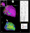
FIGURE 2
(A) Endocardial bipolar voltage map of the left ventricle with upper and lower thresholds set to 0.5 mV and 1.5 mV, respectively. Yellow tags on the map represent fascicular and Purkinje signals observed during mapping. These annotations were manually made and reviewed after mapping. Purple tags denote local abnormal ventricular activity (LAVA) sites, and light blue tags denote sites displaying late potentials (LPs). Note that septal left inferior fascicular and septal Purkinje sites colocalize with patchy bipolar scar, LAVAs, and LPs. (B) Same patient, late activation mapping displays an isochronal crowding area, which also colocalizes with fascicular and septal Purkinje sites. Inset at A displays Rf application sites. No Rf application was made near the left anterior fascicle due to the absence of electrophysiologic substrate. (C) In patients with conduction disturbance, such as left bundle branch block or fascicular block, identification of fascicular and Purkinje electrograms can occasionally be difficult. Differential pacing maneuvers can be employed. The inset shows a multicomponent electrogram with an initial sharp, high‐frequency signal that represents retrograde activation of fascicular tissue, while low low‐frequency, fractionated component represents LAVA. [Colour figure can be viewed at wileyonlinelibrary.com]
None
Outcomes and mechanisms of fascicular/Purkinje tissue ablation within ventricular scar for ventricular fibrillation (VF) patients. Ventricular tachycardia (VT)/VF free survival graphs for VT and VF groups are presented on the left side of the figure. VF burden in each study patient is presented in the middle.

References

    1. <number>1.<number>Prystowsky E. N., Padanilam B. J., Joshi S., and Fogel R. I., “Ventricular Arrhythmias in the Absence of Structural Heart Disease,” Journal of the American College of Cardiology 59, no. 20 (2012): 1733–1744, 10.1016/j.jacc.2012.01.036. - DOI - PubMed
    1. Haïssaguerre M., Shoda M., Jaïs P., et al., “Mapping and Ablation of Idiopathic Ventricular Fibrillation,” Circulation 106, no. 8 (2002): 962–967, 10.1161/01.CIR.0000027564.55739.B1. - DOI - PubMed
    1. Anderson R. D., Kumar S., Kalman J. M., et al., “Catheter Ablation of Ventricular Fibrillation,” Heart Lung & Circulation 28, no. 1 (2019): 110–122, 10.1016/j.hlc.2018.09.005. - DOI - PubMed
    1. Livia C., Sugrue A., Witt T., et al., “Elimination of Purkinje Fibers by Electroporation Reduces Ventricular Fibrillation Vulnerability,” Journal of the American Heart Association 7, no. 15 (2018), e009070, 10.1161/JAHA.118.009070. - DOI - PMC - PubMed
    1. Imnadze G. and Zerm T., “Prevention of Ventricular Fibrillation Through De‐Networking of the Purkinje System,” Pacing and Clinical Electrophysiology 42, no. 10 (2019): 1285–1290, 10.1111/pace.13782. - DOI - PMC - PubMed

Publication types