

Marginal adaptation and porosity of calcium silicate-based cements in furcation perforations: a micro-CT comparative study
- PMID: 40456808
- PMCID: PMC12130320
- DOI: 10.1038/s41598-025-03729-7
Marginal adaptation and porosity of calcium silicate-based cements in furcation perforations: a micro-CT comparative study
Abstract
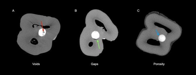
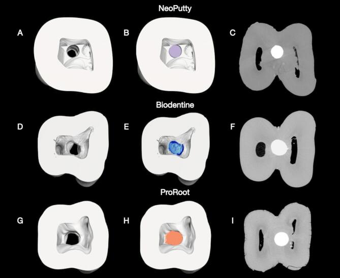
This ex-vivo study compares the adaptation, porosity, and sealing performance of ProRoot MTA, NeoPutty, and Biodentine in standardized, simulated furcation perforations created in extracted mandibular molars using clinical evaluation and high-resolution micro-computed tomography (micro-CT). Thirty-six mandibular molars with furcation perforations were randomly assigned to three groups (n = 12). Clinical evaluation assessed adaptation, porosity, and overfilling, while micro-computed tomography (micro-CT) provided quantitative data on voids and gaps. Statistical analysis used chi-square, Kruskal-Wallis, and Mann-Whitney U tests. NeoPutty and ProRoot MTA showed superior adaptation compared to Biodentine (p < 0.05). Biodentine showed higher porosity (28.44%) than ProRoot MTA (0%) and NeoPutty (8.3%) (p < 0.001). Biodentine also had the highest void volume (1.05 mm3) and gap volume (1.37 mm3), while ProRoot MTA recorded the lowest void volume (0.59 mm3), and NeoPutty had the smallest gap volume (0.85 mm3). No significant differences were observed in overfilling rates. Overall, ProRoot MTA exhibited the most consistent sealing ability, whereas NeoPutty emerged as a viable alternative due to its favorable handling and reliable adaptation. Biodentine, by contrast, showed the poorest performance in terms of structural integrity and sealing capacity. Within the limitations of this study, these findings support the use of ProRoot MTA or NeoPutty for furcation perforation repair, while suggesting more cautious use of Biodentine in such applications.
Keywords: Calcium silicate-based cement; Furcal perforations; Mandibular molars; Marginal adaptation; Micro-computed tomography; Perforation sealing; Sealer porosity.
© 2025. The Author(s).
Conflict of interest statement
Declarations. Competing interests: José Aranguren and Alejandro R. Pérez are the opinion leaders of ZARC (Zarc4endo, Gijón, Asturias, Spain), and Juan Miraglia Cantarini is the opinion leader of Dentsply (Dentsply-Sirona, Baillagues, Switzerland). The other authors deny any conflicts of interest. Ethics approval and consent to participate: The Rey Juan Carlos institutional ethics committee approved the study (protocol 1301202302823).
Figures

References
-
- Cardoso, M., Catré, D., Noites, R., Paulo, M. & Viegas, C. Animal models used in furcation perforation studies: A systematic review and comprehensive synthesis of model characteristics. Aust Endod J.44, 273–280 (2018). - PubMed
-
- Silva, L. A. B. et al. Furcation perforation: periradicular tissue response to Biodentine as a repair material by histopathologic and indirect Immunofluorescence analyses. J. Endod. 43, 1137–1142 (2017). - PubMed
-
- Mente, J., Leo, M., Panagidis, D., Saure, D. & Pfefferle, T. Treatment outcome of mineral trioxide aggregate: repair of root perforations-long-term results. J. Endod. 40, 790–796 (2014). - PubMed
-
- Gorni, F. G., Andreano, A., Ambrogi, F., Brambilla, E. & Gagliani, M. Patient and clinical characteristics associated with primary healing of iatrogenic perforations after root Canal treatment: results of a Long-term Italian study. J. Endod. 42, 211–215 (2016). - PubMed
-
- Singh, P. et al. Sealing ability of mineral trioxide aggregate, calcium phosphate cement, and glass ionomer cement in the repair of furcation perforations. Acta Med. (Hradec Kralove). 56, 97–103 (2013). - PubMed
Publication types
MeSH terms
Substances
LinkOut - more resources
Full Text Sources
Miscellaneous
