

Integrating microwave-assisted green synthesis, DFT simulations, and biological activity evaluation of copper-doped zinc oxide nanoparticles
- PMID: 40456814
- PMCID: PMC12130543
- DOI: 10.1038/s41598-025-03922-8
Integrating microwave-assisted green synthesis, DFT simulations, and biological activity evaluation of copper-doped zinc oxide nanoparticles
Abstract
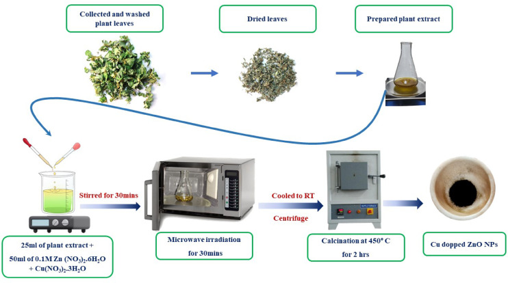
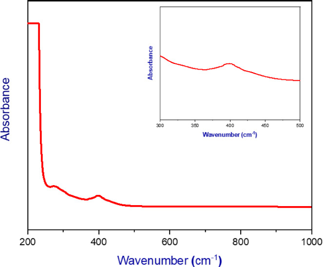
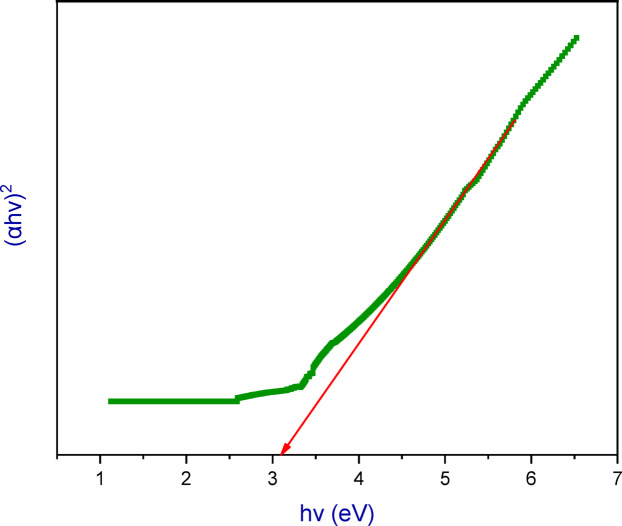
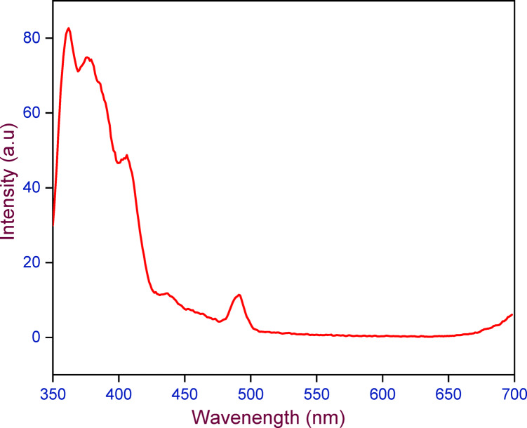
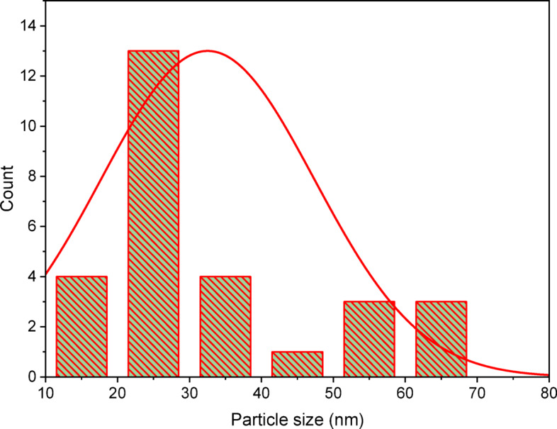
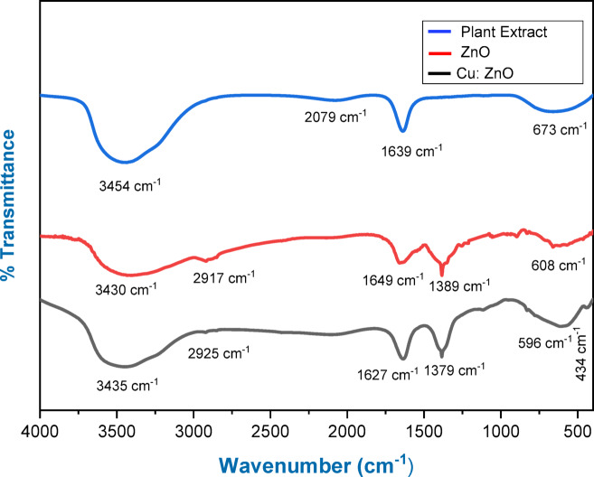
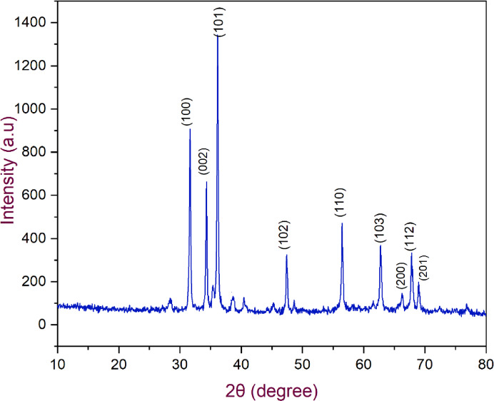
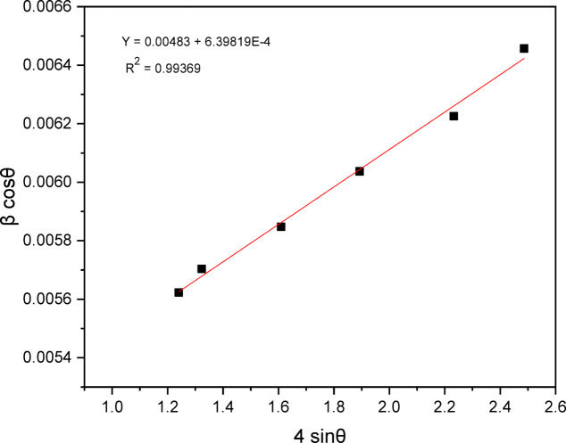
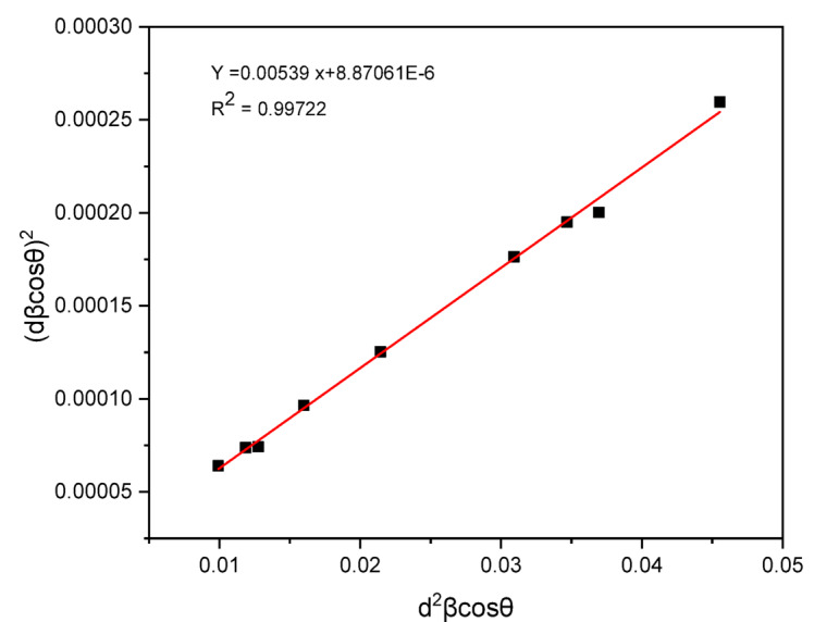
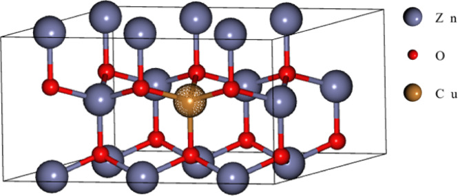
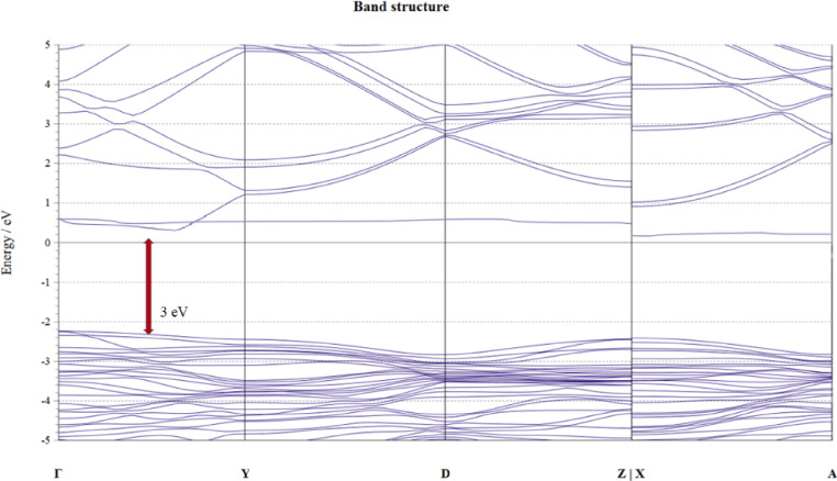
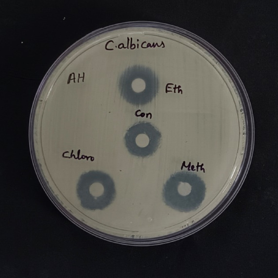
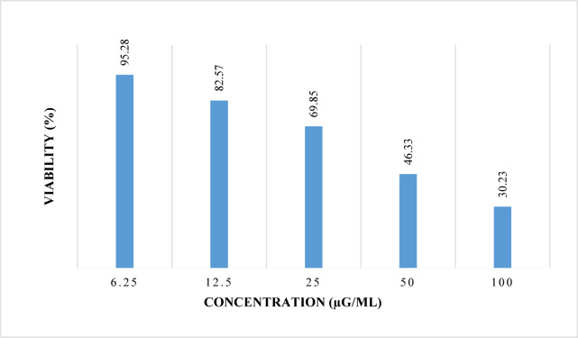
The advancement of nanotechnology and the growing demand for environmentally sustainable processes have fueled interest in green synthesis methods. In this research, copper-doped zinc oxide nanoparticles (Cu: ZnO NPs) were synthesized using a microwave-assisted approach, employing a bio-extract derived from Pistia Stratiotes (PS) leaves as a reducing agent. Comprehensive characterization through UV-Visible spectroscopy, PL, FTIR, SEM with EDS, TEM, DLS, XRD and XPS confirmed the formation, optical and structural features of the synthesized NPs. SEM and TEM images revealed spherical and nanorod-like morphologies, with particle sizes ranging from 15 nm to 65 nm and a tendency to agglomerate. Density Functional Theory (DFT) simulations using Quantum Espresso indicated a band gap narrowing to 3.0 eV after copper doping. Biologically, the Cu: ZnO NPs exhibited strong antibacterial activity against Candida albicans (16.3-17.5 mm), Staphylococcus aureus (18.4-21.5 mm), and Escherichia coli (19-21.6 mm). Additionally, the NPs showed promising anticancer potential against SK-MEL-28 melanoma cells, with an IC50 value of 30.53 µg/mL. Overall, this research demonstrates an eco-friendly and efficient route for fabricating Cu: ZnO NPs with significant antimicrobial and anticancer properties, emphasizing their potential for future biomedical applications.
Keywords: Antibacterial; Anticancer; Antifungal; Characterization; Cu-doped ZnO; Green synthesis; Microwave; Nanoparticles.
© 2025. The Author(s).
Conflict of interest statement
Declarations. Competing interests: The authors declare no competing interests. Plant Guidelines: The authors confirm that the use of plant in the present study complies with international, national and/or institutional guidelines. Permissions to collect the plants/plant parts: The plant specimen used in this research was collected with proper permissions. Source of the plant used in your study: The name of the plant and its source are mentioned in the Materials and Methods section.
Figures


References
-
- Anjum, A., Das, M. & Garg, R. Introduction to Nanotechnology: Transformative Frontier, in: R. Garg, A. Anjum (Eds.), Advances in Chemical and Materials Engineering, IGI Global, : pp. 1–35. (2024). 10.4018/979-8-3693-1094-6.ch001
-
- Singh, N. B., Kumar, B., Usman, U. L. & Susan, M. A. B. H. Nano revolution: exploring the frontiers of nanomaterials in science, technology, and society. Nano-Structures Nano-Objects. 39, 101299. 10.1016/j.nanoso.2024.101299 (2024).
-
- Ahmed, S. F. et al. Green approaches in synthesising nanomaterials for environmental nanobioremediation: technological advancements, applications, benefits and challenges. Environ. Res.204, 111967. 10.1016/j.envres.2021.111967 (2022). - PubMed
MeSH terms
Substances
LinkOut - more resources
Full Text Sources
Miscellaneous
