. 2025 Aug;39(8):1892-1904.
doi: 10.1038/s41375-025-02632-4. Epub 2025 Jun 2.

NOTCH1 signaling is dysregulated by loss of the deubiquitinase USP28 with del(11q), uncovering USP28 inhibition as novel therapeutic target in CLL

Affiliations

NOTCH1 signaling is dysregulated by loss of the deubiquitinase USP28 with del(11q), uncovering USP28 inhibition as novel therapeutic target in CLL

Alena Sophie Ehrmann et al. Leukemia. 2025 Aug.

Abstract

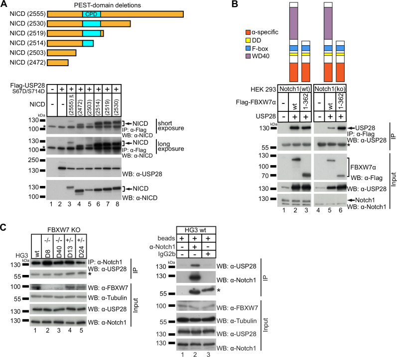
Aberrant active NOTCH1 signaling is a key pathogenic factor in chronic lymphocytic leukemia (CLL), detectable in half of patients and associated with disease progression. While some cases of active NOTCH1 signaling can be explained by mutations in NOTCH1 or its regulators, like FBXW7, alternative mechanisms remain elusive. Here, we identified the deubiquitinase USP28 as regulator of NOTCH1 signaling in CLL. Notably, USP28 is located within the frequently deleted chr11q23 region and is deleted in 90% of del(11q) patients, resulting in its decreased expression. USP28 interacts with the NOTCH1 intracellular domain (NICD) independently of FBXW7 and the NICD-PEST domain, stabilizing NICD and enhancing NOTCH1 signaling. Integrating RBPJ-occupied genes in HG3 cells, RNA-Seq of USP28WT/KO cells and gene expression from del(11q) CLL patients, we identified 15 NOTCH1 target genes specifically dysregulated by deletion of USP28 and del(11q) potentially influencing CLL pathogenesis. Pharmacological inhibition of USP28 with the small molecule AZ1 suppressed NOTCH1 activation in primary CLL cells. AZ1 combined with the BCL-2 inhibitor venetoclax reduced CLL cell viability, particularly in samples with high NOTCH1 activity. Our findings highlight USP28 as promising therapeutic target and provide a rationale for combined inhibition of USP28 and BCL-2 in CLL patients with active NOTCH1 signaling.


Conflict of interest statement

Competing interests: The authors declare no competing interests. Ethics approval: Collection and usage of primary cells from CLL patients has been approved by the Ethics Committee of Ulm University (Vote 242/20). All methods were performed in accordance with the relevant guidelines and regulations. Informed consent: All patients had given written informed consent to the collection and usage of their cells for research purposes according to the Declaration of Helsinki.

Figures

Fig. 1
Fig. 1. USP28 is deleted and downregulated by del(11q) in CLL.
A Schematic representation of genes that are affected by deletion in del(11q) CLL cases. Included genes in the region: BIRC3, ATM, USP28 and ZBTB16. B Proportion of monoallelic gene deletion of BIRC3, ATM, USP28 and ZBTB16 among 96 del(11q) cases from high-resolution SNP-array data [34]. C USP28 expression in a clinical trial cohort of CLL patients (CLL8). Comparison between patients with (n = 86) and without (n = 199) del(11q). The gene expression data was generated from CD19+ sorted CLL cell samples [35]. Lines represent the median. Statistical significance was determined by Kruskal–Wallis test followed by Dunn’s multiple comparison test.
Fig. 2
Fig. 2. USP28 and its target proteins are downregulated in del(11q) CLL patients.
Protein expression of USP28 and FBXW7 and their target proteins NICD, Cyclin-E and c-JUN in primary CLL cells with (n = 14) and without del(11q) (n = 14). Additional 8 patients and quantification of protein levels are available in Supplementary Fig. 1. Patient characteristics are displayed in Supplementary Table 1.
Fig. 3
Fig. 3. USP28 interacts with NICD independently of the NICD PEST domain and FBXW7.
A HEK293 cells were co-transfected with FLAG-USP28 S67D/S714D and different NICD variants with different levels of truncation from the side of the C-terminus (upper panel). Co-immunoprecipitation (lower panel) was performed 24 h after transfection and analyzed by western blot of which a short (upper panel) and long (second panel from top) exposure are shown. Expression of the protein derived from the transfected constructs was detected via western blot shown in the two lower panels. * marks the heavy chain of the anti-FLAG antibody used for immunoprecipitation. B NOTCH1 wt (left panel) or NOTCH1 ko (right panel) HEK293 cells were transfected with the indicated FLAG-tagged FBXW7α constructs with complete or truncated WD40 domain which is important for interaction with NICD. Additionally, untagged USP28 was transfected. Co-immunoprecipitation was performed 24 h after transfection and analyzed via western blot shown in the upper panel (IP). The expression of the protein derived from the transfected constructs is shown in the lower panels (Input). * marks the heavy fragment of the anti-FLAG antibody used for IP. C Immunoprecipitation of endogenous NOTCH1 from HG3 wt or the HG3 CRISPR/Cas9-modified FBXW7 WD40 domain knockout cell lines D8, D40, D13 and D24 [17]. Co-immunoprecipitation of USP28 was analyzed via western blot (upper panel, IP). The expression of FBXW7, USP28 and NOTCH1 in the cell lines is shown in the lower panels (Input). Specificity of the endogenous NOTCH1 precipitation was confirmed by IP reactions with only beads or an IgG2b isotype control antibody. * marks the heavy chain of antibodies used for IP. Western blots are representative for at least three independently performed experiments. IP immunoprecipitation, WB western blot, wt wild type.
Fig. 4
Fig. 4. USP28 stabilizes NICD and modulates NOTCH1 signaling activity.
NICD protein stability was assessed in (A) HEK293 cells with or without overexpression of USP28 (USP28 OE) or in (B) HG3 USP28WT/WT and HG3 USP28WT/KO cells (clones 9 and 10) after a time course of translational inhibition using cycloheximide (CHX). β-actin was used as loading control. Western blots are representative for three independently performed experiments. C NOTCH1 activity measured by luciferase reporter assays (pGL3-Hes1-Luc reporter) in HG3 USP28WT/WT clones with or without overexpression of a USP28 wt plasmid (n = 6 independently performed transfections; left) or in HG3 WT, HG3-del(11q) and HG3-del(11q) ATMKO cell clones (n = 3 clones for each condition; right). Firefly signal was normalized to expression of Renilla luciferase from a constitutively active co-transfected plasmid. Statistical significance was assessed via paired Student’s t test (left panel) or one-way ANOVA, followed by Tukey’s multiple comparisons test (right panel). Single data points depict firefly/renilla ratios measured for each single sample and lines connect untransfected with USP28 wt transfected conditions of each repetition (n = 6; left). Columns depict mean of firefly/renilla ratios measured for n = 3 clones per analyzed cell line and error bars represent standard deviation (right). OE overexpression, wt wild type, WT cell line without modifications.
Fig. 5
Fig. 5. 11q deletion and heterozygous loss of USP28 define a set of 11 dysregulated NOTCH1 target genes.
A Differential gene expression profiles (RNA-Seq) of the USP28WT/KO (clones 9 and 10) and USP28WT/WT cell lines (clones 1 and 2). Gene expression z-scores are color-coded, with blue indicating downregulation and yellow indicating upregulation. The differential expression analysis was performed on a set of pre-selected genes which were identified to be bona fide NOTCH1 targets in an RBPJ ChIP-Seq experiment performed on HG3 wt cells (Supplementary Fig. 9). B Venn diagram illustrating the intersection of: the identified RBPJ-bound, differentially expressed genes in USP28WT/KO (n = 129; blue circle) and significantly differentially expressed genes in del(11q) CLL patients from Lütge et al. [39] (n = 5037 genes in del(11q) samples differentially expressed, 27 genes overlapping with USP28WT/KO dysregulated genes; green circle). C Volcano plot showing the differential expression of NOTCH1 target genes identified by RBPJ ChIP-Seq in HG3 cells and significantly dysregulated in the USP28WT/KO cell lines as shown in panel (A), in del(11q) CLL patients of the CLL8 cohort [35]. The x-axis represents the log2 fold change of gene expression between patients with del(11q) and patients without del(11q). The y-axis shows the negative log10 of the p-value, indicating the significance of the differential expression. The dashed line gives the significance threshold of p = 0.05. Genes significantly dysregulated between del(11q) and non-del(11q) patients from Lütge et al. [39] but not in the CLL8 dataset [35] are highlighted in green. The 11 genes marked in orange are significantly differentially expressed between del(11q) and non-del11q) patients in both datasets. C clone.
Fig. 6
Fig. 6. SOX5, IRF8, KANK1, TOX2, APH1B and ZFHX3 are commonly dysregulated in del(11q) CLL.
NOTCH1 target gene expression in the data sets of A non-del(11q) (n = 199) and del(11q) (n = 86) CLL patients from the CLL8 gene expression study [35], and B non-del(11q) (n = 138) and del(11q) (n = 31) patients analyzed in Lütge et al. [39]. Statistical significance was assessed via Mann–Whitney test. Lines depict mean, data points represent single patients.
Fig. 7
Fig. 7. USP28 inhibition decreases NOTCH1 signaling activity in cell lines and NICD protein levels in primary CLL cells.
A NOTCH1 activity (pGA-981-6(12xCSL) luciferase reporter) and protein levels of NICD and USP28 in cultured mouse embryonic fibroblasts of USP28(−/−) mice and their respective USP28(+/+) controls and in HG3 WT cells upon treatment with USP28 inhibitors AZ1 and AZ2 (10 µM each; n = 3 for each treatment per cell line; left panel) or knockdown of USP28 using two different USP28 shRNA-expressing vectors (sh1 and sh3; n = 3 independent transfections per cell line; right panel). α-Tubulin was used as loading control. Statistical significance was assessed via ordinary one-way ANOVA using Šídák’s multiple comparisons test. B USP28, NICD and c-MYC protein levels in primary CLL samples (n = 18; 12 NOTCH1 WT and 6 NOTCH1 MUT) treated with DMSO or the USP28 inhibitor AZ1 (10 µM; 24 h). α-Tubulin was used as loading control and MOLT-4 cell line lysates were used as a positive control. WT wild type, MEFs mouse embryonic fibroblasts, MUT mutated.
Fig. 8
Fig. 8. USP28 inhibition decreases CLL cell viability and shows additive effect to venetoclax treatment.
A Cell viability analysis by 7-AAD staining of primary CLL cells (n = 12; 7 NOTCH1 WT and 5 NOTCH1 MUT) treated with 10 µM AZ1. Non-viable cells are quantified as the percentage of 7-AAD+ cells in each individual sample. Statistical significance was assessed via unpaired Student’s t test. B Cell viability analysis by ATP quantification (Cell-Titer Glo) of primary CLL cells (n = 30; 17 NOTCH1 WT and 13 NOTCH1 MUT; Supplementary Table 5) treated with DMSO, 10 µM AZ1, 1 µM nirogacestat (Niro) or the combination of AZ1 and nirogacestat for 24 h. Lines depict median and boxes the interquartile range, data points represent single patient samples. Statistical significance was assessed via two-way ANOVA, followed by Tukey’s multiple comparisons test. C Cell viability analysis by ATP quantification (Cell-Titer Glo) of primary CLL cells (n = 15; 10 NOTCH1 WT (4 NICD low, 6 NICD high) and 5 NOTCH1 MUT; Supplementary Table 4) treated with 1 µM ibrutinib (Ibr), 1.25 nm venetoclax (Ven), increasing doses of AZ1 (5 and 10 µM) or the combination of 10 µM AZ1 with ibrutinib or venetoclax for 24 h. Lines depict median and boxes the interquartile range, data points represent single patient samples. Statistical significance was assessed via one-way ANOVA, followed by Tukey’s multiple comparisons test. WT wild type, MUT mutated, Niro nirogacestat, Ibr ibrutinib, Ven venetoclax.

References

    1. Döhner H, Stilgenbauer S, Benner A, Leupolt E, Kröber A, Bullinger L, et al. Genomic aberrations and survival in chronic lymphocytic leukemia. N Engl J Med. 2000;343:1910–6. - PubMed
    1. Landau DA, Tausch E, Taylor-Weiner AN, Stewart C, Reiter JG, Bahlo J, et al. Mutations driving CLL and their evolution in progression and relapse. Nature. 2015;526:525–30. - PMC - PubMed
    1. Puente XS, Pinyol M, Quesada V, Conde L, Ordóñez GR, Villamor N, et al. Whole-genome sequencing identifies recurrent mutations in chronic lymphocytic leukaemia. Nature. 2011;475:101–5. - PMC - PubMed
    1. Knisbacher BA, Lin Z, Hahn CK, Nadeu F, Duran-Ferrer M, Stevenson KE, et al. Molecular map of chronic lymphocytic leukemia and its impact on outcome. Nat Genet. 2022;54:1664–74. - PMC - PubMed
    1. Baldoni S, Del Papa B, De Falco F, Dorillo E, Sorrentino C, Rompietti C, et al. NOTCH1 activation negatively impacts on chronic lymphocytic leukemia outcome and is not correlated to the NOTCH1 and IGHV mutational status. Front Oncol. 2021;11:668573. - PMC - PubMed

MeSH terms

LinkOut - more resources