Review
. 2025 Jun 2;25(1):331.
doi: 10.1186/s12886-025-04166-w.

Sequential bilateral retinal artery occlusions with promising visual prognosis in a diabetic patient: a case report and literature review

Affiliations
Review

Sequential bilateral retinal artery occlusions with promising visual prognosis in a diabetic patient: a case report and literature review

Ziyue Li et al. BMC Ophthalmol. .

Abstract

Background: Bilateral retinal artery occlusion (RAO) in patients with Type 2 diabetes mellitus is an extremely rare condition. Even with rigorous treatment, RAO often results in devastating visual impairment.

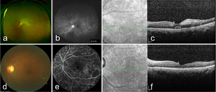
Case presentation: A 50-year-old female with Type 2 diabetes mellitus was referred to our hospital. Notably, she developed bilateral RAO sequentially within a mere five-month period. This rapid progression of the disease in both eyes is of particular interest.

Observations: Fundus examination confirmed branch retinal artery occlusion (BRAO) in her left eye and central retinal artery occlusion (CRAO) in her right eye. After timely intra-arterial thrombolysis, significant bilateral visual recovery was achieved. The patient's visual acuity in the left eye improved from 20/400 to 20/100 and in the right eye from 20/2000 to 20/33.3, indicating the effectiveness of this treatment approach in this case. Further examinations also showed positive results: the cherry-red spot in the macula disappeared, FFA indicated improved retinal artery perfusion, and OCT revealed a certain degree of restoration in the retinal structure.

Conclusion: This case highlights that metabolic disorders, like hyperlipidemia and diabetes, can be considered high-risk factors for the development of RAO. Prompt and effective intervention, such as timely intra-arterial thrombolysis, as demonstrated in this case, is crucial for preserving patients' visual prognosis.

Keywords: Bilateral retinal artery occlusion; Branch retinal artery occlusion; Central retinal artery occlusion; Intra-arterial thrombolysis; Type 2 diabetes mellitus.


Conflict of interest statement

Declarations. Ethics approval and consent to participate: This study was carried out following the institutional guidelines and ethical standards of the 1964 Declaration of Helsinki and was approved by the Institutional Review Board of Renmin Hospital of Wuhan University (WDRY2022-K278). Consent for publication: Informed consent for publication was obtained from the patient. Competing interests: The authors declare no competing interests.

Figures

Fig. 1
Fig. 1
Fundus images of the left eye at presentation and 5 months later. a Fundus photography of the left eye at presentation; b FFA image captured at 15 seconds of the left eye at presentation; c Initial OCT through the left fovea shows marked inner retinal layers edema; d Fundus photography of the left eye 5 months after symptom onset; e FFA image captured at 1 minute 46 seconds of the left eye 5 months after symptom onset; f OCT of the left eye 5 months later shows inner retinal thinning with sparing temporal to the optic disc
Fig. 2
Fig. 2
Brain MRI and CTA images of the patient. a-c Brain MRI of both admissions showed features of lacunar infarction in the left corona radiata; d-f CTA showed moyamoya-like vascular and left ICA stenosis with calcified plaque formation in the right carotid sinus and the initial segment of the right SCA. The white arrow indicates the infarct areas and calcified plaques in the vascular
Fig. 3
Fig. 3
Thrombolysis within the left central retinal artery. a-b Total cerebral angiography was performed smoothly, and the cerebral vascular flow appeared normal except for reduced visualization of the left ophthalmic artery; c-d Injection of 300,000 units of urokinase at the left CRA opening
Fig. 4
Fig. 4
Fundus images of the right eye at initial presentation and after therapeutic intervention. a-d Fundus photography, FFA image captured at 20 seconds and 1 minute 35 seconds, and OCT image of the right eye at presentation; e-h Fundus photography, FFA images captured at 20 seconds and 1 minute 22 seconds, and OCT image of the right eye 3 days after intra-arterial thrombolysis
Fig. 5
Fig. 5
Thrombolysis within the right central retinal artery. a-b Total cerebral angiography was performed smoothly, and the cerebral vascular flow appeared normal except for reduced visualization of the right ophthalmic artery; c Injection of 300,000 units of urokinase at the right CRA opening

References

    1. Hayreh SS. Acute Retinal Arterial Occlusive Disorders. Prog Retin Eye Res. 2011;30(5):359–94. 10.1016/j.preteyeres.2011.05.001. - PMC - PubMed
    1. Hayreh SS. Ocular Vascular Occlusive Disorders: Natural History of Visual Outcome. Prog Retin Eye Res. 2014;41:1–25. 10.1016/j.preteyeres.2014.04.001. - PMC - PubMed
    1. Varma DD, Cugati S, Lee AW, Chen CS. A Review of Central Retinal Artery Occlusion: Clinical Presentation and Management. Eye (London, England). 2013;27(6):688–97. 10.1038/eye.2013.25. - PMC - PubMed
    1. Park SH, Kim BJ, Kim JH, Kim SC, Kim RB, Han YS. Incidence Rates of Retinal Vascular Occlusive Diseases from 2011 to 2020 in South Korea: A Nationwide Cohort Study. BMC Ophthalmol. 2024;24(1):128. 10.1186/s12886-024-03397-7. - PMC - PubMed
    1. Callizo J, Feltgen N, Pantenburg S, Wolf A, Neubauer AS, Jurklies B, et al. Cardiovascular Risk Factors in Central Retinal Artery Occlusion: Results of a Prospective and Standardized Medical Examination. Ophthalmology. 2015;122(9):1881–8. 10.1016/j.ophtha.2015.05.044. - PubMed

MeSH terms

Substances

LinkOut - more resources