Review
. 2025 Jun 2;23(1):407.
doi: 10.1186/s12951-025-03472-1.

In situ radiochemical doping of functionalized inorganic nanoplatforms for theranostic applications: a paradigm shift in nanooncology

Affiliations
Review

In situ radiochemical doping of functionalized inorganic nanoplatforms for theranostic applications: a paradigm shift in nanooncology

Sanchita Ghosh et al. J Nanobiotechnology. .

Abstract

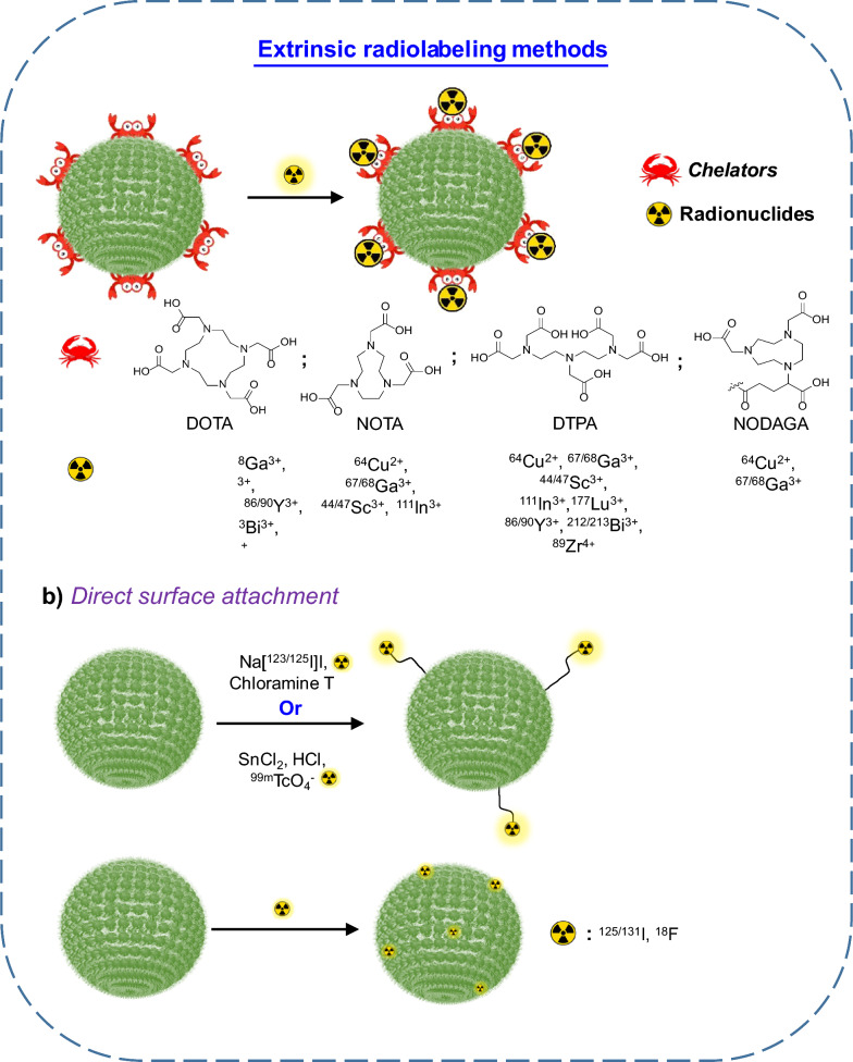
In situ radiochemical doping presents a transformative approach for synthesizing radiolabeled inorganic nanoparticles (NPs) for cancer theranostics. Traditional radiolabeling techniques rely on bifunctional chelators, which often require harsh reaction conditions that can degrade the physicochemical properties of NPs. Additionally, the enzymatic dissociation of radiometals can potentially induce in vivo toxicity. In contrast, in situ doping directly incorporates radiometals into the NP crystal lattice, significantly enhancing both radiolabeling yield and radiochemical stability. This method preserves the pharmacokinetic profiles of the radiolabeled NPs, improving their theranostic efficacy. This review provides an up-to-date overview of the progress made in the development of radiolabeled inorganic nanoplatforms through in situ doping, with a focus on their stability, physicochemical characteristics, and applications in cancer theranostics. Our findings highlight the advantages in situ doping as a more efficient and stable alternative to conventional radiolabeling methods, offering substantial potential for the development of more effective cancer theranostic agents.

Keywords: Cancer; Doping; In situ; Nanoparticles; Precision oncology; Radiation therapy; Radiolabeling; Theranostics.


Conflict of interest statement

Declarations. Competing interests: Weibo Cai declares conflict of interest with the following corporations: Portrai, Inc., rTR Technovation Corporation, and Four Health Global Pharmaceuticals Inc. All other authors declare no competing of interest.

Figures

Fig. 1
Fig. 1
Different types of nanoplatforms used for theranostic applications
Fig. 2
Fig. 2
Schematic of different extrinsic radiolabeling methods for NPs
Fig. 3
Fig. 3
Schematic of different intrinsic radiolabeling methods of NPs
Fig. 4
Fig. 4
Schematic illustration of passive and active targeting of NPs into the tumor
Fig. 5
Fig. 5
Schematic representation of how size, shape and surface charge of NPs affect the biodistribution in different organs
Fig. 6
Fig. 6
Schematic representation of internalization of NPs into cells
Fig. 7
Fig. 7
Different types of emissions from radionuclides and their penetration ranges in tissues
Fig. 8
Fig. 8
a TEM micrograph of PEG-CuS NPs (inset: distribution of particle size); b PET/CT images of U87 glioma tumor bearing mice after intravenous injection of PEG-[64Cu]CuS NPs at different time points (yellow arrow: tumor, orange arrow: bladder, red arrow: standard). Reproduced from ref. [32] with permission. Copyright 2010 ACS Publication. c TEM micrograph of [64Cu]CuAuNPs after decay (scale bar = 10 nm); d PET/CT images at 1 h, 24 h, 48 h post injection and [18F]-FDG at 1 h post injection after injection of [64Cu]CuAuNPs in tumor bearing mice. (T: tumor); e Quantitative plot of tumor uptake of [64Cu]CuAuNPs and [18F]-FDG at different time points. Reproduced from ref. [146] with permission. Copyright 2014 Wiley. f Schematic representation of synthesis of [64Cu]CIS/ZnS QDs for PET/CRET imaging; g whole body PET images after intravenous injection of [64Cu]CuCl2, GSH-[64Cu]CIS/ZnS and PEGylated GSH-[64Cu]CIS/ZnS QDs in U87MG tumor-bearing mice at different time points (white arrow indicates tumor). Reproduced from ref. [130] with permission. Copyright 2015 ACS Publication
Fig. 9
Fig. 9
a Synthetic scheme of [68Ga]Ga-C-IONP functionalized with RGD peptide using 1,4-(butanediol) diglycidyl ether linker; b TEM images of [68Ga]Ga-C-IONP; c phantom images acquired by MRI (above) and PET (below) at various concentration; d T1 weighted MRI images of tumor bearing mouse before (left) and 24 h post injection (right) of [68 Ga]Ga-C-IONP-RGD; e PET/CT images of tumor bearing mouse at 1 h post injection of [68 Ga]Ga-C-IONP-RGD; f PET/CT images of tumor bearing mouse at 1 h post injection of [68Ga]Ga-C-IONP; g biodistribution pattern of ([68Ga]Ga-C-IONP-RGD and [68Ga]Ga-C-IONP after intratumoral injection. Reproduced form ref. [75] with permission. Copyright 2016 John Wiley & Sons, Ltd
Fig. 10
Fig. 10
a Schematic representation of synthesis of [69Ge]Ge:Ga2O3-GA NPs; b PET images of Wistar rat after (i) intravenous injection of [69Ge]Ge:Ga2O3-GA NPs via tail vein and (ii) subcutaneous injection of [69Ge]Ge:Ga2O3-GA NPs for visualization of lymph node; c biodistribution pattern of Wistar rat after intravenous injection of [69Ge]Ge:Ga2O3-GA NPs. Reproduced from ref. [150] with permission. Copyright 2023 ACS Publication
Fig. 11
Fig. 11
TEM images of GdF3:Y NPs synthesized at various reaction conditions: a ellipsoidal NPs (290 °C, 20 min) and rhombic NPs synthesized at b 320 °C for 40 min and c 290 °C for 4 h; d EDS spectrum of GdF3:Y NPs. eh Using YCl3 is as the precursor, YF3 nanowires were synthesized at 320 °C after 20 min (e, f), and bypiramidal LiYF4 nanocrystals are synthesized at 320 °C for 40 min. i Optical images of GdF3:90Y/Y NPs having different amount of radioactivity (0.55–17.5 μCi); j quantification of radioluminescence of GdF3:90Y/Y NPs dispersed in aqueous medium. Reproduced from ref. [155] with permission. Copyright 2015 ACS Publication
Fig. 12
Fig. 12
a Graphical representation of one pot synthesis technique of 103Pd:Pd@Au-PEG NPs and 103Pd:Pd@198Au:Au-PEG; b HRTEM image of 103Pd:Pd@198Au:Au-PEG NPs. Reproduced from ref. [158] with permission. Copyright 2017 WILEY–VCH Verlag GmbH & Company. c Schematic representation of [103Pd]AuPdNPs preparation and 103Pd-nanogel formation; d Dissolution of 103Pd-nanogel in ethanol (left) and formation of stable droplet of 103Pd-nanogel in PBS (right); e Isolated tumor tissue after intratumoral injection of 103Pd-nanogel in mice, (i) The injected 103Pd-nanogel was visible under tumor surface, (ii) Image of separated injected gel surrounded by tumor tissue (white arrow). Reproduced from ref. [160] with permission. Copyright 2021 Wiley
Fig. 13
Fig. 13
a TEM micrograph of Sm-UCNPs; b In vivo, in situ and ex vivo imaging of Kunming mouse at 1 h post injection of Sm-UCNPs via tail vein; c SPECT images at 1 h (left) and 24 h(right) after intravenous injection of Sm-UCNPs. The arrows indicate liver (L) and spleen (S). Reproduced from ref. [133] with permission. Copyright 2013 Elsevier. D) SPECT/CT images of Kunming mouse after intravenous injection of [153Sm]Sm-PEG-UCNPs at different time points. The arrows indicate heart (He), liver (Li), spleen (Sp), kidney (Ki) and bladder (Bl). Reproduced from ref. [169] with permission. Copyright 2013 Elsevier
Fig. 14
Fig. 14
a Synthetic process of core shell 153Sm doped NaLuF4:Yb,Tm@NaGdF4 NPs; TEM images of b NaLuF4:Yb,Tm and NaLuF4:Yb,Tm@NaGdF4(153Sm) NPs; c UCL imaging and d X-ray CT imaging (focused on peritoneum) of nude mice after intravenous injection of NaLuF4:Yb,Tm@NaGdF4(153Sm) NPs; e MR images of mouse pre and post injection intravenous of NaLuF4:Yb,Tm@NaGdF4(153Sm) NPs; f SPECT/CT images after intravenous injection of NaLuF4:Yb,Tm@NaGdF4(153Sm) NPs at different time points, g Four different types of images of tumor at 1 h after intravenous injection of NaLuF4:Yb,Tm@NaGdF4(153Sm) NPs. Reproduced from ref [132] with permission. Copyright 2013 ACS Publication
Fig. 15
Fig. 15
a Schematic illustration of synthesis of [169Yb]Yb2O3-GA NPs; b SPECT/CT images of melanoma tumor bearing C57BL/6 mice after intratumoral injection of [169Yb]Yb2O3-GA NPs at different time points; c biodistribution plot of tumor bearing mice after intratumoral injection of radiolabeled NPs; d H & E stained images of both treated and nontreated mice. Reproduced from ref. [171] with permission. Copyright 2023 Springer
Fig. 16
Fig. 16
a Schematic illustration of synthesis of [188Re]ReOx-HSA NPs; b SPECT/CT images of melanoma tumor bearing C57BL/6 mice after intratumoral injection of [188Re]ReOx-HSA NPs at different time points; c tumor growth and d body weight index plot after combined radio-photothermal treatment; e H & E stained images of both treated and nontreated mice. Reproduced from ref. [176] with permission. Copyright 2025 Springer
Fig. 17
Fig. 17
TEM images of a Au nanospheres; b Au nanodisks; c Au nanorods; and d Au nanocages; e in vivo luminescence image of tumor bearing mice at 24 h post injection after injection of different types of Au NPs; f Autoradiographic impression of tumor slices at 24 h post injection of i) 198Au nanospheres, ii) 198Au nanodisks, iii) 198Au nanorods and iv) 198Au nanocubes. Reproduced from ref. [183] with permission. Copyright 2014 ACS Publication. TEM micrographs of 199Au doped gold NPs of g 5 nm and h 18 nm size after 90 days of decay; i SPECT/CT image of 4T1 tumor-bearing mouse at 24 h post injection after injection of 5 nm [199Au]AuNP-DAPTA nanoprobe (T: tumor). Reproduced from ref. [184] with permission. Copyright 2016 Wiley Publication

Similar articles

Cited by

  • Metal-based nanomedicines for cancer theranostics.
    Yu HJ, Liu JH, Liu W, Niu R, Zhang B, Xiong Y, Liu Y, Wang YH, Zhang HJ. Yu HJ, et al. Mil Med Res. 2025 Jul 30;12(1):41. doi: 10.1186/s40779-025-00627-x. Mil Med Res. 2025. PMID: 40739670 Free PMC article. Review.

References

    1. Deo S, Sharma J, Kumar S. GLOBOCAN 2020 report on global cancer burden: challenges and opportunities for surgical oncologists. Ann Surg Oncol. 2022;29:6497–500. - PubMed
    1. Kashyap BK, Singh VV, Solanki MK, Kumar A, Ruokolainen J, Kesari KK. Smart nanomaterials in cancer theranostics: challenges and opportunities. ACS Omega. 2023;8:14290–320. - PMC - PubMed
    1. Pei Z, Lei H, Cheng L. Bioactive inorganic nanomaterials for cancer theranostics. Chem Soc Rev. 2023;52:2031–81. - PubMed
    1. Zhao D, Zhang W, Yu S, Xia S-L, Liu Y-N, Yang G-J. Application of MOF-based nanotherapeutics in light-mediated cancer diagnosis and therapy. J Nanobiotechnol. 2022;20:421. - PMC - PubMed
    1. Deng S, Gu J, Jiang Z, Cao Y, Mao F, Xue Y, Wang J, Dai K, Qin L, Liu K, et al. Application of nanotechnology in the early diagnosis and comprehensive treatment of gastrointestinal cancer. J Nanobiotechnol. 2022;20:415. - PMC - PubMed

LinkOut - more resources