Review
. 2025 Jun 2;23(1):617.
doi: 10.1186/s12967-025-06611-2.

Mitochondrial dynamics reveal potential to facilitate axonal regeneration after spinal cord injury

Affiliations
Review

Mitochondrial dynamics reveal potential to facilitate axonal regeneration after spinal cord injury

Kaixuan Wang et al. J Transl Med. .

Abstract

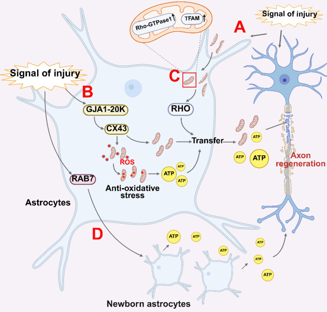
Background: Spinal cord injury (SCI) arises from traumatic damage to the spinal cord, resulting in varying degrees of sensory, motor, and autonomic dysfunction. Mitochondria, as the primary energy-producing organelles within cells, have garnered increasing attention for their critical role in promoting axonal regeneration following SCI.

Aim of review: This review aims to systematically examine the alterations in mitochondrial dynamics post-SCI and to elucidate their influence on axonal regeneration. Furthermore, the review evaluates the current challenges associated with SCI treatment and proposes potential therapeutic strategies for future research.

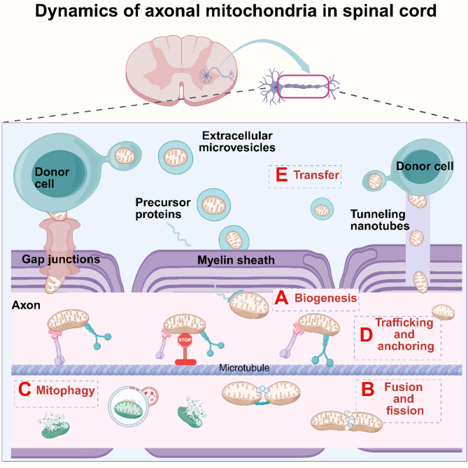
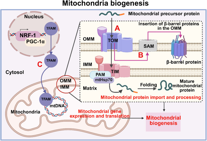
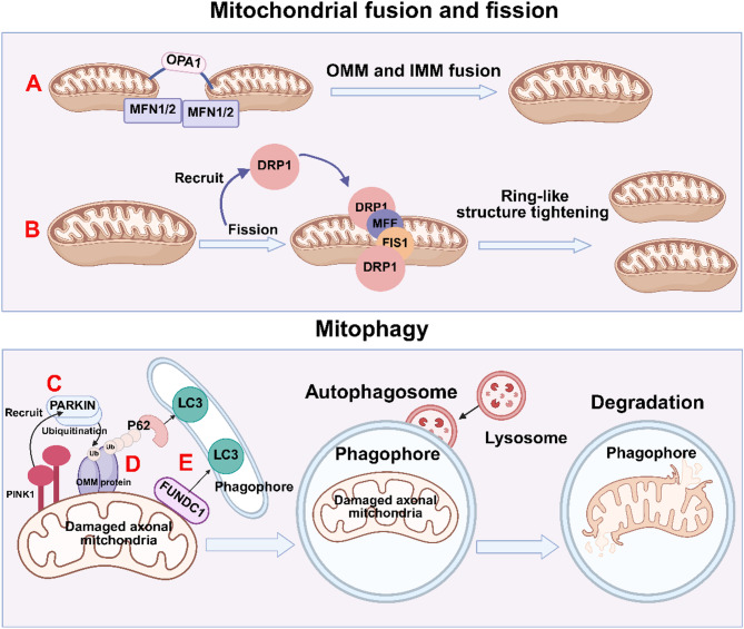
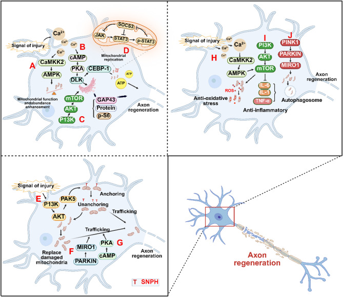
Key scientific concepts of review: The review comprehensively addresses mitochondrial dynamics, with a focus on key processes such as biogenesis, fusion and fission, mitophagy, trafficking, and anchoring. It delves into the molecular mechanisms by which signaling pathways within neurons and glial cells regulate these mitochondrial processes to facilitate axonal regeneration. Additionally, the review identifies existing challenges in SCI treatment and advocates for targeted interventions in mitochondrial dynamics as a promising therapeutic avenue, offering significant potential for advancing future research and treatment of SCI.

Keywords: Axonal mitochondria; Axonal regeneration; Dynamics; Spinal cord injury.


Conflict of interest statement

Declarations. Consent for publication: Not applicable. Ethics approval: Not applicable. Competing interests: The authors declare that they have no competing interests.

Figures

Fig. 1
Fig. 1
The intricate and dynamic behavior of mitochondria within axons in the spinal cord (A) Mitochondrial biogenesis: Cytoplasmic precursor proteins contribute significantly to mitochondrial protein synthesis via a coordinated system of synthesis and import. This mechanism operates in tandem with mitochondrial DNA replication, supporting the continuous generation of mitochondria and ensuring a sustained energy supply vital for the maintenance of damaged axons. (B) Mitochondrial fusion and fission: Fusion allows damaged mitochondria to undergo repair by merging with healthier counterparts, facilitated by fusion proteins. This fusion mechanism not only facilitates repair but also aids in regenerating mitochondria, ensuring their functionality is preserved. Conversely, the fission process, mediated by mitochondrial fission proteins, plays a vital role in ensuring an adequate number of mitochondria are available to support axonal regeneration. (C) Mitophagy: Damaged mitochondria are pinpointed and flagged as targets requiring removal. This process typically involves the attachment of autophagy-related proteins. These proteins naturally congregate on the outer membrane of damaged mitochondria, facilitating the formation of autophagosomes. Subsequently, autophagosomes merge with lysosomes, creating autophagosomal lysosomes. Within these structures, diverse degrading enzymes facilitate the breakdown of damaged mitochondria. (D) Mitochondrial trafficking and anchoring: Mitochondrial trafficking and anchoring play crucial roles in facilitating the distribution of dynamic mitochondria between the soma and axons. This process heavily relies on the support and regulation provided by the cytoskeleton, kinesins, and anchor proteins within cells. Through stable connections formed with mitochondria, these components maintain specific positions and morphologies, thereby regulating the precise localization and movement of mitochondria within the cell. (E) Mitochondrial transfer: Mitochondrial transfer from donor cells into axons to repair the regeneration function of the axon. This intricate process entails the delivery of healthy mitochondria from donor cells to the target axons through tunneling nanotubes, extracellular microvesicles, and gap junctions
Fig. 2
Fig. 2
Mitochondrial biogenesis (A) Precursor proteins are synthesized in the cytoplasm, transported through the OMM via the TOM-TIM23 complex, and directed into the mitochondrial matrix for folding and translocation, facilitated by Hsp70 and PAM. (B) Outer membrane β-barrel proteins are imported into mitochondria via the TOM complex and subsequently integrated into the outer membrane with the assistance of SAM proteins. (C) NRF-1 interacts with PGC-1α to enhance the transcriptional activity that stimulates TFAM, which binds to specific sequences on mtDNA, promoting mtDNA replication
Fig. 3
Fig. 3
Mitochondrial fusion, fission and mitophagy (A) Mitochondrial fusion and fission processes within axons are orchestrated by specific protein interactions. OMM fusion relies on the coordinated activity of proteins such as MFN1 and MFN2, while OPA1 plays a crucial role in facilitating IMM fusion. (B) Upon receiving a signal for fission, DRP1 translocates to the mitochondrial surface. There, it interacts with receptors like FIS1 and MFF situated on the OMM. This interaction leads to forming a ring-like structure that constricts around the mitochondria, resulting in an eventual division of the mitochondria into distinct entities. (C) When mitochondria become damaged, PINK1 accumulates on the mitochondrial outer membrane, recruiting PARKIN. Subsequently, Parkin ubiquitinates proteins on the mitochondrial outer membrane, initiating mitochondrial autophagy. (D) Following ubiquitination, the receptor protein p62 accumulates on the mitochondrial outer membrane, facilitating the recruitment of ubiquitinated products to autophagosomes by binding to LC3. (E) Additionally, FUNDC1 interacts with LC3 through its LC3-binding domain on the outer mitochondrial membrane, promoting the binding of damaged mitochondria to autophagosomes. Ultimately, these products are degraded by lysosomes
Fig. 4
Fig. 4
Mitochondrial trafficking, anchoring and transfer (A) LIS1 operates as a regulator for cytoplasmic dynein, binding to its motor structural domain. This interaction stimulates the assembly of active cytoplasmic dynein complexes, consequently facilitating the retrograde trafficking of mitochondria. (B) SNPH’s C- and N-terminal microtubule-binding domains engage with the mitochondrial outer membrane and microtubules, respectively. This interaction serves to anchor mitochondria to microtubules, halting their mobility effectively. (C) KIF5 functions as a kinesin, forming a complex with MIRO and TRAK, orchestrating the movement of mitochondria in an anterograde manner towards the distal axon. (D) EVs are key mediators of intercellular communication, facilitating the transfer of diverse cellular components, including structurally intact and functional mitochondria. (E) TNTs, which consist of microtubules, depend on kinesin adaptor proteins like Miro1 and TRAK for facilitating the movement of mitochondria into axons. (F) Cx43-GJC structures facilitate cytoplasmic connections between cells, forming the basis for mitochondrial transfer to the axon
Fig. 5
Fig. 5
Signaling pathways for axonal regeneration within neurons. A-D: Signaling pathways that promote mitochondrial energy metabolism in axonal regeneration. E-G: Signaling pathways that promote mitochondrial transport in axon regeneration. H-J: Signaling pathways promoting mitophagy, anti-inflammatory and anti-oxidative stress in axonal regeneration. (A) Following SCI, injury triggers Ca2+ influx into neurons, activating the CaMKK2/AMPK signaling pathway. This activation leads to an increase in mitochondrial abundance and enhances ATP synthesis function. (B) Injury-induced Ca2+ influx elevates intraneuronal Ca2+ concentration, activating the cAMP/PKA signaling pathway. Subsequently, this pathway triggers the downstream DLK/CEBP-1 signaling cascade, promoting mitochondrial replication and the synthesis of axon growth-associated proteins. (C) Activation of the PI3K/AKT/mTOR signaling pathway facilitates the synthesis of axon growth-related proteins like GAP43 and p-S6 in neurons. This activation synergizes with the cAMP/PKA/DLK signaling pathway to further support axonal regeneration. (D) Activation of the JAKA/STAT3 signaling pathway in mitochondria during mitochondrial replication inhibits SOCS3 from functioning. (E) SNPH plays a crucial role in mitochondrial anchoring, maintaining their fixed position within neurons. Following neuronal injury, the PI3K/AKT/PAK5 signaling pathway is activated to release mitochondrial anchorage, facilitating their trafficking toward the axon terminals. This process promotes the substitution of damaged mitochondria with functionally intact ones, ensuring the requisite energy supply for axonal regeneration. (F) and (G) The collaboration between the PARKIN/MIRO1 phase and the cAMP/PKA signaling pathway synergistically enhances mitochondrial trafficking towards axon terminals in neurons. (H) Following neuronal injury, calcium influx activates the CaMKK2/AMPK/mTOR signaling pathway, regulating inflammatory responses and suppressing the expression of inhibitory cytokines IL-1, IL-6, and TNF-α. (I) Neuronal injury triggers the activation of the PI3K/AKT/mTOR signaling pathway, facilitating mitochondrial clearance of intra-neuronal ROS to counteract oxidative stress. (J) Neuronal injury induces activation of the PINK/PARKIN/MIRO1 signaling pathway, which promotes mitophagy, eliminates dysfunctional mitochondria, and creates a conducive environment for axonal regeneration
Fig. 6
Fig. 6
Role played by glial cells in axonal regeneration. (A and C) Following SCI, neuronal damage signals prompt the release of damaged mitochondria from neurons. Subsequently, astrocytes phagocytose these damaged mitochondria, leading to increased expression levels of Rho-GTPase1 and TFAM within mitochondria. This activation triggers the Rho-related signaling pathway, facilitating mitochondrial transfer from astrocytes to neurons. (B) Injury-induced signaling activates the GJA1-20 K/CX43 signaling pathway in astrocytes, enabling mitochondria to mitigate oxidative stress and facilitating their transfer to neurons to sustain energy supply capacity. (D) Activation of the RAB7-related signaling pathway in astrocytes promotes astrocytic proliferation and facilitates association with peripheral astrocytes to provide ATP to neurons, thus fostering axonal regeneration

Similar articles

References

    1. Tian T, Zhang S, Yang M. Recent progress and challenges in the treatment of spinal cord injury. Protein Cell. 2023;14(9):635–52. - PMC - PubMed
    1. McDonald JW, Sadowsky C. Spinal-cord injury. Lancet. 2002;359(9304):417–25. - PubMed
    1. Jiang B, Sun D, Sun H, Ru X, Liu H, Ge S et al. Prevalence, incidence, and external causes of traumatic spinal cord injury in China: A nationally representative Cross-Sectional survey. Front Neurol. 2022;12. - PMC - PubMed
    1. Zheng B, Tuszynski MH. Regulation of axonal regeneration after mammalian spinal cord injury. Nat Rev Mol Cell Biol. 2023;24(6):396–413. - PubMed
    1. Hu X, Xu W, Ren Y, Wang Z, He X, Huang R et al. Spinal cord injury: molecular mechanisms and therapeutic interventions. Signal Transduct Target Therapy. 2023;8(1). - PMC - PubMed