Case Reports
. 2025 Jun 2;23(1):60.
doi: 10.1186/s12969-025-01115-1.

Two unique cases of eosinophilic granulomatosis with polyangiitis in childhood treated with anti-interleukin-5 therapy: infantile-onset and submandibular salivary gland involvement

Affiliations
Case Reports

Two unique cases of eosinophilic granulomatosis with polyangiitis in childhood treated with anti-interleukin-5 therapy: infantile-onset and submandibular salivary gland involvement

Evangelia Constantine et al. Pediatr Rheumatol Online J. .

Abstract

Background: ANCA-associated vasculitis is a systemic autoimmune disease involving small- and medium-sized blood vessels. Eosinophilic granulomatosis with polyangiitis (EGPA, previously Churg Strauss Syndrome) is the least common form in childhood with few cases reported. We present two unique pediatric cases, both of which were treated with anti-interleukin-5 therapy.

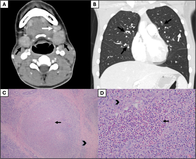
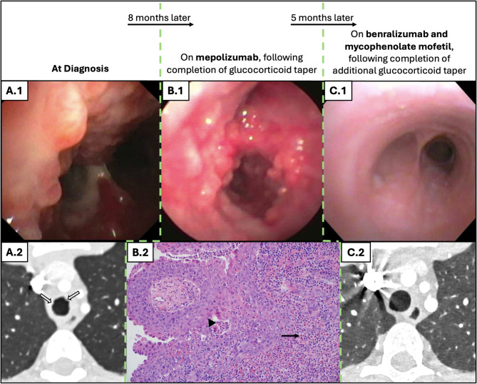
Case presentation: Case one is a 13-year-old male with asthma and allergies who presented with one month of cough and periorbital edema and subsequently developed submandibular swelling. Evaluation identified chronic sinusitis, weight loss, positive c-ANCA and anti-MPO IgG antibodies, peripheral blood eosinophilia, pulmonary eosinophilia, tracheal and pulmonary nodules, and eosinophilic infiltration of the submandibular salivary gland with granulomas and fibrosis fitting a diagnosis of EGPA. He improved with glucocorticoids and mepolizumab with a significant partial response, and eventually switched to benralizumab and mycophenolate mofetil with complete response. Case two presented at 19-months-old in acute respiratory distress with a history of reactive airway disease. EGPA diagnosis was confirmed on lung biopsy (eosinophilic capillaritis and interstitial expansion of eosinophils) in the setting of anti-MPO and p-ANCA positivity. He has done very well on mepolizumab for three years.

Conclusions: To our knowledge, this is the first reported case of submandibular salivary gland infiltrate in a child with EGPA and the youngest successfully treated patient with EGPA reported in the literature. These cases demonstrate the variation in age and disease manifestations seen in children with EGPA as well as positive responses to anti-interleukin-5 therapy. Children with EGPA may present with common or unusual complaints and require astute recognition to avoid delays in diagnosis and long-term damage.

Keywords: ANCA-associated vasculitis; Benralizumab; Churg Strauss; EGPA; Eosinophilic granulomatosis with polyangiitis; Mepolizumab; Sialadenitis; anti-IL5 therapy.


Conflict of interest statement

Declarations. Ethics approval and consent to participate: Not applicable. Consent for publication: Written consent was obtained from both patients. Competing interests: The authors declare no competing interests.

Figures

Fig. 1
Fig. 1
Salivary gland and lung inflammation in 13-year-old with EGPA (Case 1). Axial CT image of the neck (A) shows bilateral enlargement of the submandibular glands (black arrows) and cervical adenopathy (white arrow). Coronal CT image of the the chest (B) demonstrates moderate diffuse bronchial wall thickening (black arrows) and scattered nodules (white arrow). Additional nodules not shown. Salivary gland biopsy low-power view (C; H&E, 40X) shows abundant periacinar fibrosis (arrowhead), glandular atrophy, and central regions of necrosis (black arrow). Salivary gland biopsy high-power view (D; H&E, 200X) of a region of necrotizing granuloma demonstrates peripheral epithelioid histiocytes (arrowhead), central neutrophils, and eosinophils (black arrow)
Fig. 2
Fig. 2
Evolution of inflammatory nodules in trachea of 13-year-old with EGPA (Case 1). At diagnosis, diffuse nodular mucosa with mucus and inflammatory changes were seen on bronchoscopy (A.1), while axial CT image of his chest demonstrated irregularity and nodularity of the trachea (A.2; outlined arrows). Nodular changes remained on bronchoscopy after treatment with mepolizumab and completion of a prolonged glucocorticoid taper (B.1). A tracheal nodule biopsy was obtained, which showed an exemplary pattern of injury throughout with prominent acute epithelial inflammation, microabscess formation (arrowhead), and innumerable eosinophils (black arrow) (B.2; H&E, 20X). There was a drastic decrease in inflammation and mucosal changes on bronchoscopy (C.1) after changing to benralizumab and mycophenolate mofetil and completion of an additional glucocorticoid taper. The nodularity of the trachea resolved on repeat axial CT imaging of his chest (C.2). All bronchoscopy images shown were obtained at the mainstem carina
Fig. 3
Fig. 3
Renal histopathology of 13-year-old with EGPA (Case 1). (A) Renal biopsy showed only mild mesangial hypercellularity (black arrow) and matrix expansion, a pattern of injury seen in IgA nephropathy. Typical features of renal involvement by EGPA (e.g. crescentic glomerulonephritis, granuloma formation, and vascular inflammation) were distinctly absent in this case (PAS, 40X). (B) Immunofluorescence performed on the renal biopsy shows 3–4 + mesangial IgA, a typical feature in IgA nephropathy. This specimen also showed 1 + mesangial C3, commonly seen in IgA nephropathy. (C) Electron microscopy performed on the renal biopsy shows abundant mesangial deposits (white arrow), frequently seen in IgA nephropathy
Fig. 4
Fig. 4
Chest computed tomography of 19-month-old male with respiratory failure. Axial (A) and Coronal (B) images demonstrate bilateral ground glass nodularity in a bronchovascular distribution (arrows). (C) A higher power H&E illustrates prominent alveolar hemorrhage and abundant hemosiderin-laden macrophages associated with regions of lung damage. An eosinophil-predominant infiltrate is still present and, in many areas, was associated with interstitial capillary damage, small arteriolar/venular damage, and bronchiolar wall injury. (D) Representative H&E stain shows a diffusely expanded interstitium with prominent eosinophilic infiltrate. Eosinophils are also present within the alveolar spaces

References

    1. Bloom JL, Langford CA, Wechsler ME. Therapeutic advances in eosinophilic granulomatosis with polyangiitis. Rheum Dis Clin North Am. 2023;49(3):563–84. - PubMed
    1. Fina A, et al. Eosinophilic granulomatosis with polyangiitis in children: data from the French RespiRare® cohort. Pediatr Pulmonol. 2018;53(12):1640–50. - PubMed
    1. Eleftheriou D, et al. Eosinophilic granulomatosis with polyangiitis in childhood: retrospective experience from a tertiary referral centre in the UK. Rheumatology. 2016;55(7):1263–72. - PubMed
    1. Doubelt I, et al. Clinical manifestations and Long-Term outcomes of eosinophilic granulomatosis with polyangiitis in North America. ACR Open Rheumatol. 2021;3(6):404–12. - PMC - PubMed
    1. Chung SA, et al. 2021 American college of rheumatology/vasculitis foundation guideline for the management of antineutrophil cytoplasmic Antibody–Associated vasculitis. Arthritis Rheumatol (Hoboken N J). 2021;73(8):1366–83. - PubMed

Publication types

LinkOut - more resources