

Revisiting nephrin signaling and its specialized effects on the uniquely adaptable podocyte
- PMID: 40457651
- PMCID: PMC12203975
- DOI: 10.1042/BCJ20230234
Revisiting nephrin signaling and its specialized effects on the uniquely adaptable podocyte
Abstract
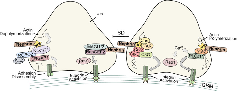
Nephrin is a transmembrane Ig-like domain-containing protein that serves as a central structural and signaling scaffold in kidney filtration. First identified in 1998 as mutated in congenital nephrotic syndrome, the recent identification of nephrin autoantibodies in acquired kidney diseases has sparked renewed interest in nephrin biology. In specialized cells known as podocytes, nephrin helps establish and maintain the slit diaphragm (SD), a unique cell-cell junction formed between interdigitating cell projections known as foot processes (FPs). Together, the SD and FP are among the first stages of renal filtration, where they are subject to numerous biochemical and mechanical stressors. Although podocytes are highly adapted to this environment, over time and with injury, this elevated strain can lead to pathological structural changes, detachment, and proteinuria. As such, the complex set of signaling mechanisms provided by nephrin are essential for controlling podocyte adaptability. Herein, we provide a thorough and up-to-date review on nephrin signaling, including a focus on cross-talk between nephrin interactors and signaling regions across podocytes. We first highlight new findings regarding podocyte structure and function, followed by an emphasis on why nephrin is among the most critical proteins for maintaining these features. We then detail a comprehensive list of known nephrin interactors and describe several of their effects, including calcium regulation, cell survival, cell polarity, phase separation-mediated actin reorganization, and SD-focal adhesion dynamics. Collectively, our emerging understanding of the broader cellular context of nephrin signaling provides important insight for clinical strategies to mitigate podocyte injury and kidney disease progression.
Keywords: cell signaling; chronic kidney disease; focal adhesion; nephrin; podocytes.
© 2025 The Author(s).
Conflict of interest statement
The authors declare that there are no competing interests associated with the manuscript.
Figures


Similar articles
-
Nephrin is necessary for podocyte recovery following injury in an adult mature glomerulus.PLoS One. 2018 Jun 20;13(6):e0198013. doi: 10.1371/journal.pone.0198013. eCollection 2018. PLoS One. 2018. PMID: 29924795 Free PMC article.
-
Multi-glycoside of Tripterygium wilfordii Hook. f. reduces proteinuria through improving podocyte slit diaphragm dysfunction in anti-Thy1.1 glomerulonephritis.J Ethnopharmacol. 2011 Jun 22;136(2):322-33. doi: 10.1016/j.jep.2011.04.046. Epub 2011 May 6. J Ethnopharmacol. 2011. Retraction in: J Ethnopharmacol. 2025 Aug 29;352:120138. doi: 10.1016/j.jep.2025.120138. PMID: 21570456 Retracted.
-
The Life of a Kidney Podocyte.Acta Physiol (Oxf). 2025 Aug;241(8):e70081. doi: 10.1111/apha.70081. Acta Physiol (Oxf). 2025. PMID: 40698593 Free PMC article. Review.
-
Signs and symptoms to determine if a patient presenting in primary care or hospital outpatient settings has COVID-19.Cochrane Database Syst Rev. 2022 May 20;5(5):CD013665. doi: 10.1002/14651858.CD013665.pub3. Cochrane Database Syst Rev. 2022. PMID: 35593186 Free PMC article.
-
Oxysterol-binding protein-like 7 deficiency leads to ER stress-mediated apoptosis in podocytes and proteinuria.Am J Physiol Renal Physiol. 2024 Sep 1;327(3):F340-F350. doi: 10.1152/ajprenal.00319.2023. Epub 2024 Jul 4. Am J Physiol Renal Physiol. 2024. PMID: 38961844 Free PMC article.
References
Publication types
MeSH terms
Substances
LinkOut - more resources
Full Text Sources
Research Materials
Miscellaneous