. 2025 Apr 17;28(5):112463.
doi: 10.1016/j.isci.2025.112463. eCollection 2025 May 16.

ANKRD17 induces pro-survival signaling pathways that enhance cellular invasion and migration during hepatocellular carcinoma tumorigenesis

Affiliations

ANKRD17 induces pro-survival signaling pathways that enhance cellular invasion and migration during hepatocellular carcinoma tumorigenesis

Vincent W Keng et al. iScience. .

Abstract

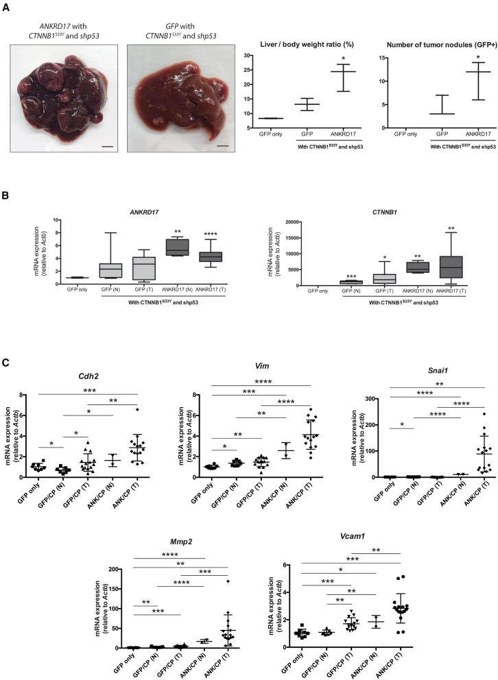
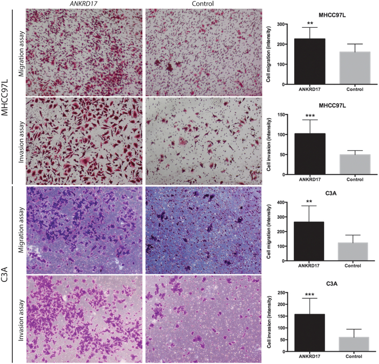
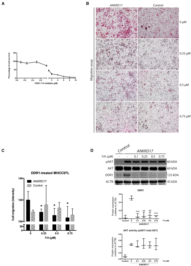
Metastasis is the primary cause of high mortality in patients with hepatocellular carcinoma (HCC) . A prior study identified ankyrin repeat domain 17 (Ankrd17) as a key gene linked to HCC metastasis. Through reverse genetics, it was observed that mouse liver tumors overexpressing ANKRD17 exhibited a higher tumor load and increased expression of endothelial-mesenchymal transition (EMT) markers. Similarly, ANKRD17 overexpression in human liver cell lines resulted in an amplified cellular motility and invasion capability, whereas knockdown studies reversed this effect. Abnormal regulation of signaling pathways was linked to increased metastasis and survival in cells overexpressing ANKRD17. Notably, the pro-metastatic discoidin domain receptor tyrosine kinase 1 (DDR1) gene was upregulated in these cells, and its suppression reduced motility and invasion without affecting AKT signaling. Clinically, higher ANKRD17 expression correlated with aggressive HCC progression. These findings suggest that ANKRD17 enhances metastatic progression in HCC by activating pro-metastatic and pro-survival pathways.

Keywords: cancer; cell biology; molecular biology.


Conflict of interest statement

The other authors declare no conflict of interest.

Figures

None
Graphical abstract
Figure 1
Figure 1
ANKRD17 enhances tumorigenic and pro-metastatic characteristics during HCC disease progression (A) ANKRD17-overexpression in a CTNNB1S33Y and shp53 predisposed background (n = 3), resulted in larger liver to body weight percentage ratios and higher tumor burden in Fah/SB11 mouse injected livers at around 356-day PHI compared to control cohorts (GFP only, n = 2; GFP in a CTNNB1S33Y and shp53 predisposed background, n = 3). Scale bars, 0.5cm. (B) Quantitative PCR (qPCR) results showing upregulated ANKRD17 and CTNNB1S33Y expression levels in liver samples from experimental and control injected Fah/SB11 mice. (C) qPCR results show significant upregulation of EMT markers in liver tumors of experimental Fah/SB11 injected mice compared with control cohorts. Arbitrary value relative to Actb mRNA levels expressed as mean ± SD. GFP, GFP only control; GFP/CP, GFP, CTNNB1S33Y, and shp53 control; ANK/CP, ANKRD17, CTNNB1S33Y, and shp53 experimental; N, peripheral normal liver; T, tumor nodule. p, unpaired Student’s t test: ∗∗∗∗, p < 0.0001; ∗∗∗, p < 0.001; ∗∗, p < 0.01; ∗, p < 0.05.
Figure 2
Figure 2
ANKRD17-overexpression enhanced cellular migration and invasion abilities in human liver cancer cell lines ANKRD17-overexpressing MHCC97L (top) and C3A (bottom) cells both exhibited significant increases in cellular motility and invasion abilities compared with control orange fluorescent protein (OFP)-transfected cells. Representative graphs of stained migrated or invaded cell intensity expressed as mean ± SD; P, unpaired Student’s t test: ∗∗∗, p < 0.001; ∗∗, p < 0.01.
Figure 3
Figure 3
ANKRD17-overexpression induces the dysregulation of several important signaling pathways (A) Liver tumors from experimental mice demonstrating dysregulated HIPPO signaling pathway with significantly higher expression of Yap1 than control cohorts. Quantitative PCR results of Yap1 relative to Actb levels expressed as mean ± SD. GFP, GFP only control; GFP/CP, GFP, CTNNB1S33Y, and shp53 control; ANK/CP, ANKRD17, CTNNB1S33Y, and shp53 experimental; N, peripheral normal liver; T, tumor nodule. (B) Representative Western blot demonstrating dysregulated AKT/PTEN signaling pathway in tumors (T) and their peripheral normal (N) tissues. Semi-quantitative analyses of AKT activity (pAKT/total AKT) relative to ACTB levels expressed as mean ± SD. (C) Representative Western blot demonstrating dysregulated AKT/PTEN signaling pathway in ANKRD17-overexpressing MHCC97L cells. Semi-quantitative analyses of AKT activity (pAKT/total AKT) relative to ACTB levels expressed as mean ± SD. (D) Representative Western blot demonstrating dysregulated STAT3 signaling pathway in ANKRD17-overexpressing MHCC97L cells. Control, OFP-overexpressing cells. Semi-quantitative analyses of STAT3 activity (pSTAT3/total STAT3) relative to ACTB levels expressed as mean ± SD. (E) Liver tumors from experimental mice demonstrated significantly higher expression of Ddr1 than control cohorts at both the transcriptional (top) and translational (bottom) levels. Western blot for DDR1 as shown in (B). Semi-quantitative analyses of Ddr1 and DDR1, relative to Actb and ACTB levels, respectively, expressed as mean ± SD. (F) Induced expression of DDR1 at both the transcriptional (top) and translational (bottom) levels in ANKRD17-overexpressing MHCC97L cells. Western blot for DDR1 as shown in (C). Semi-quantitative analyses of DDR1 expression at both the transcriptional (top) and translational (bottom) levels relative to ACTB and ACTB levels, respectively, expressed as mean ± SD. P, unpaired Student’s t test: ∗∗∗∗, p < 0.0001; ∗∗∗, p < 0.001; ∗∗, p < 0.01; ∗, p < 0.05.
Figure 4
Figure 4
Inhibition of DDR1 suppressed ANKRD17-induced cellular migration ability in HCC cancer cell line (A) Cell survival assay of DDR1 inhibitor 7rh at different concentrations to determine the sub-cytotoxic conditions for subsequent experiments. (B) Cell motility assessed by transwell migration assay using DDR1 inhibitor 7rh at 0, 0.25, 0.5, and 0.75 μM in ANKRD17-overexpressing MHCC97L cells. (C) Semi-quantitative analyses of the cellular migration results from (B) expressed as mean ± SD. (D) Representative Western blot demonstrates reduction in DDR1 levels but no alterations in AKT phosphorylation at Ser473 were detected with DDR1 inhibitor 7rh at all tested concentrations. Control, OFP-overexpressing cells. Semi-quantitative analyses of DDR1 and AKT activity (pAKT/total AKT) relative to ACTB levels expressed as mean ± SD. P, unpaired Student’s t test: ∗∗∗, p < 0.001; ∗∗, p < 0.01; ∗, p < 0.05.

References

    1. Chitapanarux T., Phornphutkul K. Risk Factors for the Development of Hepatocellular Carcinoma in Thailand. J. Clin. Transl. Hepatol. 2015;3:182–188. doi: 10.14218/jcth.2015.00025. - DOI - PMC - PubMed
    1. Karagozian R., Derdák Z., Baffy G. Obesity-associated mechanisms of hepatocarcinogenesis. Metabolism. 2014;63:607–617. doi: 10.1016/j.metabol.2014.01.011. - DOI - PubMed
    1. Montella M., D'Arena G., Crispo A., Capunzo M., Nocerino F., Grimaldi M., Barbieri A., D'Ursi A.M., Tecce M.F., Amore A., et al. Role of Sex Hormones in the Development and Progression of Hepatitis B Virus-Associated Hepatocellular Carcinoma. Int. J. Endocrinol. 2015;2015 doi: 10.1155/2015/854530. - DOI - PMC - PubMed
    1. Kew M.C. Hepatic iron overload and hepatocellular carcinoma. Liver Cancer. 2014;3:31–40. doi: 10.1159/000343856. - DOI - PMC - PubMed
    1. McKillop I.H., Moran D.M., Jin X., Koniaris L.G. Molecular pathogenesis of hepatocellular carcinoma. J. Surg. Res. 2006;136:125–135. doi: 10.1016/j.jss.2006.04.013. - DOI - PubMed

LinkOut - more resources