. 2025 May 26:2025:7334303.
doi: 10.1155/adpp/7334303. eCollection 2025.

Tacca chantrieri André Rhizome Extract Alleviates Scopolamine-Induced Cognitive Impairment and Neuroinflammation in Rats

Affiliations

Tacca chantrieri André Rhizome Extract Alleviates Scopolamine-Induced Cognitive Impairment and Neuroinflammation in Rats

Thaneeya Hawiset et al. Adv Pharmacol Pharm Sci. .

Abstract

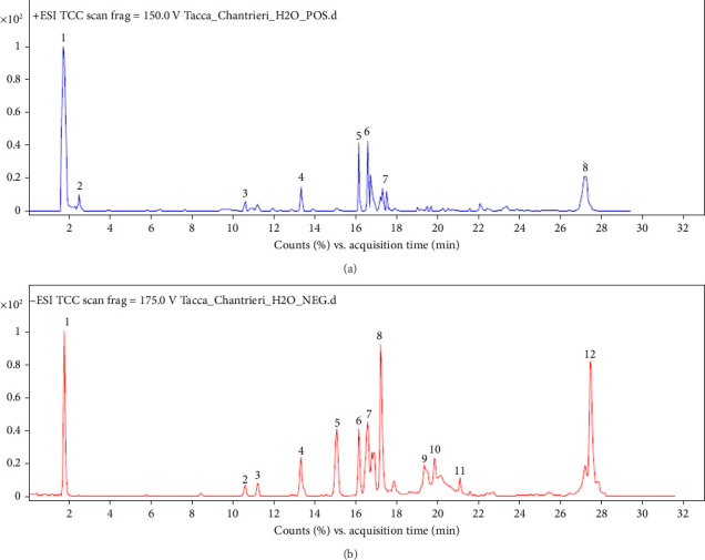
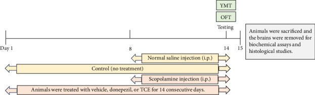
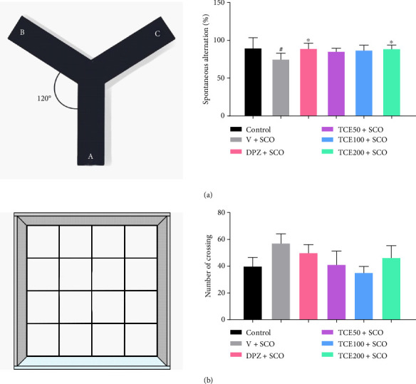
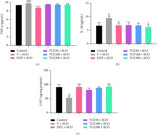
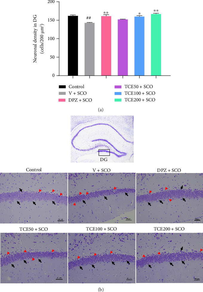
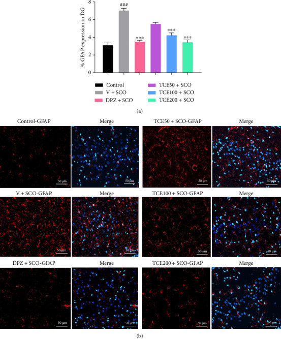
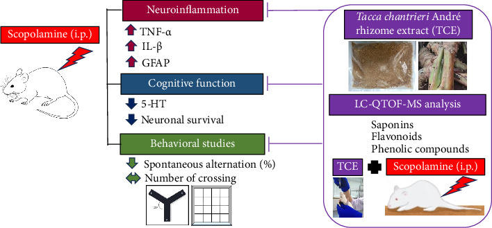
Tacca chantrieri André is a native plant from Northern Thailand with reported pharmacological effects, including antioxidant, anti-inflammatory, and neuroprotective properties. This study investigated the neuroinflammatory and cognitive-enhancing effects of Tacca chantrieri André rhizome extract (TCE) in a scopolamine-injected model, which mimics an Alzheimer's disease (AD) animal model. Animals were divided into six groups: (1) a control group, (2) a vehicle-treated group, (3) a donepezil-treated group (3 mg/kg BW) as a positive control, and (4-6) three TCE-treated groups receiving 50, 100, or 200 mg/kg BW once daily for 14 days. Starting on Day 8, animals received daily intraperitoneal injections of scopolamine (3 mg/kg BW) for 7 consecutive days to induce cognitive impairment. On day 14, behavioral tests were conducted, including the Y-maze and open field tests. On day 15, animals were euthanized, and their brains were collected for Nissl staining, immunofluorescence staining, and biochemical analyses using an ELISA kit. Our results demonstrated that TCE treatment attenuated scopolamine-induced memory deficits and neuroinflammation. Specifically, TCE administration reduced levels of proinflammatory cytokines, including tumor necrosis factor-α (TNF-α) and interleukin-1β (IL-1β), and decreased glial fibrillary acidic protein (GFAP) expression in the hippocampus. Additionally, TCE improved neuronal survival and enhanced serotonin levels, contributing to cognitive improvements. The qualitative analysis of TCE using LC-QTOF-MS identified various chemical constituents, including saponins, flavonoids, and phenolic compounds. These bioactive compounds contributed to the neuroprotective effects of TCE by modulating neuroinflammation and cognitive function. The neuroprotective effects of TCE suggested its potential as a therapeutic agent for memory impairment associated with AD.

Keywords: GFAP; Tacca chantrieri; memory; neuroinflammation; proinflammatory cytokine; scopolamine; serotonin.


Conflict of interest statement

The authors declare no conflicts of interest.

Figures

Figure 1
Figure 1
Total ion current chromatograms were monitored in (a) electrospray ionization positive mode and (b) negative mode.
Figure 2
Figure 2
The protocol and design of the experiment. The animals were administered with vehicle, donepezil, or TCE (50, 100, or 200 mg/kg BW) once a day for 14 days. On day 8, scopolamine was intraperitoneally injected into the animals daily for the following 7 days. On day 14, the behavioral tests were investigated.
Figure 3
Figure 3
Effect of TCE on scopolamine-induced memory deficit and locomotor behavior. (a) Diagram of the Y-maze test (left) and a graph showing the results of spontaneous alternation behavior (right). (b) Diagram of open field test (left) and a graph showing the results of the number of crossings (right). One-way ANOVA was used to analyze the data, and the mean ± SEM (n = 6) was presented as the outcome. #p < 0.05 versus the control group. p < 0.05 versus the vehicle with the scopolamine group.
Figure 4
Figure 4
Graphs of the effect of TCE on (a) TNF-α levels, (b) IL-1β levels, and (c) 5-HT concentration. One-way ANOVA was used to analyze the data, and the mean ± SEM (n = 6) was presented as the outcome. #p < 0.05 and ###p < 0.001 versus the control group; p < 0.05∗∗p < 0.01, and ∗∗∗p < 0.001 versus the vehicle with the scopolamine group.
Figure 5
Figure 5
Effect of TCE on neuronal density in the hippocampus. (a) Graph of neuronal density in the DG region of the hippocampus. (b) Photomicrographs of the DG sections of the hippocampus at a 20x magnification. Scale bars with a length of 20 μm accompany these images. The red arrowhead indicated the dark, shrunken, and damaged neurons, while the black arrow indicated the neurons that were still alive. One-way ANOVA was used to analyze the data, and the mean ± SEM (n = 6) was presented as the outcome. ##p < 0.01 versus the control group; p < 0.05 and ∗∗p < 0.01 versus the vehicle with the scopolamine group.
Figure 6
Figure 6
Effect of TCE on the percentage of GFAP expression in the hippocampus. (a) Percentage of GFAP expression in the DG. (b) The immunofluorescence of GFAP (red) expression in the DG, Hoechst 33258 (blue), and merged images at a 20x magnification. Scale bars measuring 50 μm accompany these images. One-way ANOVA was used to analyze the data, and the mean ± SEM (n = 6) was presented as the outcome. ###p < 0.001 versus the control group; ∗∗∗p < 0.001 versus the vehicle with scopolamine group.
Figure 7
Figure 7
A schematic diagram illustrates the possible mechanisms TCE protects against scopolamine-induced memory impairment and neuroinflammation.

Similar articles

References

    1. Revi M. Alzheimer’s Disease Therapeutic Approaches. Advances in Experimental Medicine and Biology . 2020;1195:105–116. doi: 10.1007/978-3-030-32633-3_15. - DOI - PubMed
    1. Kwon H. S., Koh S. H. Neuroinflammation in Neurodegenerative Disorders: The Roles of Microglia and Astrocytes. Translational Neurodegeneration . 2020;9(1):42–12. doi: 10.1186/s40035-020-00221-2. - DOI - PMC - PubMed
    1. Wang W. Y., Tan M. S., Yu J. T., Tan L. Role of Pro-Inflammatory Cytokines Released From Microglia in Alzheimer’s Disease. Annals of Translational Medicine . 2015;3(10):p. 136. doi: 10.3978/j.issn.2305-5839.2015.03.49. - DOI - PMC - PubMed
    1. Yang Z., Wang K. K. Glial Fibrillary Acidic Protein: From Intermediate Filament Assembly and Gliosis to Neurobiomarker. Trends in Neurosciences . 2015;38(6):364–374. doi: 10.1016/j.tins.2015.04.003. - DOI - PMC - PubMed
    1. Winkler B., Funke D., Benmimoun B., et al. Brain Inflammation Triggers Macrophage Invasion across the Blood-Brain Barrier in Drosophila during Pupal Stages. Science Advances . 2021;7(44) doi: 10.1126/sciadv.abh0050. - DOI - PMC - PubMed

LinkOut - more resources