

In Silico, In Vitro, and In Vivo Investigations of Anticancer Properties of a Novel Platinum (II) Complex and Its PLGA Encapsulated Form
- PMID: 40458703
- PMCID: PMC12127131
- DOI: 10.1155/bca/2673015
In Silico, In Vitro, and In Vivo Investigations of Anticancer Properties of a Novel Platinum (II) Complex and Its PLGA Encapsulated Form
Abstract
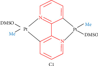
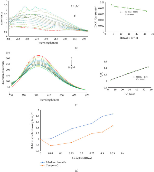
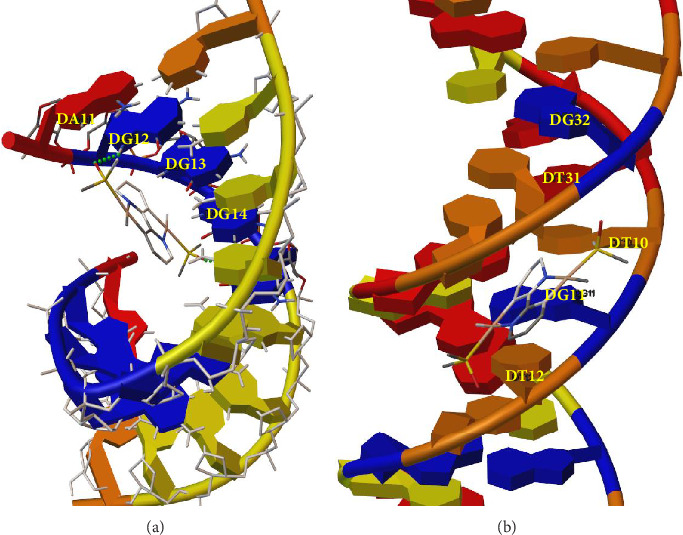
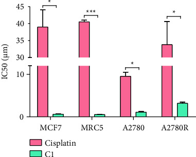
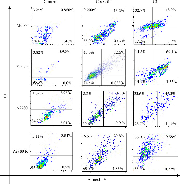
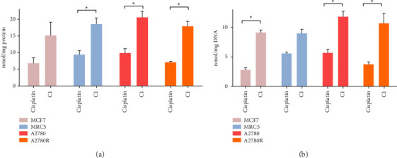
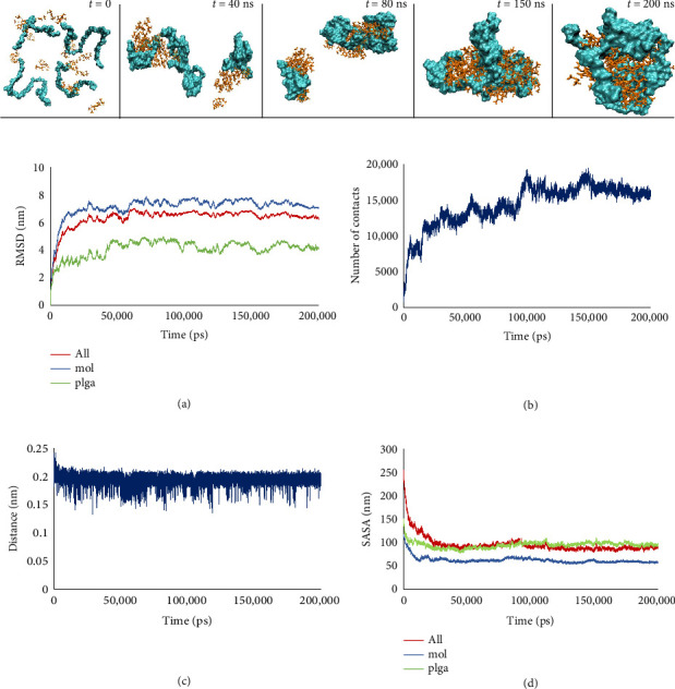
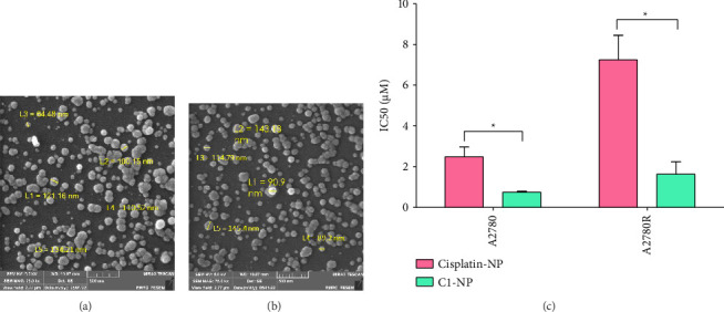
In recent years, the development of multinuclear platinum complexes has introduced a new era in platinum-based chemotherapy, offering improved cytotoxicity and the ability to overcome resistance. However, these complexes still face challenges related to water solubility, biodistribution, and targeted delivery. This study provides a comprehensive investigation of a novel platinum (II) complex, [Pt2(μ-bpy-2H) (Me)2(dmso)2] (C1), focusing on its DNA binding ability and anticancer activity. Computational and experimental approaches revealed that C1 binding to guanine bases and involvement of intercalative interactions. C1 exhibited cytotoxicity in both cisplatin sensitive and resistant cancer cell lines. To enhance the pharmacokinetic and pharmacodynamic properties of C1, it was encapsulated using poly (D, L-lactic-co-glycolic acid) (PLGA). Molecular dynamic simulations predicted the formation of stable C1/PLGA complexes during the early stages of simulation. Encapsulated C1 showed superior antitumor activity with significantly reduced side effects in tumor-bearing mouse models. In conclusion, this study highlights the novel platinum (II) complex C1 as a promising anticancer agent, especially when paired with PLGA encapsulation to improve its effectiveness and reduce side effects.
Keywords: PLGA; antitumor; apoptosis; nanoencapsulation; platinum complex.
Copyright © 2025 Zahra Shabaninejad et al. Bioinorganic Chemistry and Applications published by John Wiley & Sons Ltd.
Conflict of interest statement
The authors declare no conflicts of interest.
Figures
Similar articles
-
Anticancer Activity Assessment and DNA Binding Properties of Two Binuclear Platinum (II) Complexes using Spectroscopic and Molecular Simulation Approaches.Anticancer Agents Med Chem. 2020;20(17):2066-2073. doi: 10.2174/1871520620666200705221325. Anticancer Agents Med Chem. 2020. PMID: 32628598
-
DNA binding properties and cytotoxic effects of two double rollover cycloplatinated (II) complexes on cancer cell lines.J Inorg Biochem. 2023 Jun;243:112194. doi: 10.1016/j.jinorgbio.2023.112194. Epub 2023 Mar 16. J Inorg Biochem. 2023. PMID: 36966676
-
Novel oximato-bridged platinum(II) di- and trimer(s): synthetic, structural, and in vitro anticancer activity studies.Inorg Chem. 2012 Jul 2;51(13):7153-63. doi: 10.1021/ic300148e. Epub 2012 Jun 12. Inorg Chem. 2012. PMID: 22691006
-
[Drug discovery research in in-vivo antitumor-active azolato-bridged dinuclear Pt(II) complexes].Yakugaku Zasshi. 2012;132(3):253-9. doi: 10.1248/yakushi.132.253. Yakugaku Zasshi. 2012. PMID: 22382827 Review. Japanese.
-
Advancements in the Use of Platinum Complexes as Anticancer Agents.Anticancer Agents Med Chem. 2022;22(5):821-835. doi: 10.2174/1871520621666210805150705. Anticancer Agents Med Chem. 2022. PMID: 34353272 Review.
References
-
- Ma L., Li L., Zhu G. Platinum-Containing Heterometallic Complexes in Cancer Therapy: Advances and Perspectives. Inorganic Chemistry Frontiers . 2022;9(11):2424–2453. doi: 10.1039/D2QI00205A. - DOI
LinkOut - more resources
Full Text Sources
Research Materials
