Clinical Trial
. 2025 Sep;115(3):266-277.
doi: 10.1111/ejh.14426. Epub 2025 Jun 3.

Sensitization of Non-M3 Acute Myeloid Leukemia Blasts to All-Trans Retinoic Acid by the LSD1 Inhibitor Tranylcypromine: TRANSATRA Phase I Study

Affiliations
Clinical Trial

Sensitization of Non-M3 Acute Myeloid Leukemia Blasts to All-Trans Retinoic Acid by the LSD1 Inhibitor Tranylcypromine: TRANSATRA Phase I Study

Michael Kruszewski et al. Eur J Haematol. 2025 Sep.

Abstract

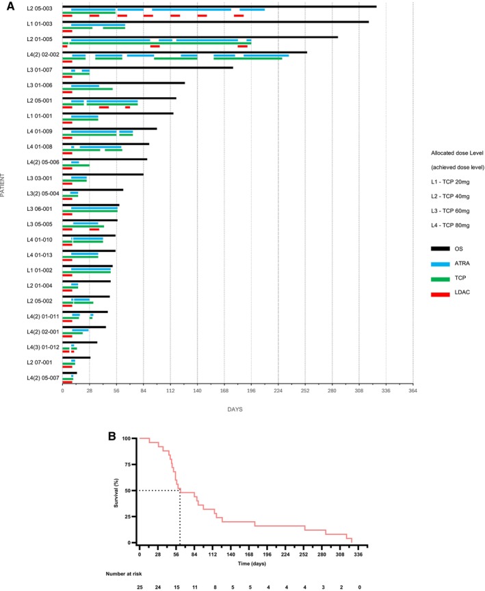
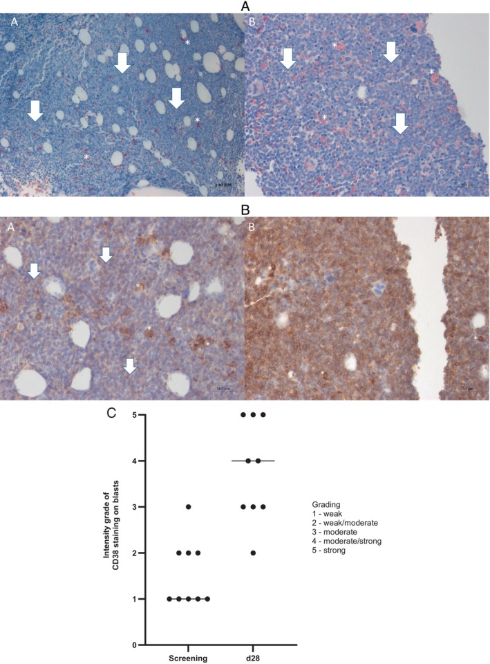
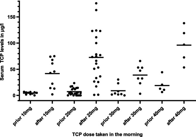
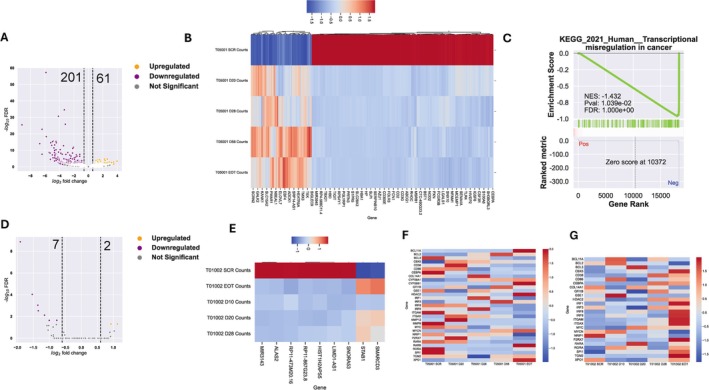
The treatment of elderly, nonfit acute myeloid leukemia (AML)/MDS patients with relapsed/refractory (R/R) disease remains challenging. As histone demethylase LSD1 (KDM1A) is a rational therapeutic target in AML, we conducted a phase I trial ("rolling-six design") with the LSD1 inhibitor tranylcypromine (TCP, dose levels [DL] 20, 40, 60, 80 mg p.o. d1-28) combined with fixed-dose ATRA (45 mg/m2 p.o. d10-28) and low-dose cytarabine (LDAC, 40 mg s.c. d1-10). The primary endpoint was dose-limiting toxicity (DLT) in the first 28 days of treatment. The aim was the determination of the maximum tolerated TCP dose (MTD). Twenty-three patients with AML and 2 with MDS were accrued. TCP was administered for a median of 39.5 days (range: 11-228). No DLTs were observed at any DL; MTD could not be established. No differentiation syndrome occurred. Two patients attained a PR; SD was achieved in 10 of 22 evaluable patients. Median OS was 62 days (range: 14-325). Accompanying studies included pharmacokinetics, serial determinations of fetal hemoglobin (HbF), detection of CD38 upregulation with treatment, as well as transcriptome changes in purified blood blasts over time. In conclusion, the combination of TCP with ATRA and LDAC was well feasible, even at the highest DL. Hence, studies with more potent LSD1 inhibitors appear warranted. Trial Registration: German Clinical Trials Register (DRKS): DRKS00006055. For further Information see https://drks.de/search/en/trial/DRKS00006055.

Keywords: CD38; LSD1; chromatin; differentiation; histone demethylase; myelodysplastic syndrome.


Conflict of interest statement

The authors declare no conflicts of interest.

Figures

FIGURE 1
FIGURE 1
(A) Treatment exposure and overall survival. Overall survival depicted next to number of days of exposure to TCP, ATRA, and LDAC. For patient 01‐013 last documented intake dates of TCP and ATRA were taken. For patient 02‐001 last documented intake date of ATRA was taken. Exact dates of last intake are unknown for both patients. Interruptions are due to protocol, adverse events, or other reasons. The allocated dose level of each patient is depicted (L1‐4); if the allocated dose level was not reached, the highest achieved dose level is shown in parenthesis. (B) Kaplan–Meier plot. Overall survival rate estimated by Kaplan–Meier method. The median OS was 62 days. The number at risk is shown below the graph.
FIGURE 2
FIGURE 2
In vivo induction of CD38 protein expression on AML blasts. The images depict formalin‐fixed, paraffin‐embedded bone marrow trephine biopsy samples obtained from patient 01‐013 (DL 4). (A) The picture shows the NACE (Naphthol‐AS‐D Chloroacetate‐esterase) labeling of formalin‐fixed paraffin embedded bone marrow trephine biopsy samples (magnification 40X). Panel A represents the pretreatment sample, while panel B shows the post‐treatment (after 1 cycle on day 28) sample. There are repressed granulopoiesis (marked with white asterisks) and massive blast cell infiltration (> 90%) (marked with white arrows) on both panels. (B) The picture shows the CD38 labeling of formalin‐fixed paraffin embedded bone marrow trephine biopsy samples (magnification 40X). Panel A represents the pretreatment sample, while panel B shows the post‐treatment (after one cycle on day 28) sample. On panel A, low CD38 expression on the blast cells (marked with white arrows) is seen, while panel B shows strong expression of CD38 on the blast cells. As an internal positive control, plasma cells have been used (marked with white asterisks). (C) IHC staining intensity in bone marrow. Immunohistochemical analysis of bone marrow biopsies, collected from nine patients both pretreatment and postfirst cycle of therapy. Staining intensity categorized into 5 groups: weak—1, weak/moderate—2, moderate—3, moderate/strong—4, and strong—5.
FIGURE 3
FIGURE 3
TCP plasma levels. TCP dose taken in the morning with patients' serum levels prior and two hours after intake with medians. Levels were measured at days 10, 20, 28, and 56. TCP was taken in the morning and evening as per protocol. Serum levels of TCP were quantified using a validated high‐performance liquid chromatography (HPLC) method coupled with tandem mass spectrometry (MS/MS).
FIGURE 4
FIGURE 4
Treatment‐induced in vivo changes in global transcriptomics (serially purified peripheral blood AML blasts) (A, B) Volcano plot and Heatmap of differentially expressed genes (DEGs) (FDR < 0.05 & Log2foldchange > 0.6) in patient 05‐001 (dose level 2, treated for 78 days). (C) GSEA enrichment of transcriptome on ‘KEGG 2021 Human’ database. (D, E) Volcano plot and Heatmap of DEGs (FDR < 0.05 & Log2foldchange > 0.6) in patient 01‐002 (dose level 1, treated for 50 days). (F, G) Regulation of known LSD1‐regulated genes in patient 05‐001 (F) and patient 01‐002 (G).

Similar articles

References

    1. Juliusson G., Lazarevic V., Hörstedt A.‐S., Hagberg O., and Höglund M., “Acute Myeloid Leukemia in the Real World: Why Population‐Based Registries Are Needed,” Blood 119, no. 17 (2012): 3890–3899, 10.1182/blood-2011-12-379 008. - DOI - PMC - PubMed
    1. Tchekmedyian R., Elson P., Gerds A. T., et al., “Analysis of Outcomes of Patients With Relapsed/Refractory Acute Myeloid Leukemia Treated in Randomized Clinical Trials,” Blood 128, no. 22 (2016): 4000, 10.1182/blood.V128.22.4000.4000. - DOI
    1. Bouligny I. M., Mehta V., Isom S., et al., “Efficacy of 10‐Day Decitabine in Acute Myeloid Leukemia,” Leukemia Research 103 (2021): 106524, 10.1016/j.leukres.2021.106524. - DOI - PMC - PubMed
    1. Stomper J., Rotondo J. C., Greve G., and Lübbert M., “Hypomethylating Agents (HMA) for the Treatment of Acute Myeloid Leukemia and Myelodysplastic Syndromes: Mechanisms of Resistance and Novel HMA‐Based Therapies,” Leukemia 35, no. 7 (2021): 1873–1889, 10.1038/s41375-021-01218-0. - DOI - PMC - PubMed
    1. Zeidan A. M., Fenaux P., Gobbi M., et al., “Prospective Comparison of Outcomes With Azacitidine and Decitabine in Patients With AML Ineligible for Intensive Chemotherapy,” Blood 140, no. 3 (2022): 285–289, 10.1182/blood.2022015832. - DOI - PMC - PubMed

Publication types

MeSH terms