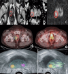
Combined 68Ga-PSMA PET/CT and mpMRI Findings Improve Tumor Localization and Biopsy Guidance in the Initial Diagnosis of Prostate Cancer
- PMID: 40459021
- PMCID: PMC12134961
- DOI: 10.4274/mirt.galenos.2024.75547
Combined 68Ga-PSMA PET/CT and mpMRI Findings Improve Tumor Localization and Biopsy Guidance in the Initial Diagnosis of Prostate Cancer
Abstract
Gallium-68 (68Ga)-prostate-specific membrane antigen (PSMA) positron emission tomography/computed tomography (PET/CT) is a relatively new imaging modality that has already proved its role in the initial staging of prostate cancer and in biochemical recurrence following definitive primary therapy. Furthermore, emerging data several ongoing studies demonstrate its potential role in the primary diagnosis of this malignancy. We present a 67-year-old male patient with increasing clinical suspicion of prostate cancer despite a previous negative prostate gland biopsy. He was referred to our nuclear medicine department for a 68Ga-PSMA PET/CT with the aim of improving tumor localization and assisting in the guidance of repeat prostate biopsy. One month before presentation of elevated prostate-specific antigen levels, he underwent multiparametric magnetic resonance imaging (mpMRI), which revealed a prostate imaging reporting and data system 4 lesion in the right lobe of the prostate gland. The MRI lesion completely matched a PRIMARY score 5 lesion registered by PET/CT. Furthermore, we detected another suspicious finding in the left lobe (PRIMARY score 4). The patient underwent PSMA PET/CT-guided MRI/ultrasonography fusion transperineal biopsy of both lesions. The latter were histologically confirmed as prostate carcinoma with a Gleason score of 3+4 =7.
68Ga-prostat-spesifik membran antijeni (PSMA) pozitron emisyon tomografisi/bilgisayarlı tomografi (PET/BT), prostat kanserinin ilk evrelemesinde ve birincil tedaviyi takiben biyokimyasal nükste rolünü kanıtlamış nispeten yeni bir görüntüleme yöntemidir. Ayrıca, devam eden birkaç çalışmadan elde edilen yeni veriler, bu malignitenin birincil tanısında potansiyel rolünü göstermektedir. Daha önce negatif prostat bezi biyopsisine rağmen artan prostat kanseri klinik şüphesi olan 67 yaşında erkek hastayı sunuyoruz. Hasta tümör lokalizasyonunu iyileştirmek ve tekrar prostat biyopsisinin rehberliğine yardımcı olmak amacıyla 68Ga-PSMA PET/BT için nükleer tıp bölümümüze sevk edildi. Bir ay önce yüksek prostat spesifik antijen seviyeleri nedeniyle multiparametrik manyetik rezonans görüntüleme (mpMRG) uygulanan hastada prostat bezinin sağ lobunda prostat görüntüleme raporlama ve veri sistemi 4 lezyon saptandı. MRG lezyonu PET/BT ile kaydedilen PRİMARY skor 5 lezyonla tamamen uyuşuyordu. Ayrıca sol lobda PRİMARY skor 4 olan bir şüpheli lezyon daha tespit ettik. Hastanın her iki lezyonundan PSMA PET/BT kılavuzluğunda MRI/ultrasonografi füzyon transperineal biyopsi alındı ve histolojik olarak Gleason skoru 3+4=7 olan prostat karsinomu doğrulandı.
Keywords: 68Ga-PSMA PET/CT; initial diagnosis of prostate cancer; mpMRI.
Copyright© 2025 The Author. Published by Galenos Publishing House on behalf of the Turkish Society of Nuclear Medicine.
Conflict of interest statement
Conflict of Interest: No conflicts of interest were declared by the authors.
Figures

References
-
- Emmett L, Papa N, Buteau J, Ho B, Liu V, Roberts M, Thompson J, Moon D, Sheehan-Dare G, Alghazo O, Agrawal S, Murphy D, Stricker P, Hope TA, Hofman MS. The PRIMARY Score: Using Intraprostatic 68Ga-PSMA PET/CT Patterns to Optimize Prostate Cancer Diagnosis. J Nucl Med. 2022;63(11):1644–1650. doi: 10.2967/jnumed.121.263448. - DOI - PMC - PubMed
-
- Fendler WP, Eiber M, Beheshti M, Bomanji J, Calais J, Ceci F, Cho SY, Fanti S, Giesel FL, Goffin K, Haberkorn U, Jacene H, Koo PJ, Kopka K, Krause BJ, Lindenberg L, Marcus C, Mottaghy FM, Oprea-Lager DE, Osborne JR, Piert M, Rowe SP, Schöder H, Wan S, Wester HJ, Hope TA, Herrmann K. PSMA PET/CT: joint EANM procedure guideline/SNMMI procedure standard for prostate cancer imaging 2.0. Eur J Nucl Med Mol Imaging. 2023;50(5):1466–1486. doi: 10.1007/s00259-022-06089-w. - DOI - PMC - PubMed
-
- EAU Guidelines. Edn. presented at the EAU Annual Congress Milan 2023. ISBN 978-94-92671-19-6.
LinkOut - more resources
Full Text Sources
Miscellaneous
