

Visualization of Molluscum Contagiosum Virus in FFPE Skin Sections Using NanoSuit-CLEM: Ultrastructural Evidence of Viral Spread via Skin Barrier Disruption
- PMID: 40459079
- PMCID: PMC12131199
- DOI: 10.1002/iid3.70212
Visualization of Molluscum Contagiosum Virus in FFPE Skin Sections Using NanoSuit-CLEM: Ultrastructural Evidence of Viral Spread via Skin Barrier Disruption
Abstract
Background: Molluscum contagiosum (MC) is a common viral skin infection caused by members of the Poxviridae family. It primarily affects children, sexually active adults, and immunocompromised individuals. Although MC spreads through direct contact and auto-inoculation, the precise mechanisms by which the virus penetrates the skin barrier remain poorly understood.
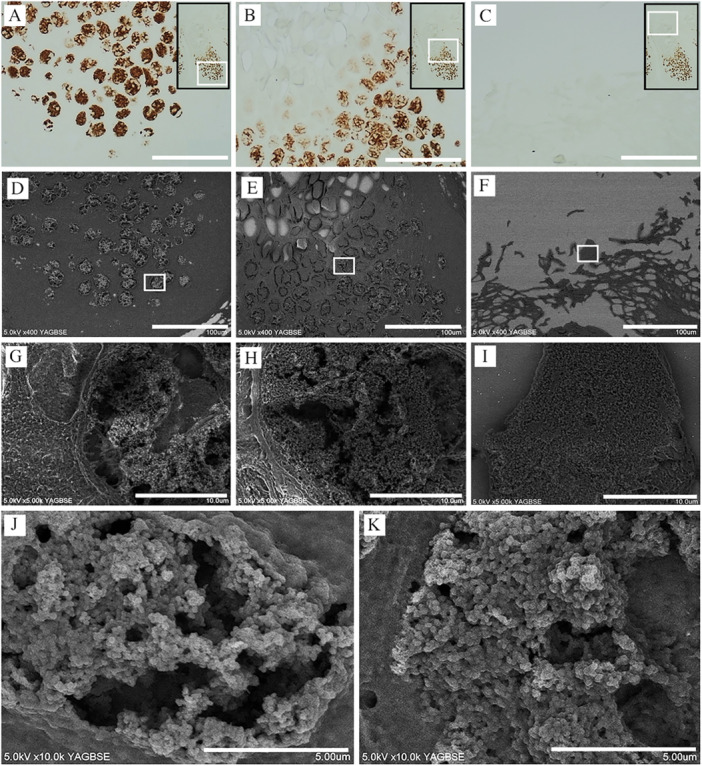
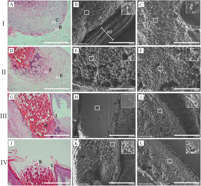
Methods: We applied NanoSuit-correlative light and electron microscopy (NanoSuit-CLEM) to formalin-fixed paraffin-embedded (FFPE) skin sections to visualize MC virus particles in situ with high resolution. Melan-A immunohistochemistry using 3,3'-diaminobenzidine (DAB) with osmium staining was performed to identify Henderson-Patterson bodies.
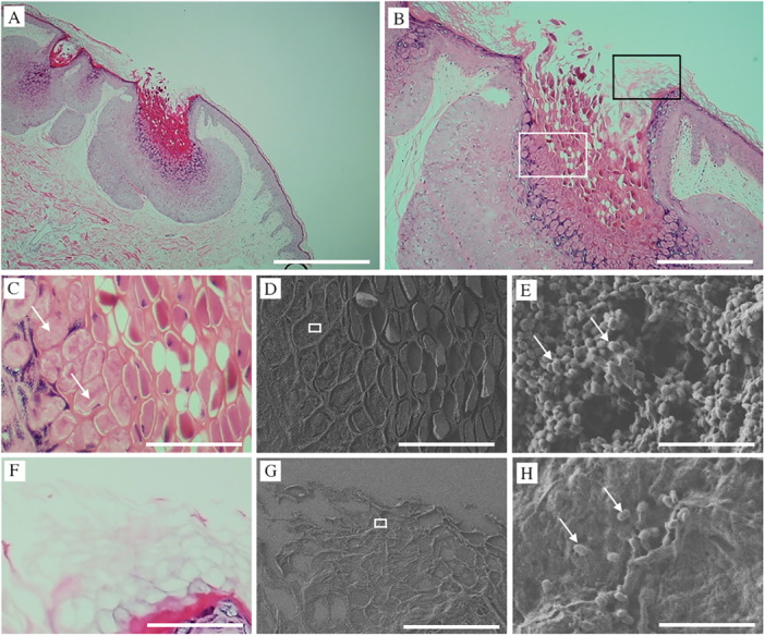
Results: Ultrastructural analysis revealed that MC virus particles were densely localized in the stratum corneum but did not invade deeper epithelial layers in intact skin. However, in areas of epidermal disruption, such as detached or damaged stratum corneum, the virus was observed penetrating into lower layers. While Melan-A immunostaining successfully detected Henderson-Patterson bodies, it failed to identify mature MC virus particles. In contrast, NanoSuit-CLEM combined with Mayer's hematoxylin and lead staining enabled detailed visualization of mature viral particles and their distribution within the stratum corneum.
Conclusions: These findings provide direct ultrastructural evidence that MC virus entry occurs through compromised skin, underscoring the crucial role of the stratum corneum in barrier function. This study highlights the importance of preventing mechanical skin injury, such as scratching or shaving, to limit MC transmission. NanoSuit-CLEM offers a powerful new tool for studying viral pathogenesis in archival tissue samples.
Keywords: Molluscum contagiosum; NanoSuit‐CLEM; skin; viral particles.
© 2025 The Author(s). Immunity, Inflammation and Disease published by John Wiley & Sons Ltd.
Conflict of interest statement
The authors declare no conflicts of interest.
Figures

References
-
- Scholz J., Rösen‐Wolff A., Bugert J., et al., “Epidemiology of Molluscum Contagiosum Using Genetic Analysis of the Viral DNA,” Journal of Medical Virology 27 (1989): 87–90. - PubMed
-
- Postlethwaite R., “Molluscum Contagiosum,” Archives of Environmental Health: An International Journal 21 (1970): 432–452. - PubMed
-
- Leung A. K. C., Barankin B., and Hon K. L. E., “Molluscum Contagiosum: An Update,” Recent Patents on Inflammation and Allergy Drug Discovery 11 (2017): 22–31. - PubMed
-
- Gerlero P. and Hernández‐Martín Á., “Actualización Sobre El Tratamiento De Moluscos Contagiosos En Los Niños,” Actas Dermo‐sifiliográficas 109 (2018): 408–415. - PubMed
-
- Chen X., Anstey A. V., and Bugert J. J., “Molluscum Contagiosum Virus Infection,” Lancet Infectious Diseases 13 (2013): 877–888. - PubMed
Publication types
MeSH terms
Substances
Grants and funding
LinkOut - more resources
Full Text Sources
