. 2025 Jul 22;99(7):e0030525.
doi: 10.1128/jvi.00305-25. Epub 2025 Jun 3.

COP I vesicles facilitate classical swine fever virus proliferation by transporting fatty acid synthase from the Golgi apparatus to the endoplasmic reticulum

Affiliations

COP I vesicles facilitate classical swine fever virus proliferation by transporting fatty acid synthase from the Golgi apparatus to the endoplasmic reticulum

Liang Zhang et al. J Virol. .

Abstract

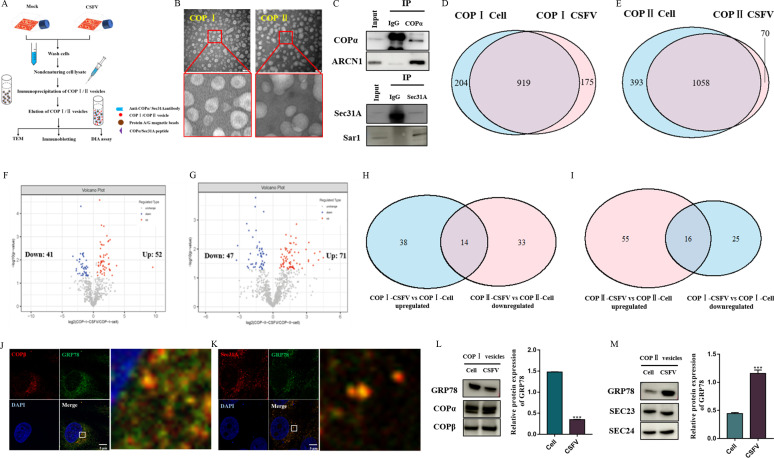
Classical swine fever virus (CSFV) is an enveloped, positive-sense, single-stranded RNA virus in the Flaviviridae family that remodels the cell's endomembrane for its own propagation. The early secretory pathway is exploited by viruses for their lifecycle, but the mechanism underlying this hijacking of the early secretory pathway in CSFV infection remains unknown. Here, we observed that disrupting the functions of the early secretory pathway organelles, the Golgi apparatus, the endoplasmic reticulum (ER), and coatomer protein I (COP I) vesicles and coatomer protein II (COP II) vesicles resulted in a significant inhibition of CSFV propagation. Further, we revealed that COP I vesicles were required for CSFV RNA replication, but not for the formation of viral replication complexes. The results support the hypothesis that participation of COP I vesicles in viral RNA replication involves their capacity for cargo trafficking. Intact COP I vesicles were isolated and subjected to data-independent acquisition quantitative proteomics analysis to identify the differences in the proteomes of COP I vesicles. This analysis revealed an increase in fatty acid synthase (FASN), a critical factor for CSFV RNA replication, within COP I vesicles, while its presence in COP II vesicles decreased in CSFV-infected cells. Meanwhile, blocking COP I vesicle formation resulted in decreased levels of FASN in the ER, impairing CSFV RNA replication. Collectively, we provide evidence that COP I vesicles mediate FASN trafficking from the Golgi apparatus to the ER to facilitate CSFV RNA replication, which advances our understanding of the role of the early secretory pathway in CSFV proliferation.IMPORTANCEClassical swine fever is a highly contagious disease caused by the classical swine fever virus (CSFV) that infects domestic pigs and wild boars and results in significant economic losses to the swine industry. The early secretory pathway in host cells has often been hijacked by viruses for viral genome replication, assembly, and release of virions. Here, our data revealed that the function of early secretory pathway organelles such as the endoplasmic reticulum (ER) and the Golgi apparatus, and the membrane-bound transport intermediates, COP I vesicles and COP II vesicles, that facilitate transport, were involved in CSFV proliferation in PK-15 cells. Our findings demonstrate that COP I vesicles significantly promote CSFV RNA replication by trafficking fatty acid synthase from the Golgi apparatus to the ER. Our data suggest that manipulation of early secretory pathway function in target host cells could provide a promising strategy for a novel anti-CSFV therapeutic.

Keywords: classical swine fever virus; coatomer protein I; coatomer protein II; early secretory pathway; fatty acid synthase.


Conflict of interest statement

The authors declare no conflict of interest.

Figures

Fig 1
Fig 1
Disrupting the functions of early secretory pathway organelles inhibits CSFV infection. (A) CCK-8 assays were conducted to measure the viability of cells after treatment with BFA and tunicamycin. (B) PK-15 cells were treated with or without 100 nM BFA for 24 h, cells were fixed in 4% paraformaldehyde, and immunofluorescence staining was performed using an anti-GM130 antibody. Scale bars: 5 µm. (C) PK-15 cells were treated with or without 10 µM tunicamycin for 24 h, cells were fixed in 4% paraformaldehyde, and immunofluorescence staining was performed using an anti-Sec61 antibody. Scale bars: 5 µm. (D and E) PK-15 cells were co-treated with 1 MOI CSFV and 10 µM tunicamycin or 100 nM BFA for 48 h, and then cell and culture supernatants were collected for determination of CSFV RNA copy numbers and virus titers by RT-qPCR and TCID50/mL, respectively. (F) PK-15 cells were co-treated with 1 MOI CSFV and 10 µM tunicamycin or 100 nM BFA for 48 h, and then cells were fixed in 4% paraformaldehyde and stained with anti-E2 antibody (green). Scale bars, 200 µm.
Fig 2
Fig 2
COP I and COP II positively regulate CSFV propagation. (A and B) CCK-8 assays were conducted to measure the cell viability after treatment with GCA and Exo-1. (C and D) PK-15 cells were pretreated with 300 nM GCA or 5 µM Exo-1 for 24 h and then infected with 1 MOI CSFV. After 48 h, the cells and culture supernatants were harvested for determining CSFV RNA copy numbers and virus titers by RT-qPCR and TCID50/mL, respectively. (E and F) Cells were transfected with NTsiRNA, siCOPA-1, siCOPA-2, siCOPA-3, siCOPD-1, siCOPD-2, and siCOPD-3 for 48 h. Cells were harvested, and COPα and ARCN1 levels were quantitated by Western blot. (G and H) Cells transfected with NTsiRNA, siCOPA-3, and siCOPD-1 were infected with 1 MOI CSFV. Then, cells and culture supernatants were harvested for measurement of CSFV RNA copy numbers and virus titers by RT-qPCR and TCID50/mL. (I) NTsiRNA-, siCOPA-3-, or siCOPD-1-transfected cells were infected with 1 MOI of CSFV. After 48 h, the cells were fixed in 4% paraformaldehyde and stained with anti-E2 antibody (green). Scale bars, 200 µm. (J) CCK-8 assays were conducted to measure the viability of cells after treatment with H89. (K and L) PK-15 cells were pretreated with various concentrations of H89 for 24 h and then infected with 1 MOI of CSFV. After 48 h, cell and culture supernatants were harvested for measuring CSFV RNA copy numbers and virus titers by RT-qPCR and TCID50/mL, respectively. (M and N) Cells were transfected with NTsiRNA, siSec23, and siSec24 for 48 h and then harvested for determination of Sec23 and Sec24 levels by Western immunoblot assay. (O and P) NTsiRNA-, siSec23-2-, and siSec24-2-transfected cells were infected with 1 MOI of CSFV, and after 48 h, the cells and culture supernatants were harvested for measurement of CSFV RNA copy numbers and virus titers by RT-qPCR and TCID50/mL, respectively. (Q) NTsiRNA-, siSec23-2-, and siSec24-2-transfected cells were infected with 1 MOI of CSFV, and after 48 h, the cells were fixed in 4% paraformaldehyde and stained with anti-E2 antibody. Scale bars, 200 µm. (R) CSFV (1 MOI)-infected cells were treated with GCA, Exo-1, and H89 or transfected with siCOPA, siCOPD, siSec23, and siSec24 for 48 h, and the distribution of the KDEL was viewed under confocal microscopy. (S) CSFV (1 MOI)-infected cells were treated with GCA, Exo-1, and H89 or transfected with siCOPA, siCOPD, siSec23, and siSec24 for 48 h, and the distribution of the ERGIC53 was viewed under confocal microscopy. Scale bars: 5 µm.
Fig 3
Fig 3
COP I is required for CSFV RNA replication. (A) NTsiRNA-, siCOPA-3-, and siCOPD-1-transfected cells were infected with 10 MOI of CSFV in FBS-free medium for 1 h at 4°C. Unbound virions were washed away with pre-cooled citrate buffer (pH = 3). Total cells were collected for CSFV RNA copy number measurement by RT-qPCR. (B) NTsiRNA-, siCOPA-3-, and siCOPD-1-transfected cells were infected with 10 MOI CSFV (MOI = 10) in FBS-free medium for 1 h at 4°C to allow virion binding. Cells were then washed with pre-cooled citrate buffer (pH = 3) to remove unbound virions and cultured for another 2 h at 37°C. The cells were washed and collected for CSFV RNA copy number determination by RT-qPCR. (C and D) PK-15 cells were infected with 1 MOI of CSFV for 2 h, then the medium was discarded, and the fresh medium containing GCA or Exo1 was added. Samples were collected after 8 h incubation for CSFV RNA copy number determination by RT-qPCR. (E) The pEGFP-NS5B-transfected cells were infected with 1 MOI of CSFV for 48 h. Cells were stained for the COP I vesicle marker, COPβ, the ER marker, Sec61, and nucleocapsids. Scale bars: 5 µm.
Fig 4
Fig 4
Proteomics characterization of isolated COP I and COP II vesicles. (A) Schematic representation of the experimental timeline for isolation and purification of COP I and COP II vesicles. (B) The isolated COP I and COP II vesicles were analyzed by negatively stained TEM. (C) The isolated COP I and COP II vesicles were analyzed by Western blot. (D) Venn diagram showing proteins in common identified in COP I vesicles from CSFV-infected and noninfected cells. (E) Venn diagram of proteins in common identified in COP II vesicles from CSFV-infected and noninfected cells. (F) Volcano plot showing differences in protein abundance from COP I vesicles isolated from CSFV-infected and noninfected cells. (G) Volcano plot showing protein abundance differences in COP II vesicles between CSFV-infected and noninfected cells. (H) Venn diagram of common proteins in upregulated proteins of COP I vesicles (CSFV vs noninfected) and downregulated proteins of COP II vesicles (CSFV vs noninfected) (I) Venn diagram of common proteins in downregulated proteins of COP I vesicles (CSFV vs noninfected) and upregulated proteins of COP II vesicles (CSFV vs noninfected). (J) Colocalization of GRP78 and COPβ. Cells were infected with CSFV (MOI = 1) for 48 h and stained for GRP78, COPβ, and nucleocapsid. Scale bars: 5 µm. (K) Colocalization of GRP78 and Sec31A. Cells were infected with CSFV (MOI = 1) for 48 h and stained for GRP78, Sec31A, and nucleocapsid. Scale bars: 5 µm. (L) The GRP78 levels in isolated COP I vesicles from CSFV-infected and uninfected cells were analyzed by Western blot. (M) The GRP78 levels in isolated COP II vesicles from CSFV-infected and uninfected cells were analyzed by Western blot.
Fig 5
Fig 5
COP I vesicles regulate FASN trafficking from the Golgi apparatus to the ER. (A) Colocalization of FASN and COPβ. Cells were infected with CSFV (MOI = 1) for 48 h and stained for FASN, COPβ, and nucleocapsid. Scale bars: 5 µm. (B) Colocalization of FASN and Sec31A. Cells were infected with CSFV (MOI = 1) for 48 h and stained for FASN, Sec31A, and nucleocapsid. Scale bars: 5 µm. (C) The FASN levels in isolated COP I vesicles from CSFV-infected and uninfected cells were analyzed by Western blot. (D) The FASN levels in isolated COP II vesicles from CSFV-infected and uninfected cells were analyzed by Western blot. (E) NTsiRNA- and siCOPA-3-transfected cells were infected with or without CSFV (MOI = 1). After 48 h, the cells were stained for FASN, Sec61, and nucleocapsid. Scale bars: 5 µm. (F) NTsiRNA-, siFASN-1-, siFASN-2-, and siFASN-3-transfected cells were collected for protein extraction with RIPA lysis, and the FASN expression levels were analyzed by Western blot. (G and H) NTsiRNA- and siFASN-1-transfected cells were infected with 1 MOI CSFV. After 48 h, cells and culture supernatants were collected for CSFV RNA copy numbers and virus titers detection by RT-qPCR and TCID50/mL, respectively. (I) NTsiRNA- and siFASN-1-transfected cells were infected with CSFV (MOI = 10) in the FBS-free medium for 1 h at 4°C. Unbound virions were then washed away using a pre-cooled citrate buffer solution (pH = 3). Total cells were collected for CSFV RNA copy number detection by RT-qPCR. (J) NTsiRNA- and siFASN-1-transfected cells were infected with CSFV (MOI = 10) in the FBS-free medium for 1 h at 4°C to allow virion binding. Cells were then washed with pre-cooled citrate buffer solution (pH = 3) to remove unbound virions and cultured for another 2 h at 37°C. The cells were washed and collected for CSFV RNA copy number detection by RT-qPCR. (K) NTsiRNA- and siFASN-1-transfected cells were infected with CSFV (MOI = 1), and CSFV RNA copy numbers were detected by RT-qPCR after 10 h.
Fig 6
Fig 6
CSFV infection induces morphological alterations of early secretory pathway organelles. (A and B) Electron micrographs of cells infected with or without CSFV (MOI = 10). Boxed areas indicate the Golgi apparatus (1A) or ER (1B), respectively. Scale bars, 500 nm. (C) Cells were infected with or without CSFV (10 MOI) for 48 h and stained for a Golgi apparatus marker GM130 and nucleocapsid. Scale bars: 5 µm. (D) Cells were infected with or without CSFV (10 MOI) for 48 h and stained for an ER marker Sec61 and nucleocapsid. Scale bars: 5 µm. (E) Cells were infected with or without CSFV (10 MOI) for 48 h and stained for the ERGIC marker ERGIC53 and nucleocapsid. Scale bars: 5 µm. (F) Cells were infected with or without CSFV (10 MOI) for 48 h and stained for a COP I vesicle marker COPβ and nucleocapsid. Scale bars: 5 µm. (G) Cells were infected with or without CSFV (10 MOI) for 48 h and stained for a COP II vesicle marker Sec31A and nucleocapsid. Scale bars: 5 µm.

Similar articles

References

    1. Moennig V. 2015. The control of classical swine fever in wild boar. Front Microbiol 6:1211. doi: 10.3389/fmicb.2015.01211 - DOI - PMC - PubMed
    1. Ji W, Guo Z, Ding N-Z, He C-Q. 2015. Studying classical swine fever virus: making the best of a bad virus. Virus Res 197:35–47. doi: 10.1016/j.virusres.2014.12.006 - DOI - PubMed
    1. Barlowe CK, Miller EA. 2013. Secretory protein biogenesis and traffic in the early secretory pathway. Genetics 193:383–410. doi: 10.1534/genetics.112.142810 - DOI - PMC - PubMed
    1. Budnik A, Stephens DJ. 2009. ER exit sites--localization and control of COPII vesicle formation. FEBS Lett 583:3796–3803. doi: 10.1016/j.febslet.2009.10.038 - DOI - PubMed
    1. Gomez-Navarro N, Miller E. 2016. Protein sorting at the ER-Golgi interface. J Cell Biol 215:769–778. doi: 10.1083/jcb.201610031 - DOI - PMC - PubMed

MeSH terms

Substances

LinkOut - more resources