. 2025 Aug;174(1):177-190.
doi: 10.1007/s11060-025-05050-9. Epub 2025 Jun 3.

A white matter-centered approach to investigate recurrence pathways in high-grade gliomas: a single-center retrospective study

Affiliations

A white matter-centered approach to investigate recurrence pathways in high-grade gliomas: a single-center retrospective study

Salvatore Marino et al. J Neurooncol. 2025 Aug.

Abstract

Background and aim: High-grade gliomas (HGGs) are aggressive primary brain tumors with inevitable recurrence. This single-center retrospective study investigates whether the anatomical proximity of HGGs to major white matter tracts influences progression and recurrence. The study explores the association between tumor location and recurrence type-local, remote, or ependymal-and whether recurrences align with adjacent white matter tracts.

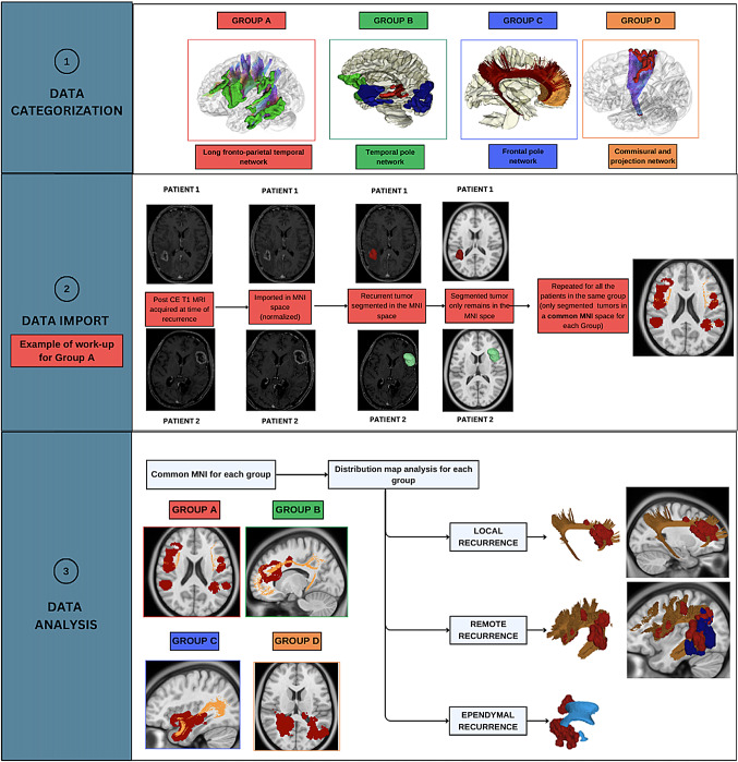
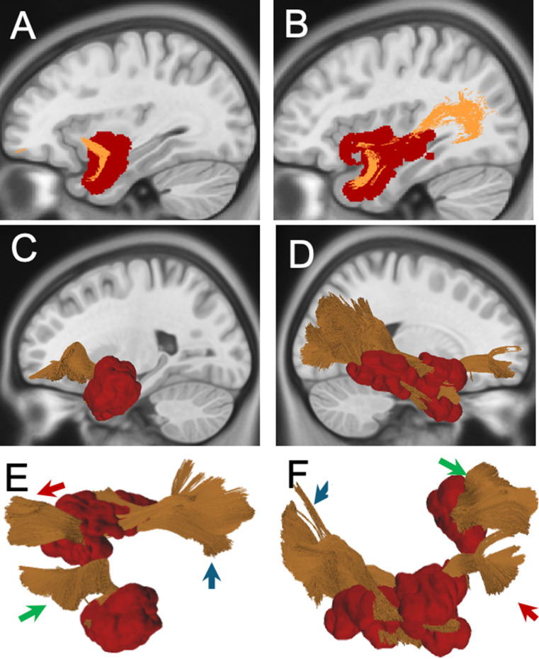
Methods: The study included patients with histopathologically confirmed recurrent HGGs who underwent reoperation. Primary tumors were categorized into four anatomical subgroups using a connectivity-based framework from the HCP 1065 Atlas: Subgroup A: Long Fronto-Temporo-Parietal Network Subgroup B: Temporal Pole Network (further divided into B1, B2, and B3 based on connectivity patterns) Subgroup C: Frontal Pole Network Subgroup D: Commissural and Projection Networks (further divided into D1 and D2). Recurrences were classified via post-contrast T1-weighted MRI as local, remote, ependymal. The Tract-to-Region Connectome (T-R-C) assessed the volumetric overlap between recurrence maps and main white matter bundles.

Results: Of 41 patients, a significant correlation emerged between tumor subgroup and recurrence type (p = 0.0003). Subgroup A predominantly showed remote recurrences (68%), while B2, B3, C, and D2 had mainly local recurrences. Subgroup D1 had a predominance of ependymal recurrences (66.7%). Local and remote recurrences largely conformed to adjacent white matter distributions, with variations in timing of recurrence and survival observed across different groups.

Conclusion: Our analysis, focused on exploring the spatial aspects of recurrence in relation to white matter anatomy, suggests that HGG recurrence patterns are strongly influenced by anatomical location and white matter architecture. Certain anatomical areas show a predisposition toward specific recurrence patterns. Recognizing these spatial dynamics may guide more precise surgical strategies, radiotherapy targeting, and recurrence risk assessment.

Keywords: High grade glioma; Recurrence; White matter.


Conflict of interest statement

Declarations. Ethical approval: The Institutional Review Board (IRB) of Fondazione Policlinico Gemelli IRCCS approved this study (ID 7028). Competing interests: The authors declare no competing interests.

Figures

Fig. 1
Fig. 1
Data Categorization: Patients were classified into different groups based on the location of the primary tumor, as detailed in the text. Group A: Tumors located along the territory of the SLF/AF complex. Group B: Tumors located along the territory of the ILF and Sagittal Stratum (B1), Uncinate Fasciculus (B2), and Hippocampal Formation (B3). Group C: Tumors located in the territory of the Cingulate Bundle and Forceps Minor. Group D: Tumors located in the territory of the Commissural pathways (mainly callosal radiation, D1) and Projection pathways (mainly the corticospinal tract, D2). Data Import: For each patient, the T1-MDC scan acquired at the time of radiological diagnosis of recurrence was normalized to the Montreal Neurological Institute (MNI) space using the integrated normalization software in DSI Studio (DSI Studio, http://dsi-studio.labsolver.org). The registration quality was visually inspected in all cases, with manual adjustments performed when necessary. Recurrences were segmented from the original MRI and aligned to the MNI space. Recurrences from patients within the same subgroup were sequentially imported into a common MNI space. This process resulted in a “Distribution Map” for each group, integrating all recurrences of patients belonging to the same group within the MNI space. The primary white matter tracts associated with each subgroup (e.g., AF/SLF for Group A) were generated using the ICBM 152 Atlas template on the MNI space with Distribution Map. Each recurrence was first assessed individually and classified as either “contiguous” or “non-contiguous” for local recurrences and “consonant” or “dissonant” for remote recurrences. After individual classification, recurrences were assessed collectively, and volumetric overlap between each subgroup’s Distribution Map and the corresponding white matter bundles was visually inspected and quantified using the Tract-to-Region Connectome function in DSI Studio. Separate Distribution Maps were generated for local and remote recurrences, as well as for ependymal recurrences within each group. Distribution maps containing fewer than two recurrences per side were excluded from the analysis, even if depicted
Fig. 2
Fig. 2
(AD) Sagittal and axial 2D scan showing Distribution Map of Group A. In red are highlighted the recurrences belonging to this group segmented into the MNI space. Specifically, local recurrences arose in all cases from the antero-inferior aspect of the surgical cavity toward the temporal termination of the AF. Regarding remote recurrences, two occurred at the level of the posterior temporal lobe from a primitive located at the SMG/AG; four at the level of the SMG/AG from a primitive located to the posterior temporal lobe; five at the level of fronto-parietal operculum, namely pars triangularis, pars opercularis and subcentral gyrus from a primitive located at the SMG/AG. (E-F, G-H) 3D rendering of the recurrences in the left and right hemispheres. In red, recurrences, in blu original tumor, in yellow AF/SLF
Fig. 3
Fig. 3
(A-B) 2D distribution map of Group B. Sagittal scan of the left side is presented on the left, with the recurrences segmented into the MNI space (in red) and UF in orange. Sagittal scan of the right side is presented on the right, with the recurrences segmented into the MNI space (in red) and UF and ILF in orange. (C-D) 3D rendering of the Group B distribution map: On the left, the anatomical relationship between recurrences and the uncinate fasciculus (UF) in the left hemisphere is shown. On the right, the anatomical relationships are illustrated between recurrences, and the UF/ILF in the right hemisphere. (E-F) 3D-rendering of the left uncinate fasciculus (green arrow), the right uncinate fasciculus (red arrow), and the right inferior longitudinal fasciculus (blue arrow) and recurrences (in red)
Fig. 4
Fig. 4
(A-B) 2D Distribution map of Group C. Sagittal scan of the left side is presented on the left, with the recurrences segmented into the MNI space (in red) and CB in orange. Sagittal scan of the right side is presented on the right, with the recurrences segmented into the MNI space (in red) and CB in orange. In all cases, recurrences were local, arose from the posterior margin of the surgical cavity and progressed along the cingulate gyrus. In four out of six cases, the recurrences also involved the GunuCC; (CF) 3D rendering of the Group C distribution map: On the left, the anatomical relationship between recurrence and Cingulate Bundle in the left hemisphere is shown. On the right, the anatomical relationships are illustrated between recurrences, and the Cingulate Bundle in the right hemisphere
Fig. 5
Fig. 5
2D Distribution Map of ependymal recurrences. (A) Axial scan is presented, with the recurrences segmented (BD) into the MNI space in red. 3D-rendered map illustrating the distribution of ependymal recurrences, with recurrences shown in red and their anatomical relationship to the lateral ventricles highlighted in blue. Regardless of location, the main characteristic associated with an ependymal route of spread was a “ventricolocentric” shape of the tumor at onset, marked by an enhancing nodule directed toward, or in direct contact with the sub-ependymal layer at onset and FLAIR alteration extending toward the ventricle. The primary ventricular walls involved in tumor recurrences were the atrium and temporal horn
Fig. 6
Fig. 6
(A) Kaplan-Meier analysis of time to first recurrence across recurrence types showed no statistically significant differences. The Log-Rank Test yielded χ² = 1.466 (p = 0.481), and the Wilcoxon Test produced χ² = 1.336 (p = 0.513). While descriptive trends suggest earlier recurrence in ependymal cases, these differences were not statistically significant. (B) Kaplan-Meier analysis of time to first recurrence across tumor subgroups revealed a statistically significant difference based on the Log-Rank Test (χ² = 11.449, p = 0.022). However, the Wilcoxon Test did not confirm this significance (χ² = 5.392, p = 0.249). (C) Kaplan-Meier analysis of overall survival across recurrence types indicated a trend toward statistical significance, though it did not meet conventional thresholds. The Log-Rank Test yielded χ² = 4.610 (p = 0.100), while the Wilcoxon Test produced χ² = 4.519 (p = 0.104). (D) Kaplan-Meier analysis of overall survival across tumor subgroups demonstrated statistically significant differences. The Log-Rank Test resulted in χ² = 14.081 (p = 0.007), and the Wilcoxon Test yielded χ² = 10.773 (p = 0.029)

References

    1. Menna G, Marinno S, Valeri F et al (2024) Diffusion tensor imaging in detecting gliomas sub-regions of infiltration, local and remote recurrences: a systematic review. Neurosurg Rev 47(1):301. 10.1007/s10143-024-02529-3. Published 2024 Jul 2 - PubMed
    1. Tu Z, Xiong H, Qiu Y, Li G, Wang L, Peng S (2021) Limited recurrence distance of glioblastoma under modern radiotherapy era. BMC Cancer 21(1):720. Published 2021 Jun 22. 10.1186/s12885-021-08467-3 - PMC - PubMed
    1. Langhans M, Popp I, Grosu AL et al (2023) Recurrence analysis of glioblastoma cases based on distance and dose information. Radiother Oncol 183:109600. 10.1016/j.radonc.2023.109600 - PMC - PubMed
    1. Marino S, Menna G, Di Bonaventura R et al (2023) The extracellular matrix in glioblastomas: A glance at its structural modifications in shaping the tumoral Microenvironment-A systematic review. Cancers (Basel) 15(6):1879. 10.3390/cancers15061879. Published 2023 Mar 21 - PMC - PubMed
    1. Spillmann AA, Amberger VR, Schwab ME (1997) High molecular weight protein of human central nervous system Myelin inhibits neurite outgrowth: an effect which can be neutralized by the monoclonal antibody IN-1. Eur J Neurosci 9(3):549–555. 10.1111/j.1460-9568.1997.tb01631.x - PubMed

MeSH terms

LinkOut - more resources