. 2025 Jun 3;16(1):5146.
doi: 10.1038/s41467-025-60372-6.

A soft robotic total artificial hybrid heart

Affiliations

A soft robotic total artificial hybrid heart

Maziar Arfaee et al. Nat Commun. .

Abstract

End-stage heart failure is a deadly disease. Current total artificial hearts (TAHs) carry high mortality and morbidity and offer low quality of life. To overcome current biocompatibility issues, we propose the concept of a soft robotic, hybrid (pumping power comes from soft robotics, innerlining from the patient's own cells) TAH. The device features a pneumatically driven actuator (septum) between two ventricles and is coated with supramolecular polymeric materials to promote anti-thrombotic and tissue engineering properties. In vitro, the Hybrid Heart pumps 5.7 L/min and mimics the native heart's adaptive function. Proof-of-concept studies in rats and an acute goat model demonstrate the Hybrid Heart's potential for clinical use and improved biocompatibility. This paper presents the first proof-of-concept of a soft, biocompatible TAH by providing a platform using soft robotics and tissue engineering to create new horizons in heart failure and transplantation medicine.


Conflict of interest statement

Competing interests: The authors declare no conflict of interest.

Figures

Fig. 1
Fig. 1. The Hybrid Heart design.
The Hybrid Heart is a soft robotic, pulsatile TAH that enables a soft contractile motion similar to the human heart. It is actuated by a pneumatic actuator (septum) positioned between the ventricles, surrounded by wires that wrap around both septum and each ventricle in a shape of (∞).
Fig. 2
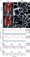
Fig. 2. In vitro test results of the hybrid heart.
All tests were performed in the double mock circulatory loop. a Hybrid Heart in front view (left images) and the view inside the ventricle, captured by a laparoscope (right images), during systole and diastole. b On top, pressure curves measured during hybrid heart operation in the mock circulatory loop, followed by pressure curves of the native heart below. AOP: aortic pressure, PAP: pulmonary artery pressure, LAP: left atrial pressure, RAP: right atrial pressure. c Flow curves measured during hybrid heart operation in the mock circulatory loop, followed by flow curves of native heart below,. AO: blood flow in aorta, PA: blood flow in pulmonary artery.
Fig. 3
Fig. 3. In vitro characterization of hybrid heart’s preload and afterload sensitivity.
All tests were performed in the double mock circulatory loop at different heart rates of 60, 70, and 80 BPM. Data are presented as mean ± SD of n = 5 cycles. a) The relation between varying left preload (4-20 mmHg) versus left cardiac output. b) The relation between varying right preload (4–20 mmHg) versus right cardiac output. c) The relation between varying left afterload (MAoP) (60–120 mmHg) versus left cardiac output. d) The relation between varying right afterload (MPAP) (15–35 mmHg) versus right cardiac output. e) Hybrid Heart’s reaction to left-to-right preload imbalance of ~12 mmHg. f) Hybrid Heart’s reaction to right-to-left preload imbalance of ~12 mmHg.
Fig. 4
Fig. 4. Test results of the Hybrid Heart in vivo experiment in an acute goat experiment.
All data corresponds to the period during which the Hybrid Heart was providing all the blood flow in the animal, without additional support of the cardiopulmonary bypass. Data are presented as mean ± SD of n = 20 cycles with shaded error bars. a Systemic pressures measured during the in vivo experiment. AOP: aortic pressure, LVP: intra ventricular pressure of the left ventricle, LAP: left atrial pressure. b Aortic flow during the animal experiment. c Pressures of the pulmonary circulation measured during the animal experiment. PAP: pulmonary artery pressure, RVP: intra ventricular pressure of the right ventricle, RAP: right atrial pressure. d Pulmonary flow during the acute goat experiment. e Screen capture of the monitor during the animal experiment. Red line shows the blood pressure measured in the iliac artery (100/46 mmHg, mean 58 mmHg). f) Photo of the Hybrid Heart implanted in the goat.
Fig. 5
Fig. 5. The biocompatible inner lining for the Hybrid Heart, in vitro studies on coated and uncoated TPU-coated nylon.
a Schematic overview of the different grafts used in the in-vitro and in-vivo rat studies. b Chemical structures of Nylon, TPU, PCL-BU, heparin and BU-HBP. c water contact angles of TPU-coated nylon material with and without PCL-BU coating with or without 5 or 20 mol% BU-HBP. Data are presented as mean ± SD of n = 3 groups, each containing 3 samples. d Fluorescence of solution taken from TPU-coated nylon coated with PCL-BU with or without 5, 10 or 20 mol% BU-HBP. Data are presented as mean ± SD of n = 3 samples. e Cytotoxicity determined from LDH assay of TPU-coated nylon material with and without PCL-BU coating with or without 5 mol% BU-HBP. Data are presented as mean ± SD of n = 4 samples. f SEM images of platelets that adhered to the TPU-coated nylon materials with and without PCL-BU coating with or without 5 or 20 mol% BU-HBP either with heparin functionalization or without. We tested n = 3 samples for each material type, representative images are shown in this figure.
Fig. 6
Fig. 6. Pre-clinical assessment of the grafts.
a Schematic drawing of the graft design with cross-sectional view as well. b Picture of fabricated graft. c Picture of implanted graft in rat aorta. d Assessment of the grafts in terms of occlusion based on hind leg movement and explantation.
Fig. 7
Fig. 7. Integration test with implantable control system.
a Left and right ventricular output flow (top panel) as a result of pressure pulses provided to the septum (bottom panel). b (all panels show 10-beat moving average values). Two vertical solid lines indicate two instances where input power is manually decreased to demonstrate the effect of electrical input power on hybrid heart output. SV = stroke volume, CO = cardiac output, HR = heart rate, P = electrical power. c Components of the experimental setup, showing how the septum of Hybrid Heart (HH) is connected inline with the sealed air circulation of the implantable control system. The ventricles of the Hybrid Heart are connected to a mock circulatory loop. A horizontal dashed line demarcates the division between the internal and external components of the transcutaneous energy transfer (TET) system.

References

    1. Bragazzi, N. L. et al. Burden of heart failure and underlying causes in 195 countries and territories from 1990 to 2017. Eur. J. Preventive Cardiol.28, 1682–1690 (2021). - PubMed
    1. Tatum, R., Briasoulis, A., Tchantchaleishvili, V. & Massey, H. T. Evaluation of donor heart for transplantation. Heart Fail Rev.27, 1819–1827 (2022). - PMC - PubMed
    1. Tsao, C. W. et al. Heart Disease and Stroke Statistics—2022 update: a report from the American Heart Association. Circulation145, e153–e639 (2022). - PubMed
    1. Dal Sasso, E., Bagno, A., Scuri, S. T. G., Gerosa, G. & Iop, L. The biocompatibility challenges in the total artificial heart evolution. Annu Rev. Biomed. Eng.21, 85–110 (2019). - PubMed
    1. Cianchetti, M., Laschi, C., Menciassi, A. & Dario, P. Biomedical applications of soft robotics. Nat. Rev. Mater.3, 143–153 (2018).

Substances

LinkOut - more resources