. 2025 Jun 3;16(1):432.
doi: 10.1038/s41419-025-07752-4.

Oncometabolite fumarate facilitates PD-L1 expression and immune evasion in clear cell renal cell carcinoma

Affiliations

Oncometabolite fumarate facilitates PD-L1 expression and immune evasion in clear cell renal cell carcinoma

Yi Gao et al. Cell Death Dis. .

Abstract

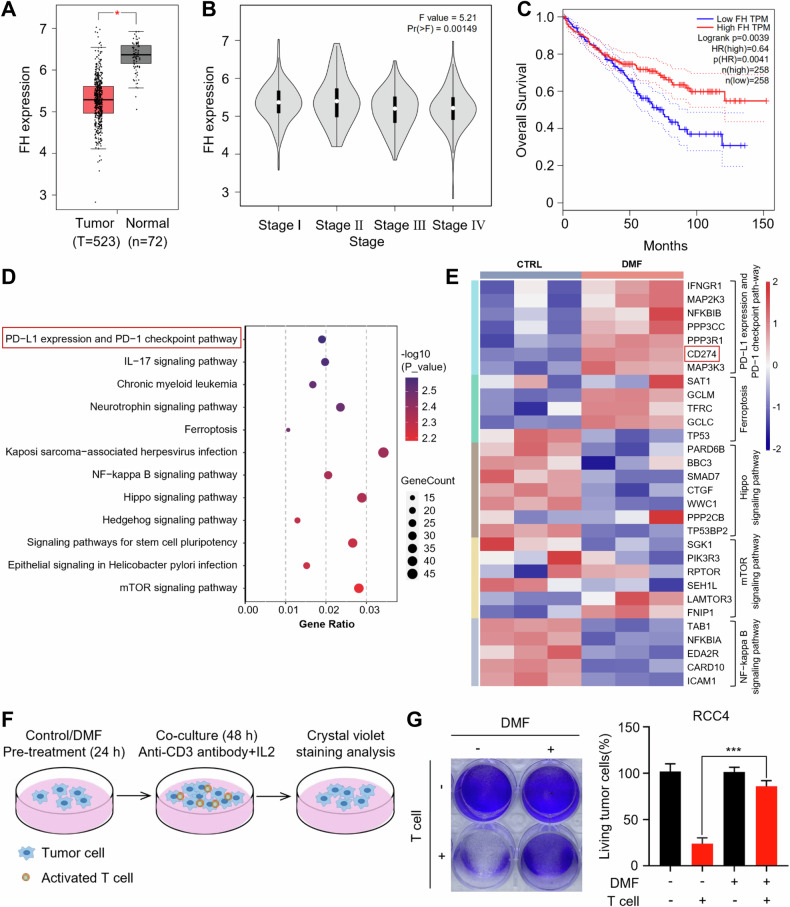
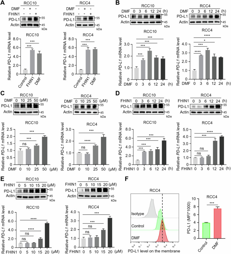
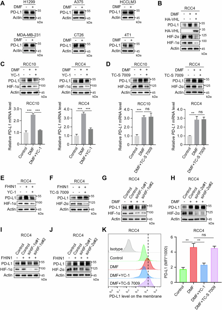
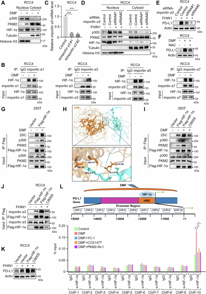
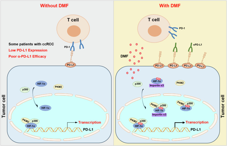
Clear cell renal cell carcinoma (ccRCC) is the most common subtype of renal cell carcinoma (RCC), with a rising incidence worldwide. However, the mechanisms by which ccRCC evades immune surveillance remain incompletely understood. Our findings indicate that fumarate hydratase (FH) expression is significantly downregulated in ccRCC, resulting in fumarate accumulation, which is correlated with a poor prognosis in ccRCC patients. RNA sequencing analysis suggests that dimethyl fumarate (DMF), an FDA-approved fumarate analogue, may impact tumor immunity. Our further investigation reveals that both DMF and the FH inhibitor (FHIN1) can promote immune evasion in ccRCC by upregulating PD-L1. Pre-treatment of tumor cells with DMF notably inhibits the cytotoxic effect of T cells. Mechanistically, fumarate induces PD-L1 expression through succination of HIF-1α at C800, facilitating its interaction with importin α3, p300, and PKM2, which promotes HIF-1α nuclear localization and transcriptional activity. Moreover, combining DMF with PD-L1 blockade therapy significantly enhances the efficacy of immunotherapy and prolongs the survival of tumor-bearing mice. Taken together, our study elucidates a mechanism by which FH downregulation promotes immune evasion through the fumarate-HIF-1α/p300/PKM2-PD-L1 axis, providing a novel target, drug, and strategy to improve immunotherapy for ccRCC.


Conflict of interest statement

Competing interests: The authors declare no competing interests.

Figures

Fig. 1
Fig. 1. Accumulation in fumarate triggers alterations of immune response in ccRCC.
A Differences in the expression of FH between ccRCC and normal samples are shown using the GEPIA platform. *P < 0.05. B FH expression levels in normal samples and four different tumor stages of ccRCC. *P < 0.05. C Kaplan–Meier curves for overall survival (OS) of ccRCC patients with low and high FH expression levels. D, E RNA-seq analysis was performed on RCC4 cells with and without DMF treatment, using three biologically independent samples per group. All listed genes showed significant differences (P < 0.05). F Schematic diagram depicting the determination of the effect of DMF on T cell-mediated killing of RCC4 cells. G Control and DMF–treated RCC4 cells were cocultured with activated T cell for 48 h and then subjected to crystal violet staining. The quantification was shown on the Right. Data represent mean ± SD, n = 3. ***P < 0.001.
Fig. 2
Fig. 2. Fumarate upregulates the expression of PD-L1 via transcriptional regulation in ccRCC.
A Western blot and qRT-PCR analyses of PD-L1 in RCC10 and RCC4 cells with or without DMF (50 μM, 12 h) or FHIN1 (20 μM, 24 h) were performed. ***P < 0.001. B Western blot and qRT-PCR analyses of PD-L1 expression in RCC10 and RCC4 cells treated with 50 μM DMF for various durations as indicated. *P < 0.05, ***P < 0.001, ****P < 0.0001. C Western blot and qRT-PCR analyses of PD-L1 expression in RCC10 and RCC4 cells treated with increasing concentrations of DMF (0–50 μM) for 12 h. ns nonsignificant, ***P < 0.001, ****P < 0.0001. D Western blot and qRT-PCR analyses evaluated PD-L1 expression in RCC10 and RCC4 cells treated with 20 μM FHIN1 at 3 h, 6 h, 12 h, and 24 h. ns, nonsignificant, ***P < 0.001, ****P < 0.0001. E Western blot and qRT-PCR analyses were used to assess PD-L1 expression levels in RCC10 and RCC4 cells after treatment with FHIN1 concentrations ranging from 0 to 20 μM for 24 h. ns nonsignificant, ***P < 0.001, ****P < 0.0001. F Flow cytometry analysis of membrane PD-L1 expression in RCC4 cells under DMF treatment. Representative histograms and summarized mean fluorescent intensity (MFI) are shown. Values are means ± SD from n = 3 independent experiments. Statistical differences were determined by Student’s t-test. ***P < 0.001.
Fig. 3
Fig. 3. Fumarate facilitates PD-L1 expression via HIF-1α.
A Western blot analysis was performed to assess the protein levels in four human tumor cell lines (non-small cell lung cancer H1299, melanoma A375, liver cancer HCCLM3, breast cancer MDA-MB-231) and two mouse tumor cell lines (colorectal cancer CT26, breast cancer 4T1) following treatment with 50 μM DMF for 12 h. B The expression levels of HIF-1α, HIF-2α, and PD-L1 in RCC4 cells with or without transfection of VHL-expressing plasmids, were examined by Western blot after treatment with DMF (50 μM, 12 h). C Western blot and qRT-PCR analyses were performed to assess the PD-L1 levels in RCC10 and RCC4 cells treated with DMF, in the absence and presence of 5 μM YC-1, an inhibitor of HIF-1α. ***P < 0.001. D Western blot and qRT-PCR analyses were performed to assess PD-L1 levels in RCC10 and RCC4 cells treated with DMF, with or without 50 μM TC-S 7009, an inhibitor of HIF-2α. ns nonsignificant, **P < 0.01, ***P < 0.001. E Western blot analyses were performed to assess the PD-L1 levels in RCC4 cells treated with FHIN1, both in the absence and presence of 5 μM YC-1, which is an inhibitor of HIF-1α. F Western blot analyses were performed to assess PD-L1 levels in RCC4 cells treated with FHIN1 with or without 50 μM TC-S 7009, which is an inhibitor of HIF-2α. G After transfection with two siRNA fragments designed to knock down HIF-1α expression, RCC4 cells were treated with DMF, followed by the detection of PD-L1 expression levels through western blot. H After transfection of RCC4 cells with two HIF-2α-knockdown siRNAs, treatment with DMF was followed by detection of PD-L1 expression using western blot. I After transfection with two siRNA fragments designed to knockdown HIF-1α expression, RCC4 cells were treated with FHIN1, followed by the detection of PD-L1 expression levels through western blot. J After transfection of RCC4 cells with two HIF-2α-knockdown siRNAs, treatment with FHIN1 was followed by detection of PD-L1 expression using western blot. K Following pretreatment with YC-1 or TC-S 7009, the membrane PD-L1 expression in DMF-treated RCC4 cells was analyzed using flow cytometry. Representative histograms and summarized mean fluorescent intensity (MFI) are shown. Values are means ± SD from n = 3 independent experiments. Statistical differences were determined by Student’s t-test. ns nonsignificant, **P < 0.01.
Fig. 4
Fig. 4. Fumarate-induced PD-L1 expression depends on HIF-1α/p300/PKM2 transcriptional complex.
A, B Western blot and qRT-PCR analyses were performed to evaluate the PD-L1 levels in RCC10 and RCC4 cells treated with DMF/FHIN1, both in the absence and presence of 20 μM A485 or 100 nM CCS1477, an inhibitor of p300. ***P < 0.001, ****P < 0.0001. C, D Western blot and qRT-PCR analyses were performed to assess PD-L1 levels in RCC10 and RCC4 cells that were treated with DMF/FHIN1, with or without the addition of 0.3 μM PKM2-IN-1 or 1 μM Shikonin, an inhibitor of PKM2. ***P < 0.001, ****P < 0.0001. E, F Western blot analyses were performed to assess the PD-L1 levels in RCC4 cells treated with FHIN1, both in the absence and presence of 20 μM A485 or 100 nM CCS1477, an inhibitor of p300. G, H Western blot analyses were performed to assess PD-L1 levels in RCC4 cells that were treated with FHIN1, with or without the addition of 0.3 μM PKM2-IN-1 or 1 μM Shikonin, an inhibitor of PKM2. I After pre-treatment with or without YC-1, CCS1477, and PKM2-IN-1, DMF-treated RCC4 cells were cocultured with activated T cells for 48 h, followed by crystal violet staining. The quantification was shown on the Right. Data represent mean ± SD, n = 3. **P < 0.01, ***P < 0.001.
Fig. 5
Fig. 5. Fumarate promotes PD-L1 expression through succination and activation of HIF-1α.
A Western blot analysis was conducted to examine the cytosolic and nuclear localization of p300, PKM2, and HIF-1α in RCC4 cells treated with DMF (50 μM, 12 h). B RCC4 cells were transfected with importin α1, importin α3, importin α5, and importin α7, both in the presence and absence of DMF. The binding interactions between Flag-HIF-1α and the four nuclear transport proteins were analyzed by co-immunoprecipitation (co-IP) and Western blot. C qRT-PCR analysis of importin α3 transcription levels in RCC4 cells transfected with control or importin α3-targeting siRNAs. D After transfection with two siRNA fragments specifically designed to knock down importin α3 expression, RCC4 cells were treated with FHIN1 (20 μM, 24 h), followed by western blot analysis to determine the cytosolic and nuclear localization of p300, PKM2, and HIF-1α. E Following transfection of RCC4 cells with two siRNA fragments designed to knock down importin α3 expression, the cells were treated with FHIN1 (20 μM, 24 h), and PD-L1 expression levels were analyzed by western blot. F The effect of N-acetyl-L-cysteine (NAC) on DMF-induced PD-L1 degradation was determined by Western blot. RCC4 cells were pretreated with 5 mM NAC for 1 h before being treated with DMF (50 μM, 12 h). G The succination status of Flag-HIF-1α in HEK-293T cells transfected with Flag-HIF-1α, with or without DMF treatment, was evaluated by co-IP analysis, as well as its binding interactions with p300 and PKM2. H Computational molecular docking simulation predicts the interaction between HIF-1α and p300. I HEK-293T cells were transfected with Flag-HIF-1α WT or C800S mutant in the presence or absence of DMF. Succination levels of Flag-HIF-1α WT and C800S mutant were analyzed by co-IP and western blot, as well as their binding interactions with p300 and PKM2. J In the presence or absence of FHIN1, RCC4 cells were transfected with Flag-HIF-1α wild-type or C800S mutant. The binding interactions between Flag-HIF-1α wild-type and C800S mutant with importin α3 were analyzed by co-IP and western blot. K In the presence or absence of FHIN1, RCC4 cells were transfected with Flag-HIF-1α WT or C800S mutant, and PD-L1 expression levels were analyzed by western blot. L Chromatin immunoprecipitation (ChIP) analysis of the PD-L1 promoter in RCC4 cells using anti-HIF-1α monoclonal antibody (mAb) was conducted. The experiments were performed in triplicates and repeated three times.
Fig. 6
Fig. 6. DMF synergizes with anti-PD-L1 antibodies to inhibit the tumor growth in renal cancer.
A A schematic representation of the animal experiment process is shown. B Female BALB/c mice underwent treatment with a control, DMF, αPD-L1, or a combination of DMF + αPD-L1. Tumors were subsequently resected from each treatment group as indicated, with n = 6 mice per group. C Tumor growth of RAG cells in female BALB/c mice was determined. Statistical differences were determined by ordinary one-way ANOVA. *P < 0.05, ****P < 0.0001. D The weight of tumors resected from each group of mice that received different treatments as indicated was analyzed. Data represent mean ± SD, n = 6 mice per group. Statistical differences were determined by ordinary one-way ANOVA. **P < 0.01, ***P < 0.001, ****P < 0.0001. E Immunohistochemistry showed CD8+ T cell infiltration and granzyme B expression in the RAG tumor tissues as indicated (scale bars, 20 μm). F Quantifications of images in (E). Data represent mean ± SD from six independent samples of each group. Statistical differences were determined by ordinary one-way ANOVA. ***P < 0.001, ****P < 0.0001.
Fig. 7
Fig. 7. Fumarate upregulates PD-L1 expression through succination and activation of HIF-1α, thereby promoting immune evasion in ccRCC.
In some patients with ccRCC, low levels of PD-L1 expression result in suboptimal clinical responses to anti-PD-L1 therapy (left panel). However, treatment with DMF upregulates PD-L1 expression, relying on the HIF-1α/p300/PKM2 transcriptional complex. Specifically, DMF induces succination of HIF-1α, which then directly interacts with the PD-L1 promoter region, regulated by the nuclear transport receptor importin α3. Importantly, combining DMF with anti-PD-L1 therapy enhances the efficacy of immunotherapy.

Similar articles

Cited by

References

    1. Frew IJ, Thoma CR, Georgiev S, Minola A, Hitz M, Montani M, et al. pVHL and PTEN tumour suppressor proteins cooperatively suppress kidney cyst formation. EMBO J. 2008;27:1747–57. - PMC - PubMed
    1. Li Y, Lih TM, Dhanasekaran SM, Mannan R, Chen L, Cieslik M, et al. Histopathologic and proteogenomic heterogeneity reveals features of clear cell renal cell carcinoma aggressiveness. Cancer Cell. 2023;41:139–63.e17. - PMC - PubMed
    1. Larkin J, Goh XY, Vetter M, Pickering L, Swanton C. Epigenetic regulation in RCC: opportunities for therapeutic intervention? Nat Rev Urol. 2012;9:147–55. - PubMed
    1. Zhang X, Li S, He J, Jin Y, Zhang R, Dong W, et al. TET2 suppresses VHL deficiency-driven clear cell renal cell carcinoma by inhibiting HIF signaling. Cancer Res. 2022;82:2097–109. - PubMed
    1. Tumkur Sitaram R, Landstrom M, Roos G, Ljungberg B. Significance of PI3K signalling pathway in clear cell renal cell carcinoma in relation to VHL and HIF status. J Clin Pathol. 2021;74:216–22. - PubMed

MeSH terms