

Desbuquois dysplasia and cardiovascular complications: a retrospective cohort study
- PMID: 40461715
- PMCID: PMC12134024
- DOI: 10.1007/s00431-025-06231-4
Desbuquois dysplasia and cardiovascular complications: a retrospective cohort study
Abstract
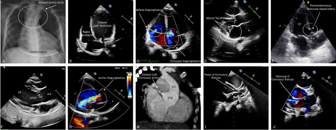
Desbuquois dysplasia (DBQD) is a rare autosomal recessive chondrodysplasia characterized by distinct skeletal abnormalities and multisystem involvement. Cardiac manifestations, such as aortic root dilatation and mitral valve prolapse, have also been reported, likely due to impaired proteoglycan production. This study aims to enhance the understanding of clinical management and cardiac implications in patients with DBQD, contributing to the broader knowledge of this rare condition. This research was conducted at Hacettepe University İhsan Doğramacı Children's Hospital, a tertiary reference center for all pediatric subspecialties. A single-center, descriptive, retrospective cohort study was performed. Demographic characteristics, genetic mutations, echocardiographic findings, and measurements of patients with Desbuquois dysplasia were documented. A total of nine patients, including five females (55%) were included in the study. The median age of the patients was 11 years (range 3.6-23.6 years), the median body weight was 15 kg (6-64 kg), and the median height was 94 cm (63-130 cm). The median follow-up period was 7.7 years (range 2.9-15.4 years). All patients had homozygous or compound heterozygous pathogenic variants in the CANT1 gene. The most common cardiac findings included mitral valve prolapse (seven patients, 77%), ascending aortic dilatation (seven patients, 77%), aortic root enlargement (six patients, 66%), small atrial septal defect (ASD) (five patients, 55%), bicuspid aortic valve (two patients, 22%), and ventricular septal defect (VSD) (one patient, 11%). Additionally, coronary-cameral fistula, a rare finding in the general population, was observed in one patient. The median individual Z scores for the sinus valsalva (SVS) in patients with aortic dilatation were 4.9 (range 2.7-7.5), while the median Z score in the ascending aorta was 5 (range 2.3-8.5).
Conclusion: Aortic root and ascending aorta dilatation as well as mitral valve prolapse are frequently observed in patients with DBQD. ASD, VSD, and bicuspid aorta are less common. Aortopathy develops early and can progress to a severe stage. Early detection of cardiac abnormalities and timely initiation of medical treatment may significantly improve the long-term prognosis of the disease.
What is known: • Desbuquois dysplasia (DBQD) is a rare autosomal recessive chondrodysplasia characterized by distinct skeletal abnormalities and multisystem involvement. Cardiac manifestations, such as aortic root dilatation and mitral valve prolapse, have also been reported, likely due to impaired proteoglycan production.
What is new: • The most frequently observed findings include aortic root and ascending aortic dilatation as well as mitral valve prolapse. Aortopathy develops early and can progress to severe disease. Early detection of cardiac abnormalities and timely initiation of medical treatment may significantly improve long-term prognosis.
Keywords: Aortic root dilatation; Aortopathy; Cardiovascular manifestations; Desbuquois dysplasia.
© 2025. The Author(s).
Conflict of interest statement
Declarations. Competing interests: The authors declare no competing interests.
Figures
References
-
- LeMerrer M, Young ID, Stanescu V, Maroteaux Des P (1991) “Desbuquois syndrome”. Eur. J. Pediatr 150(11): 793–796 10.1007/BF02026714. - PubMed
-
- Gillessen-Kaesbach G, vd (1995) “Desbuquois syndrome: three further cases and review of the literature”. Clin. Dysmorphol 4(2): 136–144 - PubMed
-
- Kim O-H, vd. (2010) “A variant of Desbuquois dysplasia characterized by advanced carpal bone age, short metacarpals, and elongated phalanges: report of seven cases”. Am. J. Med. Genet. A 152A(4): 875–885 10.1002/ajmg.a.33347. - PubMed
-
- Piussan C, Maroteaux P, Castroviejo I, Risbourg ve B (1975) “[Bone dysplasia with dwarfism and diffuse skeletal alterations]”. Arch. Fr. Pediatr 32(6): 541–550 - PubMed
-
- Shohat M, vd (1994) “Desbuquois syndrome: clinical, radiographic, and morphologic characterization”. Am. J. Med. Genet 52(1): 9–18 10.1002/ajmg.1320520104. - PubMed
MeSH terms
LinkOut - more resources
Full Text Sources
