Review
. 2025 Nov;54(11):2433-2442.
doi: 10.1007/s00256-025-04958-7. Epub 2025 Jun 3.

When it is not sacroiliitis

Affiliations
Review

When it is not sacroiliitis

Anthony De Leeuw et al. Skeletal Radiol. 2025 Nov.

Abstract

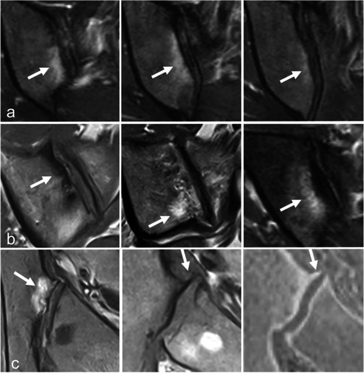
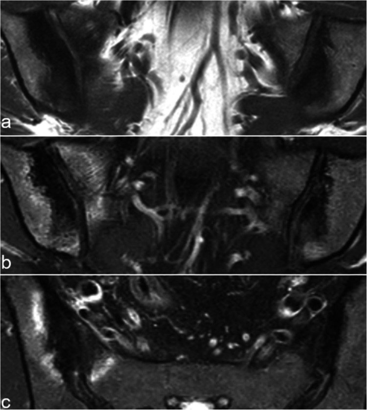
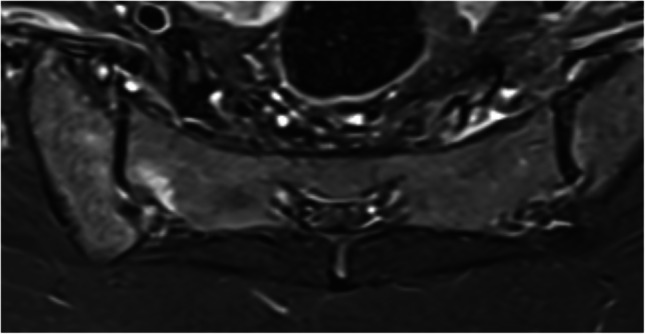
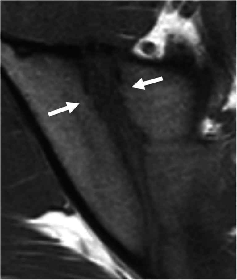
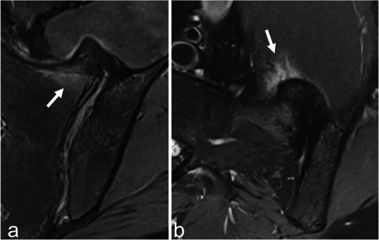
Magnetic resonance imaging of the sacroiliac joints (SIJ) is now frequently performed to detect subchondral inflammatory and structural changes in patients with early axial spondyloarthritis (SpA). However, similar changes can also occur in various other conditions, which may lead to the overdiagnosis of axial SpA. The aim of this article is to review the key imaging features of the most common disorders that may mimic inflammatory sacroiliitis, including mechanical changes and osteoarthritis, osteitis condensans ilii and pregnancy-related changes, other strain related changes, anatomical variants, pediatric SIJs, hyperostosis, infectious sacroiliitis, SAPHO syndrome, hyperparathyroidism, and sacral stress fractures.

Keywords: MRI; Osteoarthritis; Sacroiliac joint; Sacroiliitis; Spondyloarthritis.


Conflict of interest statement

Declarations. Ethics approval and consent to participate: Not applicable. Competing interest: The authors declare no competing interests.

Figures

Fig. 1
Fig. 1
Mechanical changes at the anterior aspect of the middle third of a right SIJ. a Edematous changes in the iliac bone (arrow) on three contiguous coronal oblique T2-weighted images in a 41-year-old woman. b Sclerotic, fatty, and mixed subchondral changes (arrows) on coronal oblique T1-, T1-, and fat-saturated (FS) T2-weighted images in three different patients, respectively. c Subchondral bone marrow edema (arrow) on an axial oblique FS T2-weighted image and osteophytes (arrow) on T1- and StarVIBE T1-weighted images in the same patient as in a
Fig. 2
Fig. 2
A 37-year-old woman with OCI. Coronal oblique a T1- and b FS T2-weighted images. c Axial oblique FS T2-weighted image show bilateral extensive hypointense subchondral sclerosis on the iliac side, which is well delineated, and surrounded by a mild rim of BME on the right side. Similar changes are observed on the sacral side. These findings predominate at the anterior aspect of the middle third of the SIJ
Fig. 3
Fig. 3
A 21-year-old woman, 9 days post-partum. Coronal oblique T1- (a) and T2 (b)-weighted images, axial oblique T1-weighted image (c). On the right side, mechanical changes related to pregnancy are observed, with subchondral sclerosis surrounded by mild BME; on the left side, there is extensive and pronounced subchondral BME with significant infiltration of the adjacent soft tissues, indicative of infectious sacroiliitis
Fig. 4
Fig. 4
Figures of the anatomical variants of the SIJs on axial oblique images. a Normal shape, b dysmorphic joint affecting the posterior part of the joint on the right side and more diffuse on the left side, c unfused sacral ossification center on the right side and isolated synostosis on the left side, d accessory SIJ on the right side and iliosacral complex on the left side, e semicircular defect on the right side and crescent-like iliac bony plate on the left side, f bipartite iliac bony plate on both sides
Fig. 5
Fig. 5
A 38-year-old woman with bilateral dysmorphic SIJ on axial oblique FS T2-weighted image: the central aspect of the joint is affected on the right side, the posterior aspect of the joint on the left side. Both are associated with subchondral edematous changes
Fig. 6
Fig. 6
A 41-year-old woman with a right accessory SIJ showing edematous and sclerotic changes of the facing bones on axial oblique FS T2-weighted image
Fig. 7
Fig. 7
A 17-year-old man with irregularities of the SIJ mimicking erosions on coronal oblique T1-weighted image
Fig. 8
Fig. 8
A 68-year-old man with hyperostosis of the SIJ. Slight BME at the base of the bridge (arrow) and in the adjacent soft tissues (arrow) on coronal oblique (a) and axial oblique (b) FS T2-weighted images
Fig. 9
Fig. 9
A 31-year-old man with chronic kidney disease. Extensive subchondral bone resorption on the iliac side of both SIJs (arrows) without adjacent BME or sclerosis on coronal FS T2-weighted image
Fig. 10
Fig. 10
A 72-year-old woman with bilateral sacral insufficiency fractures. Sacral BME is observed on the coronal oblique FS T2-weighted image (a) whereas thin hypointense fracture lines (arrows) paralleling the SIJs are better demonstrated on the coronal oblique T1-weighted image (b)

References

    1. Rudwaleit M, Jurik AG, Hermann KGA, Landewé R, Van Der Heijde D, Baraliakos X, et al. Defining active sacroiliitis on magnetic resonance imaging (MRI) for classification of axial spondyloarthritis: a consensual approach by the ASAS/OMERACT MRI group. Ann Rheum Dis. 2009;68(10):1520–7. - PubMed
    1. Lambert RGW, Bakker PAC, Van Der Heijde D, Weber U, Rudwaleit M, Hermann KGA, et al. Defining active sacroiliitis on MRI for classification of axial spondyloarthritis: update by the ASAS MRI working group. Ann Rheum Dis. 2016;75(11):1958–63. - PubMed
    1. Maksymowych WP, Lambert RG, Østergaard M, Pedersen SJ, Machado PM, Weber U, et al. MRI lesions in the sacroiliac joints of patients with spondyloarthritis: an update of definitions and validation by the ASAS MRI working group. Ann Rheum Dis. 2019;78(11):1550–8. - PubMed
    1. Rennie WJ, Cotten A, Jurik AG, Lecouvet F, Jans L, Omoumi P, et al. Standardized reporting of spine and sacroiliac joints in axial spondyloarthritis MRI: from the ESSR-Arthritis Subcommittee. Eur Radiol. 2024;35(1):360–9. - PubMed
    1. Carneiro BC, Rizzetto TA, Silva FD, Da Cruz IAN, Guimarães JB, Ormond Filho AG, et al. Sacroiliac joint beyond sacroiliitis—further insights and old concepts on magnetic resonance imaging. Skeletal Radiol. 2022;51(10):1923–35. - PubMed

MeSH terms

LinkOut - more resources