

Evaluation of the tubular penetration of two different types of nanoparticle root canal sealers over apically separated files: a scanning electron microscopic study (in vitro study)
- PMID: 40462057
- PMCID: PMC12135275
- DOI: 10.1186/s12903-025-06263-0
Evaluation of the tubular penetration of two different types of nanoparticle root canal sealers over apically separated files: a scanning electron microscopic study (in vitro study)
Abstract
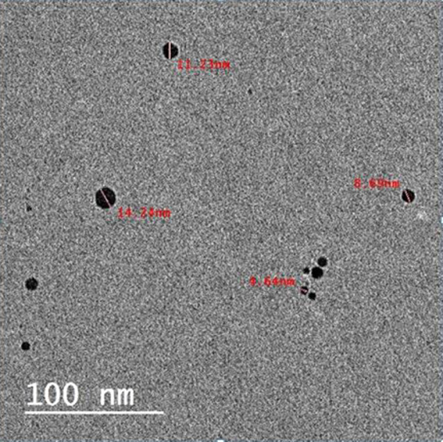
Background: The separation of root canal instruments can significantly impact the quality of canal obturation and the long-term success of endodontic treatment. Combining the benefits of nanoparticles and ultrasonic activation aim to enhance tubular sealer penetration and achieve proper sealing to improve outcome in such complex cases.
Aim of the study: This study aimed to evaluate tubular penetration of two nanoparticles modified root canal sealers compared to conventional sealers using ultrasonic activation over an apically separated file analyzed by scanning electron microscopy (SEM).
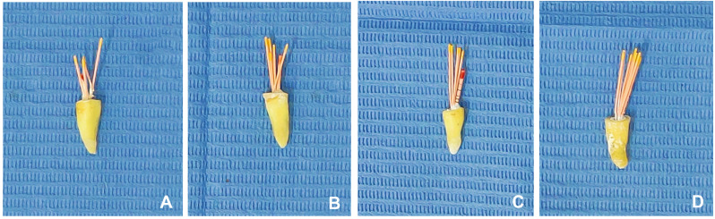
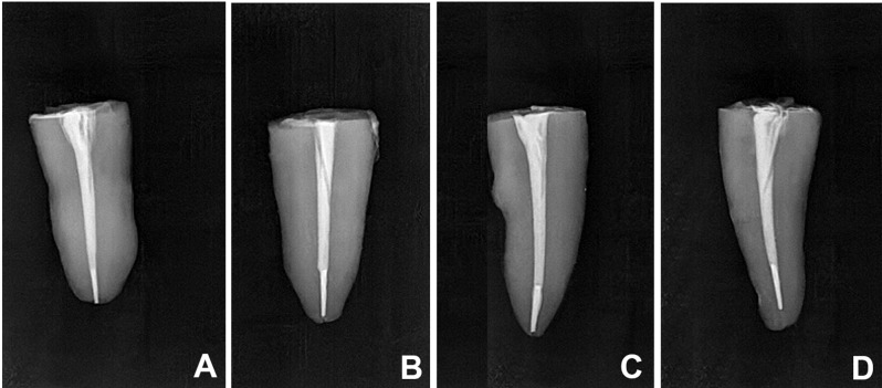
Materials and methods: Forty extracted single canaled mandibular premolar teeth were decoronated and prepared using M-Pro (IMD, Shanghai, China) nickel titanium rotary files which were intentionally separated at apical third. Teeth were randomly divided into four equal groups based on the type of sealer used. Group I: Adseal (Rs) (Meta Biomed, Cheongju, Korea) resin-based sealer, group II: Adseal resin-based sealer modified with silver nanoparticles (Rs/Ag), group III: Ceraseal (BC) (Meta Biomed, Cheongju, Korea) bioceramic sealer and group IV: Ceraseal bioceramic sealer modified with bioactive glass nanoparticles (BC/BG). The tested sealers were applied in all root specimens using lentulo-spiral size #25 (Dentsply Maillefer, Ballaigues, Switzerland) except the BC group where the sealer was injected into the root using plastic delivery tips provided by the manufacturer. All sealers were ultrasonically activated then obturated using cold lateral compaction technique. All samples were sectioned longitudinally in a buccolingual direction and analyzed for tubular sealer penetration using SEM at the area of separated file.
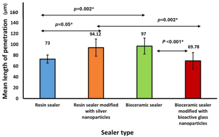
Results: The maximum mean tubular sealer penetration depth, measured in microns, was evident in the BC sealer group (97.00 ± 14.78) followed by the Rs/Ag group (94.12 ± 15.89) with no significant difference between them (p = 1.00). However, a significant difference (p < 0.05) was found when comparing these groups to the Rs group (73.00 ± 7.53) and the BC/BG group (69.78 ± 15.19). No significant difference (p = 1.00) was observed between the Rs and BC/BG groups.
Conclusions: Under the parameters of this in vitro study, conventional bioceramic sealer and resin-based sealer modified with silver nanoparticles exhibit more tubular penetration depth over apically separated file compared to conventional resin-based sealer and bioceramic sealer modified with bioactive glass nanoparticles.
Keywords: Apically separated file; Bioactive glass nanoparticles; Bioceramic sealer; Resin sealer; Silver nanoparticles; Tubular sealer penetration.
© 2025. The Author(s).
Conflict of interest statement
Declarations. Ethics approval and consent to participate: This study was approved by the Institutional Review Board of the Faculty of Dentistry, Alexandria University (IORG:0008839, approval no. 0622-02/2023). All subjects gave their informed consent for the inclusion of their extracted teeth. All methods were performed in accordance with the Declaration of Helsinki and the ethical guidelines adopted by the Research Ethics Committee of the Faculty of Dentistry, Alexandria University. No human participants were involved in the study. Consent for publication: Not applicable. Competing interests: The authors declare no competing interests.
Figures


Similar articles
-
Root canal cleanliness and debris extrusion following retreatment of thermoplastic injection technique and bioceramic-based root canal sealer.Clin Oral Investig. 2024 Oct 23;28(11):608. doi: 10.1007/s00784-024-06005-6. Clin Oral Investig. 2024. PMID: 39438302
-
Bioactivity and Element Composition of Three Endodontic Root Canal Sealers.J Contemp Dent Pract. 2025 Jan 1;26(1):62-70. doi: 10.5005/jp-journals-10024-3791. J Contemp Dent Pract. 2025. PMID: 40254872
-
Comparative Assessment of Novel Collagen Cross-linking Agents on Push-out Bond Strength of Two Different Sealers: An In Vitro Study.J Contemp Dent Pract. 2022 Nov 1;23(11):1122-1127. doi: 10.5005/jp-journals-10024-3439. J Contemp Dent Pract. 2022. PMID: 37073935
-
Influence of ultrasonic activation of 4 root canal sealers on the filling quality.J Endod. 2014 Jul;40(7):964-8. doi: 10.1016/j.joen.2013.11.016. Epub 2014 Jan 3. J Endod. 2014. PMID: 24935544
-
Comparative Evaluation of Apical Leakage in Root Canal Obturation Using AH Plus Sealer, Bioceramic Sealer, and Bioceramic Sealer Incorporated With Chitosan Nanoparticles: An In Vitro Study.Cureus. 2024 Dec 9;16(12):e75359. doi: 10.7759/cureus.75359. eCollection 2024 Dec. Cureus. 2024. PMID: 39781117 Free PMC article.
References
-
- Balguerie E, van der Sluis L, Vallaeys K, Gurgel-Georgelin M, Diemer F. Sealer penetration and adaptation in the dentinal tubules: a scanning electron microscopic study. J Endod. 2011;37(11):1576–9. - PubMed
-
- Madarati AA, Hunter MJ, Dummer PM. Management of intracanal separated instruments. J Endod. 2013;39(5):569–81. - PubMed
-
- McGuigan M, Louca C, Duncan H. Clinical decision-making after endodontic instrument fracture. Br Dent J. 2013;214(8):395–400. - PubMed
-
- Spili P, Parashos P, Messer HH. The impact of instrument fracture on outcome of endodontic treatment. J Endod. 2005;31(12):845–50. - PubMed
-
- Panitvisai P, Parunnit P, Sathorn C, Messer HH. Impact of a retained instrument on treatment outcome: a systematic review and meta-analysis. J Endod. 2010;36(5):775–80. - PubMed
MeSH terms
Substances
LinkOut - more resources
Full Text Sources
Research Materials
