Review
. 2025 Jun 3;24(1):161.
doi: 10.1186/s12943-025-02337-3.

Reprogrammed immuno-metabolic environment of cancer: the driving force of ferroptosis resistance

Affiliations
Review

Reprogrammed immuno-metabolic environment of cancer: the driving force of ferroptosis resistance

Sramana Bhowmick et al. Mol Cancer. .

Abstract

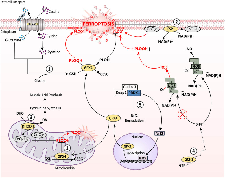
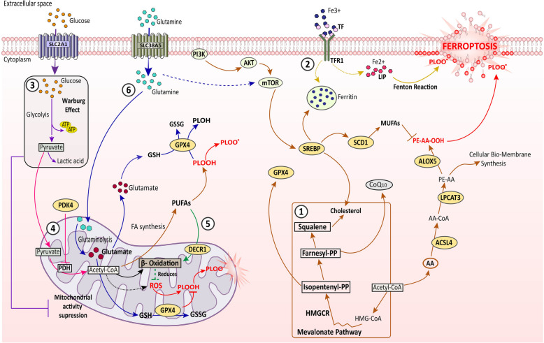
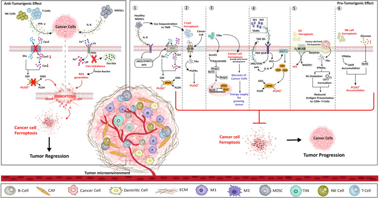
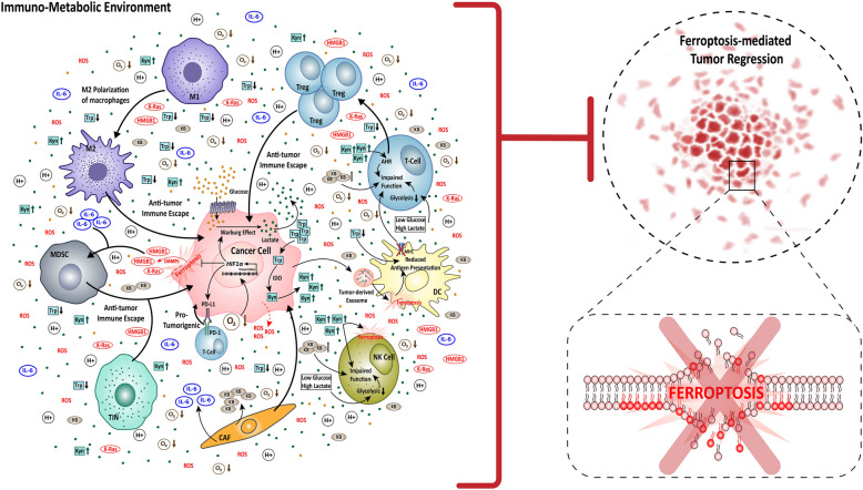
Ferroptosis, the non-apoptotic, iron-dependent form of cell death is an unavoidable outcome and byproduct of cellular metabolism. Reactive oxygen species generation during metabolic activities transcends to Fe2+-induced lipid peroxidation, leading to ferroptosis. Cancer cells being highly metabolic are more prone to ferroptosis. However, their neoplastic nature enables them to bypass ferroptosis and become ferroptosis-resistant. The capability of cancer cells to reprogram its metabolic activities is one of its finest abilities to abort oxidative damage, and hence ferroptosis. Moreover, the reprogrammed metabolism of cancer cells, also associates with the radical trapping antioxidant systems to enhance the scavenging of ferroptosis and thereby tumor progression. Additionally, the TME, which is an inevitable part and regulator of carcinogenesis, presents an intricate cooperation with tumor metabolism to build an immuno-metabolic environment to regulate the sustenance of cell proliferation and survival. This review focuses on the current understanding of ferroptosis in carcinogenesis and its resistance acquired by cancer cells via several modulators including the radical trapping antioxidant systems, the reprogrammed metabolism, the TME, and intertwined role of cancer metabolism and tumor immunity. The reprogrammed metabolism section further comprehends the functional role of lipids, iron and glucose metabolism against ferroptosis defense separately. The affiliation of TME in ferroptosis regulation is further sectioned with reference to different immune cells present within the TME such as tumor-associated macrophages, tumor-infiltrating neutrophils, myeloid-derived suppressor cells, T-cells, natural killer cells, dendritic cells, and B-cells, modifying the TME in both pro and anti-tumorigenic manner. Subsequently, this review also discusses the convergence of immuno-metabolic environment in ferroptosis regulation, and eventually brings up research gaps in this context providing consequential and significant questions to explore for better understanding of the immuno-metabolic environment's role in driving ferroptosis resistance for anti-cancer treatment progress.

Keywords: Ferroptosis; Ferroptosis resistance; Metabolic reprogramming; Reprogrammed immune metabolic environment; Tumor microenvironment.


Conflict of interest statement

Declarations. Ethics approval and consent to participate: Not applicable. Consent for publication: Not applicable. Competing interests: The authors declare no competing interests.

Figures

Fig. 1
Fig. 1
Outlook of ferroptosis with cellular RTA systems. ROS-mediated lipid-peroxidation is the prime cause of ferroptosis induction in the cell. Cells undergoing metabolic activity collaterally produce ROS, thereby inducing ferroptosis. To prevent this, cells employ several RTA systems; ① The GPX4 (cytoplasmic and mitochondrial) system, it utilizes GSH synthesised from cysteine to neutralize the ROS via a cycle of oxidation and reduction of GSH; ② The FSP1 (plasma membrane) system, and ③ the DHODH (mitochondrial) system, both utilizes CoQ10 to convert it into CoQ10H2 (non-toxic form), and subsequently reduce the ROS production. In addition, the FSP1 system uses NAD(P)H to neutralize ROS; ④ the GCH1/BH4 (cytoplasmic) system, in here GCH1 catalyzes the synthesis of the antioxidant BH4, which then binds to NOS and utilizes NAD(P)H to reduce O2 and produce NO thereby attenuating ROS generation; ⑤ PRDX1 system, it promotes the Nrf2 activity by inhibiting the Keap1-Cullin- 3 complex. PRDX1 bind to the Keap1-Cullin- 3, which is an inhibitor to Nrf2, thereby releasing and enabling Nrf2 entry into nucleus to activate of GPX4 gene transcription. All of these systems thereby effectively reduce PLOOH, and PLOO• generation, ultimately preventing ferroptosis. BH4: tetrahydrobiopterin, CoQ10: Coenzyme Q10, CoQ10H2: ubiquinol- 10, DHODH: dihydroorotate dehydrogenase, DHO: dihydroorotate, FSP1: ferroptosis suppressor protein 1, GCH1: GTP cyclohydrolase 1, GPX4: glutathione peroxidase 4, GSH: reduced glutathione, GSSG: oxidized glutathione, GTP: guanosine triphosphate, NADH: nicotinamide adenine dinucleotide (hydrogen), (reduced form), NAD(P)H: nicotinamide adenine dinucleotide phosphate, NO: nitric oxide, NOS: nitric oxide synthase, Nrf2: nuclear factor, OA: orate, PLOH: phospholipid alcohol, PLOOH: phospholipid peroxide, PLOO•: phospholipid hydroperoxyl radical, PRDX1: Peroxiredoxin- 1, ROS: reactive oxygen species, SLC3 A2: light chain subunit of System Xc (cystine/glutamate antiporter), SLC7 A11: light chain subunit of System Xc (cystine/glutamate antiporter)
Fig. 2
Fig. 2
Involvement of cellular metabolism in ferroptosis regulation. ① the lipid metabolic pathways (brown arrows), this includes both ferroptosis inducing as well as ferroptosis suppressing conditions. On one end, PI3 K/Akt/mTOR mediated SREBP-SCD1 axis activation, promoting to MUFA generation, as well as the mevalonate pathway transduced cholesterol/CoQ10 axis functioning, both suppresses ferroptosis in cancer cells. On the other end, PUFA (AA) activation by ACSL4, esterification by LPCAT3, and thereby lipid peroxidation by ALOX5 promotes ferroptosis induction. ② the iron metabolism (yellow arrows), regulate ferroptosis via both pro-ferroptotic and anti-ferroptosis incidences. TF bound with circulating Fe3 + ions binds to TFR1 and is internalized, then either it contributes to LIP which promotes Fenton reaction mediated PLOOH and PLOO• generation, and ferroptosis, or can be stored as ferritin limiting redox active iron accumulation induced ferroptosis. ③ the glucose metabolism (purple arrows), which mainly exerts ferroptosis suppression. SLC2 A1 facilitated glucose uptake escalates Warburg effect, which reduces mitochondrial function-mediated ROS generation. ④ PDK4 action (pink arrows), it helps ferroptosis reduction in cancer cells by inhibiting PDH to catalyze pyruvate to acetyl-CoA. This acetyl-CoA would otherwise contribute to mitochondrial activity linked ROS generation, and PUFA synthesis. ⑤ DECR1 action (green arrows), it exerts anti-ferroptotic condition by accelerating β-oxidation of PUFAs to reduce ROS generation. ⑥ glutamate metabolism (blue arrows), SLC38 A5 assists the import of glutamine in the cell cytoplasm, which then enters into mitochondria, and undergo glutaminolysis to generate glutamate, which is then utilized by the GPX4 enzyme to reduce lipid peroxidation, and thereby reducing ferroptosis. Additionally, glutamate also activates mTOR signaling indirectly to further strengthen the lipid metabolic pathways for ferroptosis resistance. AA: arachidonic acid, AA-CoA: arachidonic acid-Coenzyme A, Acetyl-CoA: acetyl- Coenzyme A, ACSL4: Acyl-CoA synthetase long-chain family 4, ALOX5: arachidonate lipoxygenases 5, CoQ10: Coenzyme Q10, DECR1: dienoyl-CoA-reductase1, GPX4: glutathione peroxidase 4, GSH: reduced glutathione, GSSG: oxidized glutathione, HMGCR: HMG-CoA reductase, LIP: labile iron pool, LPCATs: lysophosphatidylcholine acyltransferase, MUFA: monounsaturated fatty acid, PLOH: phospholipid alcohol, PLOOH: phospholipid peroxide, PLOO•: phospholipid hydroperoxyl radical, PDH: pyruvate dehydrogenase, PDK4: pyruvate dehydrogenase kinase 4, PE-AA-OH: arachidonic acid lipid peroxide, PUFA: polyunsaturated fatty acid, SCD- 1: Stearoyl-CoA desaturase- 1, SLC2 A1: glucose transporter (GLUT1), SLC38 A5: amino acid transporter (glutamine) SREBP: sterol regulatory element-binding protein, TF: transferrin, TFR1: transferrin receptor 1. Direct lines: direct impact on the target molecule, Dotted lines: indirect impact on the target molecules, Black arrows: general cellular mechanisms irrespective of metabolic regulation of ferroptosis, Red arrows: ferroptosis commencing signal
Fig. 3
Fig. 3
Ferroptosis regulation by TME. The TME comprising of multiple immune cells regulate ferroptosis of cancer cells in both anti-tumorigenic as well as pro-tumorigenic. The anti-tumorigenic effect is induced only by T-Cells, NK cells, and MDSCs cells (left panel). Both T-cells and NK cells secretes IFN-γ, which thereby inhibits the system Xc/GSH/GPX4 axis, thereby inducing ferroptosis in cancer cells. The MDSCs secretes IL- 6 and cause iron imbalance in the cancer cells, resulting in Fenton reaction-mediated ROS generation, lipid peroxidation and ultimately ferroptosis in cancer cells. Alternatively, the pro-tumorigenic regulation of ferroptosis is supported by MDSCs, T-Cells, TINs, TAMs, DCs, and NK cells (right panel). ① MDSCs secretes IL- 6 to activate JAK2/STAT3 pathway which then activates system Xc/GSH/GPX4 axis to resist ferroptosis in cancer cells. Additionally, MDSCs sequesters Cys in the TME and make the T-Cells undergo ferroptosis. ② T-cells ferroptosis is moreover facilitated by cancer cell regulated CD36-mediated FA uptake in T-cells, which further favours PUFA generation leading to peroxidation of lipids. ③ TINs aids ferroptosis resistance in cancer cells by Acod1-mediated release of Keap1 inhibition on Nrf2 that promotes the transcriptional activation of the GPX4 and GcLc antioxidant gene to enhance ROS scavenging in the cells. In addition, TINs also induce ferroptosis of cancer cells at early stage and thereby necrosis of cancer cells to provide energy to proliferating cancer cells. ④ TAMs secrete TGF-β1, which activates HLF/GGT1 axis to enhance GSH production and reduce ferroptosis prevalence in cancer cells. The HLF activation in TINs further releases IL- 6 creating a positive feedback loop of TGF-β1 signaling via JAK/STAT axis. ⑤ DCs itself undergo ferroptosis via NOX2-mediated ROS generation, and also losses its function by the effect of tumor-derived exosome induced PPARα signaling, which reduces the antigen presenting capacity by LD accumulation and FA oxidation. ⑥ NK cells also themselves undergo ferroptosis within the TME, PPARα-mediated lipid accumulation and, the glucose deprived TME- Nrf2 inhibition collectively cause lipid peroxide accumulation, and thereby ferroptosis of NK cells. Acod1: aconitate decarboxylase 1, CAF: cancer associated fibroblast, Cys: cysteine, Cys2: Cystine, CD36: Clusters of differentiation 36 (fatty acid transporter), DC: dendritic cells, ECM: extracellular matrix, FA: fatty acid, GcLc: glutamate-cysteine ligase catalytic subunit, GGT1: gamma-glutamyl transferase 1Glu: glutamate, GPX4: glutathione peroxidase 4, GSH: glutathione, GSSG: oxidized glutathione, HLF: hepatic leukemia factor, IFN- γ: interferon-γ, IL- 6: interleukin- 6, JAK2: janus kinase 2, Keap1: kelch-like ECH-associated protein 1, LD: lipid droplet, LIP: labile iron pool, MDSCs: myeloid-derived suppressor cells, M1: macrophage type 1, M2: macrophage type 2, NADH: nicotinamide adenine dinucleotide (hydrogen), (reduced form), NAD(P)H: nicotinamide adenine dinucleotide phosphate NK: natural killer cells, NOX2: NADPH-oxidase Nrf2: nuclear factor erythroid 2-related factor, PLOH: phospholipid alcohol, PLOO•: phospholipid hydroperoxyl radical, PPARα: peroxisome proliferator-activated receptor alpha, PUFAs: polyunsaturated fatty acids, ROS: reactive oxygen species, TIN: tumor-infiltrating neutrophils TGF-β1: transforming growth factor-beta1, TGF-β1R: transforming growth factor-beta receptor type 1, STAT3: signal transducer and activator of transcription 3, SLC3 A2: light chain subunit of System Xc- (cystine/glutamate antiporter), SLC7 A11: light chain subunit of System Xc- (cystine/glutamate antiporter), Blue lipid bilayer: T-cell membrane, Yellow lipid bilayer: DC membrane, Black arrows: TME cell signaling, Red arrows: ferroptosis commencing signal
Fig. 4
Fig. 4
The network of Immuno-metabolic environment in ferroptosis resistance. The immuno-metabolic environment corresponds to the cross-talk between cancer cells, stromal cells like CAFs, and immune cells like T-cell, Tregs, DCs, M1 macrophages, M2 macrophages, MDSCs, and TINs. The cancer cells take up glucose and Trp from the TME, to produce lactate via Warburg effect and Kyn rich by Trp degradation, thereby making the TME acidic and Kyn-rich. Additionally, the CAFs release KBs in the TME, increasing the KB content high, and due to lower levels of oxygen, the TME is inherently hypoxic. All these four factors (high acid, Kyn and KB content with low oxygen) collectively promotes cancer progression. Parallelly, the cancer cells undergoing ferroptosis also favours pro-tumorigenic condition via the release of DAMPs like HMGB1 and K-Ras. All these factors together result in inhibition of ferroptosis-mediated tumor regression by promoting anti-tumor immune escape of the cancer cells. Mechanistically, the glucose deprived, lactate and Kyn rich condition impairs the function of T-cells, DC, NK, cells by generating Tregs, reducing antigen presenting capacity, inducing ferroptosis, respectively and additionally intensifies M2 polarization of macrophages for anti. The hypoxic condition activates HIF1α transcription, leading to HIF1α signaling upregulation, which thereby increases Warburg effect and PD-L1/PD- 1 signaling, and decreases ferroptosis in cancer cells. The DAMPs released by cancer cells as well as IL- 6 released by CAFs, activates MDSCs, thereby promoting anti-tumor immune escape. The TINs also promote cancer progression. Overall, the immuno-metabolic environment created by the cancer-non-cancer cell cross talk promotes cancer by inhibiting ferroptosis-mediated tumor regression and ferroptosis of cancers. AHR: aryl hydrocarbon receptor, CAF: cancer associated fibroblast, DAMPs: damage-associated molecular patterns, DC: dendritic cells, HIF1α: hypoxia-inducible factor 1-alpha, HMGB1: high mobility group box 1, IDO: indoleamine 2,3-dioxygenase, IL- 6: interleukin- 6 KB: ketone bodies, K-Ras: Kirsten rat sarcoma viral oncogene homolog, Kyn: kynurenine, MHC: Major histocompatibility complex, MDSC: myeloid-derived suppressor cells, M1: macrophage type 1, M2: macrophage type 2, NK: natural killer cells, ROS: reactive oxygen species, PD- 1: programmed cell death protein 1, PD-L1: programmed death-ligand 1, TIN: tumor infiltrating neutrophil, Treg: regulatory T-cells, Trp: tryptophan, Narrow arrows: signaling, Broad arrows: activation of the molecule/cell, Dotted arrows: indirect effect

References

    1. Tuomela K, Ambrose AR, Davis DM. Escaping Death: How Cancer Cells and Infected Cells Resist Cell-Mediated Cytotoxicity. Front Immunol. 2022;13: 867098. 10.3389/fimmu.2022.867098/bibtex. - PMC - PubMed
    1. Tang D, Kang R, Berghe TV, Vandenabeele P, Kroemer G. The molecular machinery of regulated cell death. Cell Res. 2019;29:347–64. 10.1038/S41422-019-0164-5. - PMC - PubMed
    1. Peng F, Liao M, Qin R, Zhu S, Peng C, Fu L, Chen Y, Han B. Regulated cell death (RCD) in cancer: key pathways and targeted therapies. Signal Transduct Targeted Ther. 2022;7(17):1–66. 10.1038/s41392-022-01110-y. - PMC - PubMed
    1. Zhang C, Liu X, Jin S, Chen Y, Guo R. Ferroptosis in cancer therapy: a novel approach to reversing drug resistance. Mol Cancer. 2022. 10.1186/S12943-022-01530-Y. - PMC - PubMed
    1. Sever R, Brugge JS. Signal Transduction in Cancer. Cold Spring Harb Perspect Med. 2015. 10.1101/cshperspect.a006098. - PMC - PubMed

LinkOut - more resources