. 2025 Jun 3;16(1):285.
doi: 10.1186/s13287-025-04381-8.

MiR221/222 in the conditioned medium of adipose-derived stem cells attenuates particulate matter and high-fat diet-induced cardiac apoptosis

Affiliations

MiR221/222 in the conditioned medium of adipose-derived stem cells attenuates particulate matter and high-fat diet-induced cardiac apoptosis

Ya-Chun Chen et al. Stem Cell Res Ther. .

Abstract

Background: Air pollution and obesity are crucial risk factors for cardiovascular disease (CVD), with epidemiological evidence indicating that air pollution exacerbates obesity-induced cardiac damage. Treatment with adipose-derived stem cells (ADSCs) attenuates cardiac damage by releasing paracrine factors. However, the effects of ADSCs on air pollution- and obesity-induced cardiomyocyte apoptosis and the related mechanisms are still unclear.

Methods: Palmitic acid (PA) and a high-fat diet (HFD) were used to cause obesity, and particulate matter (PM) was used to simulate air pollution in the study. We studied the impact of conditioned medium from adipose-derived stem cells (ADSC-CM) on the apoptosis of PA + PM-treated H9c2 cells and HFD + PM-treated mouse cardiomyocytes and the underlying mechanisms involved.

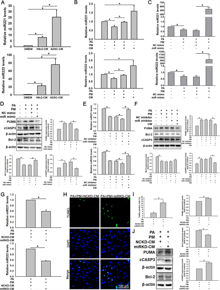
Results: The levels of apoptosis-related proteins (PUMA and cleaved caspase-3) were significantly increased in PA + PM-treated H9c2 cells and HFD + PM-treated mouse cardiomyocytes, whereas the antiapoptotic protein Bcl-2 expression was reduced. However, ADSC-CM treatment effectively reduced the PUMA and cleaved caspase-3 expression but increased the Bcl-2 expression. ADSC-CM significantly reduced PA + PM- and HFD + PM-induced cardiomyocyte apoptosis, as detected by the TUNEL assay. RT-qPCR revealed that PA + PM and HFD + PM significantly reduced miR221/222 levels, whereas ADSC-CM treatment increased miR221/222 levels. Furthermore, knockout (KO) and transgenic (TG) mice were used to demonstrate that miR221/222 in ADSC-CM ameliorated cardiac apoptosis that was induced by HFD + PM treatment. Furthermore, PA + PM treatment increased the reactive oxygen species (ROS) production, which triggered mitochondrial fission and contributed to apoptosis. However, ADSC-CM effectively reduced ROS levels and regulated mitochondrial fission, alleviating cellular apoptosis.

Conclusions: Our findings demonstrated that ADSC-CM attenuated PA + PM-induced cardiomyocyte apoptosis by modulating miR221/222 levels and suppressing ROS production.

Keywords: miR221/222; Conditioned medium from cultured adipose-derived stem cell (ADSC-CM); High-fat diet (HFD); Mitochondrial fission; Palmitic acid (PA); Particulate matter (PM); Reactive oxygen species (ROS).


Conflict of interest statement

Declarations. Ethics approval and consent to participate: The studies involving experimental animals were performed according to the guidelines of the Institutional Animal Care and Use Committee, National Taiwan University College of Medicine, and College of Laboratory Animal Center (Project title: To study the effects of particulate matter 2.5 and high fat on cardiac injury: the role of mitochondrial dynamics and mitophagy; Approval number: 20210334; Date of approval: Aug 1, 2022). ADSCs were purchased from LONZA (Basel, Switzerland), and the cells were used with permission for research applications. Consent for publication: Not applicable. Competing interests: The authors have no conflicts of interest.

Figures

Fig. 1
Fig. 1
Effects of ADSC-CM on PA + PM-induced H9c2 cell apoptosis. A H9c2 cells were treated with PA at various concentration (0, 50, 100, and 200 µM) for 24 h. An MTT method measured cell viability. B H9c2 cells were incubated with various doses (0, 10, 20, and 40 µg/mL) of PM for 24 h. An MTT method measured cell viability. C H9c2 cells were co-treated with the defined concentratin of PA and PM for 24 h. The expression levels of PUMA, Bcl-2, and cCASP3 were checked by Western blot. D H9c2 cells were incubated with PA (50 µM), PM (20 µg/mL), or PA (50 µM) + PM (20 µg/mL) for 24 h. An MTT method measured cell viability. E H9c2 cells were treated with PA (50 µM), PM (20 µg/mL), or PA (50 µM) + PM (20 µg/mL) for 24 h. F H9c2 cells were treated with 50 µM PA, 20 µg/mL PM, and ADSC-CM for 24 h. An MTT method measured cell viability. G H9c2 cells were treated with 50 µM PA, 20 µg/mL PM, and ADSC-CM for 24 h and then examined for apoptosis via a TUNEL method (TUNEL-positive cells: green; nuclei: blue). Scale bar = 100 μm. H The bar graph depicts the quantitative analysis results of counting the number of TUNEL-positive cells. I H9c2 cells were incubated with 50 µM PA, 20 µg/mL PM, and ADSC-CM for 24 h. Cell death was investigated by detecting annexin V-FITC and propidium iodide (PI) staining and analyzing by flow cytometry. J H9c2 cells were incubated with 50 µM PA, 20 µg/mL PM, and ADSC-CM for 24 h. Western blot examined the PUMA, Bcl-2, and cCASP3 expression levels. Experiments were conducted at least five times, and the quantitative data were presented as mean ± SEM. *P < 0.05 is defined as statistically significant. PA: palmitic acid; PM: particulate matter; CM: ADSC-CM; cCASP3: cleaved caspase-3
Fig. 2
Fig. 2
Effects of ADSC-CM on PA + PM-induced H9c2 cell apoptosis through miR221/222. A MiR221/222 levels were determined in DMEM, H9c2-CM, and ADSC-CM by qPCR. B H9c2 cells were incubated with 50 µM PA, 20 µg/mL PM, and ADSC-CM for 24 h. The expression levels of miR221/222 were evaluated by qPCR. C, D H9c2 cells were transfected with the corresponding negative control (NC) or miR221/222 mimics for 24 h and then incubated with 50 µM PA and 20 µg/mL PM for 24 h. MiR221/222 expression levels were examined by qPCR (C), and PUMA, Bcl-2, and cCASP3 protein expression levels were investigated by Western blot (D). E, F H9c2 cells were transfected with a scramble sequence or miR221/222 inhibitors for 24 h and then treated with 50 µM PA, 20 µg/mL PM, and ADSC-CM for 24 h. The miR221/222 expression levels were assessed by qPCR (E). PUMA, Bcl-2, and cCASP3 expression levels were investigated by Western blot (F). G ADSCs were transfected with the corresponding negative control (NC) or miR221/222 inhibitors for 24 h. These cells were then incubated in a standard culture medium for another 24 h, and NCKD-CM and miRKD-CM were collected, respectively. The levels of miR221/222 were measured by qPCR. H Apoptosis was assessed via a TUNEL method (TUNEL-positive cells: green; nuclei: blue). Scale bar = 100 μm. I Quantification data of TUNEL-positive cells is shown in the bar graphs. J Western blot measures the PUMA, Bcl-2, and cCASP3 expression levels. Experiments were conducted at least five times, and the quantitative data were presented as mean ± SEM. *P < 0.05 is defined as statistically significant. NC mimic: negative control mimic; miR mimic: miR221/222 mimic; NC inhibitor: negative control inhibitor; miR inhibitor: miR221/222 inhibitor; NCKD-CM: CM obtained after the knockdown of the negative control in ADSCs; miRKD-CM: CM obtained after the knockdown of miR221/222 in ADSCs
Fig. 3
Fig. 3
Effects of ADSC-CM on high-fat diet (HFD) + PM-induced apoptosis in B6 and KO mice via miR221/222. A Determination of miR221/222 levels in heart tissues from B6 and KO mice by qPCR. B Mice were fed a HFD for 4 weeks and were injected with or without PM and CM at the third and fourth weeks. The levels of miR221/222 in heart tissues were determined by qPCR. C Mice were weighed before the start of the experiment and weekly thereafter. D Representative photographs of transthoracic echocardiographic morphology in M mode. EH Measurements of the ejection fraction (EF), fractional shortening (FS), diastolic left ventricular posterior wall thickness (LVPWd), and systolic left ventricular posterior wall thickness (LVPWs). I Representative images of mitral valve waveforms from transthoracic echocardiography. JM Measurements of the left ventricular isovolumetric contraction time (IVCT), left ventricular isovolumetric relaxation time (IVRT), mitral valve E/A ratio, and stroke volume (SV). N Representative images of HE-stained cardiac tissue (scale bar = 2 mm). O Apoptosis was assessed in cardiac tissue using a TUNEL assay. The positive reaction product is brown. The nuclei were counterstained with hematoxylin solution. P The location of PUMA expression in cardiac tissue was investigated by immunohistochemical staining. The positive reaction product is brown. The nuclei were counterstained with hematoxylin solution. Q PUMA, Bcl-2, and cCASP3 expression levels were examined by Western blot. Experiments were conducted at least six times, and the quantitative data were presented as mean ± SEM. *P < 0.05 is defined as statistically significant. B6: C57B/L6 mice; KO: miR221/222 knockout mice; HFD: high-fat diet
Fig. 4
Fig. 4
Effects of miR221/222 on apoptosis of high-fat diet (HFD) + PM-treated TG mice. A Mice were fed an HFD for 4 weeks and injected with or without PM and CM at the third and fourth weeks. The levels of miR221/222 were determined by qPCR. B Mice were weighed before the start of the experiment and weekly thereafter. C Representative M-mode images of transthoracic echocardiography. DG Measurements of the EF, FS, LVPWd, and LVPWs. H Representative images of mitral valve waveforms from transthoracic echocardiography. IL Measurements of the IVCT, IVRT, mitral E/A ratio, and SV. M Representative images of HE-stained cardiac tissues (scale bar = 2 mm). N Apoptosis was examined in cardiac tissues using a TUNEL assay. The positive reaction product is brown. The nucleus was counterstained with hematoxylin solution. Scale bar = 100 µm. O Immunohistochemistry was used to detect the location of PUMA expression in cardiac tissue. The positive reaction product is brown. The nuclei were counterstained with hematoxylin solution. P Western blot examined the PUMA, Bcl-2, and cCASP3 expression levels. These experiments were conducted at least six times, and the quantitative data were presented as mean ± SEM. *P < 0.05 is defined as statistically significant. TG: miR221/222 transgenic mice
Fig. 5
Fig. 5
Effects of ADSC-CM on ROS production in PA- and PM-treated H9c2 cells. AE H9c2 cells were incubated with 50 µM PA, 20 µg/mL PM, and 100 nM MitoQ for 24 h. MitoSOX Red staining measures mitochondrial ROS levels using fluorescent microscopy (A) and flow cytometry (B). Apoptosis was detected using a TUNEL method (TUNEL-positive cells: green; nuclei: blue) (C). The statistical graph depicts the quantification of TUNEL-positive cell count (D). The PUMA, Bcl-2, and cCASP3 expression levels were investigated by Western blot (E). F, G H9c2 cells were treated with 50 µM PA, 20 µg/mL PM, and CM for 24 h. They were then treated with MitoSOX Red and detected by fluorescent microscopy (F) and flow cytometry (G). HI H9c2 cells were incubated with the corresponding negative control (NC) or miR221/222 mimics for 24 h and then incubated with 50 µM PA and 20 µg/mL PM for 24 h. They were treated with MitoSOX Red and checked via fluorescence microscopy (H) or flow cytometry (I). J, K ADSCs were transfected with miR221/222 inhibitors and then miRKD-CMs were collected. H9c2 cells were co-treated with 50 µM PA, 20 µg/mL PM, and DMEM or CM from H9c2, ADSCs, and miRKD (H9c2-CM, ADSC-CM, and miRKD-CM) for 24 h. The cells were then treated with MitoSOX Red and detected by fluorescent microscopy (J) and flow cytometry (K). L The SOD1, SOD2, and SOD3 expression levels were analyzed using Western Blot. The experiments were conducted at least five times, and the quantitative data were presented as mean ± SEM. *P < 0.05 is defined as statistically significant. Scale bar = 100 µm
Fig. 6
Fig. 6
Effects of ADSC-CM on PA- and PM-induced mitochondrial fission in H9c2 cells and the relationship between mitochondrial fission and ROS production. A, B H9c2 cells were treated with 50 µM PA, 20 µg/mL PM, and CM for 24 h. ΔΨm was assessed by JC-1 staining via fluorescence microscopy (A; scale bar = 100 µm) and flow cytometry (B). C ATP assay evaluated mitochondrial function. D Mitochondrial morphology was examined using MitoTracker. Scale bars = 20 µm (upper), 10 µm (lower). E Western blot analyzed p-DRP1 and FIS1 expression. F, G TUNEL assay (F; green: TUNEL-positive; blue: nuclei) and quantification (G) assessed Mdivi-1 (10 µM) pretreatment effects on apoptosis. H Western blot examined p-DRP1, PUMA, and cCASP3 expression. IK Effects of MitoQ on ΔΨm and ATP levels were assessed by JC-1 staining (I; scale bar = 100 µm), flow cytometry (J), and ATP assay (K). L, M MitoTracker staining and Western blot assessed the effects of MitoQ (100 nM) on mitochondrial morphology. Scale bars = 20 µm (upper), 10 µm (lower) (L) and protein expression (M) in H9c2 cells treated with PA and PM. NR H9c2 cells were treated with H2O2 (100 µM) and CM for 24 h. The changes of ΔΨm (N, O), ATP levels (P), mitochondrial morphology (Q), and protein expression (R) were analyzed. S, T mROS levels were assessed by MitoSOX Red staining (S; scale bar = 100 µm) and flow cytometry (T) after Mdivi-1 and PA + PM treatment. UW Cells transfected with negative control (NC) or miR221/222 mimics for 24 h and then treated with PA and PM for 24 h. ΔΨm was examined by JC-1 (U; scale bar = 100 µm) and flow cytometry (V); ATP levels were measured (W). X Western blot assessed expression of p-DRP1, FIS1, and SOD2. The experiments were conducted at least five times, and the quantitative data were presented as mean ± SEM. *P < 0.05 is defined as statistically significant
Fig. 7
Fig. 7
A graphical abstract summarizing the study findings. This schematic illustration summarizes the effects of obesity and air pollution on cardiomyocyte apoptosis and the potential protective role of ADSC-CM. In vitro, PA treatment and PM exposure simulated obesity and air pollution, respectively. In vivo, an HFD and PM were combined to replicate these conditions. This study revealed that ADSC-CM could reduce PA/HFD + PM-induced cardiomyocyte apoptosis in vitro and in vivo studies

References

    1. Li T, Providencia R, Mu N, Yin Y, Chen M, Wang Y, et al. Association of metformin monotherapy or combined therapy with cardiovascular risks in patients with type 2 diabetes mellitus. Cardiovasc Diabetol. 2021;20:30. - PMC - PubMed
    1. Li T, Mu N, Yin Y, Yu L, Ma H. Targeting AMP-activated protein kinase in aging-related cardiovascular diseases. Aging Dis. 2020;11:967–77. - PMC - PubMed
    1. Sacks FM, Lichtenstein AH, Wu JHY, Appel LJ, Creager MA, Kris-Etherton PM, et al. Dietary fats and cardiovascular disease: a presidential advisory from the american heart association. Circulation. 2017;136:e1–23. - PubMed
    1. Wali JA, Jarzebska N, Raubenheimer D, Simpson SJ, Rodionov RN, O’Sullivan JF. Cardio-metabolic effects of high-fat diets and their underlying mechanisms-a narrative review. Nutrients. 2020;12:1505. - PMC - PubMed
    1. Savini I, Catani MV, Evangelista D, Gasperi V, Avigliano L. Obesity-associated oxidative stress: strategies finalized to improve redox state. Int J Mol Sci. 2013;14:10497–538. - PMC - PubMed

MeSH terms