A 19-Month-Old Girl With Acute Choledocholithiasis: A Case Report
- PMID: 40462816
- PMCID: PMC12130344
- DOI: 10.7759/cureus.83433
A 19-Month-Old Girl With Acute Choledocholithiasis: A Case Report
Abstract
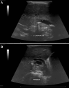
Cholelithiasis, or gallstone(s), is a leading cause of healthcare utilization in the United States. It is more common in adults but can occur in the pediatric population as well. The following is a case report of choledocholithiasis in a 19-month-old girl. A 19-month-old girl with abdominal pain presented to the emergency department (ED) after being found to have cholelithiasis on an outpatient abdominal ultrasound (US). Three days prior to presentation, the patient was seen by her pediatrician for fussiness, decreased oral intake, and non-bloody, non-bilious emesis. She was diagnosed with a suspected urinary tract infection (UTI) and prescribed amoxicillin-clavulanate for empiric treatment. The following day, the patient returned to her pediatrician for worsening abdominal pain; she was given one dose of intramuscular ceftriaxone and scheduled for outpatient abdominal US. Her past medical history is significant for omphalocele status post-surgical correction, several congenital cardiac defects, bilateral small kidneys, and poor weight gain. The patient has a normal chromosomal microarray and no family history of hepatobiliary/pancreatic disease. In the ED, the patient was afebrile and hemodynamically stable. Physical examination was significant for mild hepatomegaly, mild abdominal tenderness without peritoneal signs, and the presence of a well-healed surgical scar on the abdomen with an underlying abdominal hernia. Laboratory tests were significant for leukocytosis of 14.5×10³/microliter (mcL), elevated gamma-glutamyl transferase (GGT) of 305 unit/L (U/L), aspartate aminotransferase (AST) of 86 U/L, alanine aminotransferase (ALT) of 343 U/L, total bilirubin of 2.3 milligram/deciliter (mg/dL), direct bilirubin of 1.6 mg/dL, and lipase of 1,726 U/L. Abdominal US revealed several gallstones and mild to moderate intra- and extrahepatic biliary ductal dilatation likely due to a stone in the distal common bile duct (CBD). Pediatric surgery and gastroenterology recommended admission for pain management and magnetic resonance cholangiopancreatography (MRCP). After admission, the patient was started on ursodiol and piperacillin/tazobactam. MRCP showed a common hepatic duct measuring 13 mm and a 9×5 mm stone in the distal common bile duct. Due to a lack of available outside facilities with the capability to perform endoscopic retrograde cholangiopancreatography (ERCP) in a pediatric patient, medical management was pursued. Throughout her admission, the patient improved clinically, laboratory studies became normal, and pain was controlled. Repeat US showed persistent biliary dilation with cholelithiasis. The patient was cleared for discharge on ursodiol and amoxicillin-clavulanate and close follow-up with pediatrician, pediatric surgeon, and pediatric gastroenterologist. Follow-up US performed two weeks after discharge showed interval resolution of intra- and extrahepatic biliary duct dilatation and cholelithiasis without evidence of cholecystitis. Abdominal pain accounts for 5%-10% of all pediatric ED visits, and although cholelithiasis and choledocholithiasis are rare in the pediatric population, as this case demonstrates, it is an important differential diagnosis. Observation is the recommended management for asymptomatic patients as most cases spontaneously resolve. Patients with clinical signs or laboratory abnormalities can be treated medically, with ERCP, or with cholecystectomy.
Keywords: abdominal pain; cholecystitis; choledocholithiasis; cholelithiasis; gallstone pancreatitis; omphalocele.
Copyright © 2025, Gonzalez Isoba et al.
Conflict of interest statement
Human subjects: Consent for treatment and open access publication was obtained or waived by all participants in this study. Conflicts of interest: In compliance with the ICMJE uniform disclosure form, all authors declare the following: Payment/services info: All authors have declared that no financial support was received from any organization for the submitted work. Financial relationships: All authors have declared that they have no financial relationships at present or within the previous three years with any organizations that might have an interest in the submitted work. Other relationships: All authors have declared that there are no other relationships or activities that could appear to have influenced the submitted work.
Figures


Similar articles
-
Vesicoureteral Reflux.2024 Apr 30. In: StatPearls [Internet]. Treasure Island (FL): StatPearls Publishing; 2025 Jan–. 2024 Apr 30. In: StatPearls [Internet]. Treasure Island (FL): StatPearls Publishing; 2025 Jan–. PMID: 33085409 Free Books & Documents.
-
The role of per oral cholangiopancreatoscopy (POCPS) in complicated pancreaticobiliary disease.Acta Med Indones. 2015 Apr;47(2):169-71. Acta Med Indones. 2015. PMID: 26260560
-
Confronting Cholelithiasis: A Case Series of Patients With Sickle Cell Disease and Gallstones.Cureus. 2025 Mar 14;17(3):e80558. doi: 10.7759/cureus.80558. eCollection 2025 Mar. Cureus. 2025. PMID: 40225473 Free PMC article.
-
Cholelithiasis and cholecystitis.J Long Term Eff Med Implants. 2005;15(3):329-38. doi: 10.1615/jlongtermeffmedimplants.v15.i3.90. J Long Term Eff Med Implants. 2005. PMID: 16022643 Review.
-
Duplication of the extrahepatic bile duct: A case report and review of the literatures.Medicine (Baltimore). 2018 Feb;97(8):e9953. doi: 10.1097/MD.0000000000009953. Medicine (Baltimore). 2018. PMID: 29465584 Free PMC article. Review.
References
-
- Burden of gastrointestinal, liver, and pancreatic diseases in the United States. Peery AF, Crockett SD, Barritt AS, et al. https://doi.org/10.1053/j.gastro.2015.08.045. Gastroenterology. 2015;149:1731–1741. - PMC - PubMed
-
- Increasing gallstone disease prevalence and associations with gallbladder and biliary tract mortality in the US. Unalp-Arida A, Ruhl CE. https://doi.org/10.1097/HEP.0000000000000264. Hepatology. 2023;77:1882–1895. - PubMed
-
- Biliary lithiasis with choledocolithiasis in children. Bălănescu RN, Bălănescu L, Drăgan G, Moga A, Caragaţă R. https://pubmed.ncbi.nlm.nih.gov/26713832/ Chirurgia (Bucur) 2015;110:559–561. - PubMed
-
- Tanaja J, Lopez RA, Meer JM. https://www.ncbi.nlm.nih.gov/books/NBK470440/ Treasure Island, FL: StatPearls Publishing; 2024. Cholelithiasis.
-
- Ceftriaxone administration associated with lithiasis in children: guilty or not? A systematic review. Louta A, Kanellopoulou A, Alexopoulou Prounia L, et al. https://doi.org/10.3390/jpm13040671 J Pers Med. 2023;13 - PMC - PubMed
Publication types
LinkOut - more resources
Full Text Sources
Miscellaneous
