. 2025 May 24;7(3):fcaf182.
doi: 10.1093/braincomms/fcaf182. eCollection 2025.

Intra-amniotic infection with Ureaplasma parvum causes serovar-dependent white matter damage in preterm fetal sheep

Affiliations

Intra-amniotic infection with Ureaplasma parvum causes serovar-dependent white matter damage in preterm fetal sheep

Dima Abdu et al. Brain Commun. .

Abstract

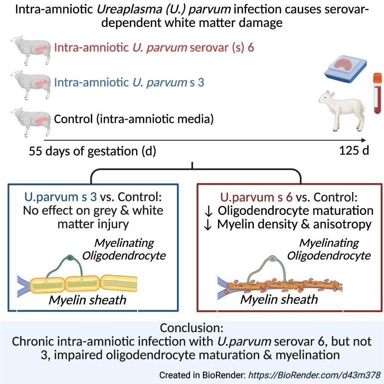
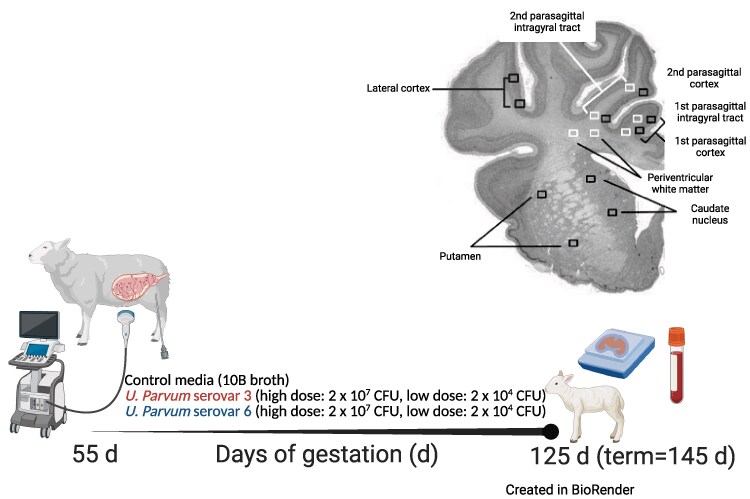
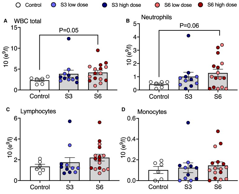
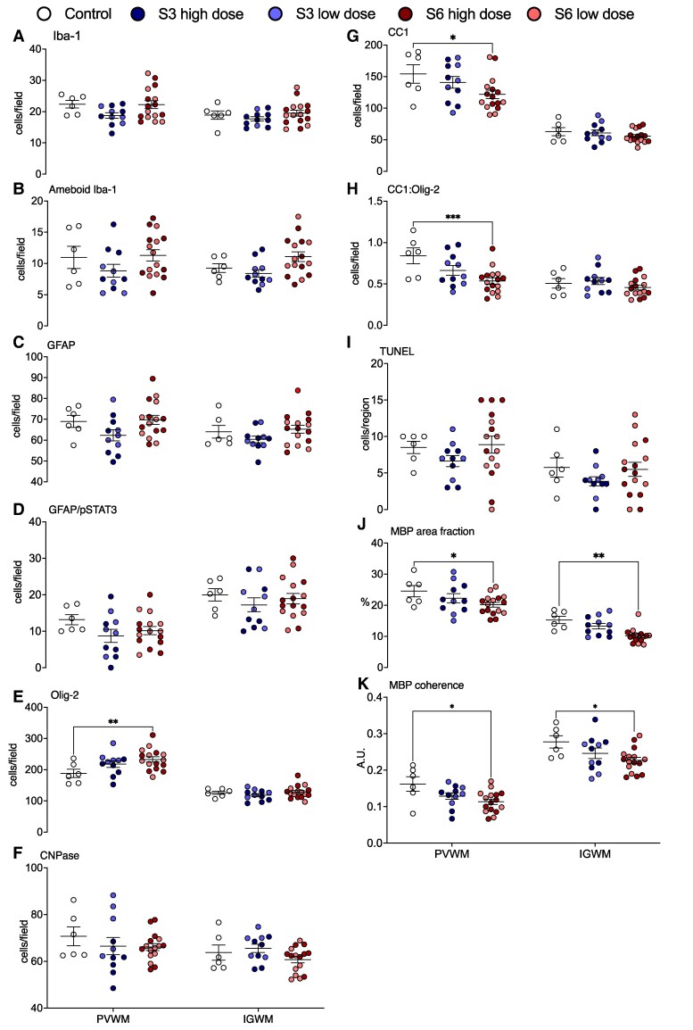
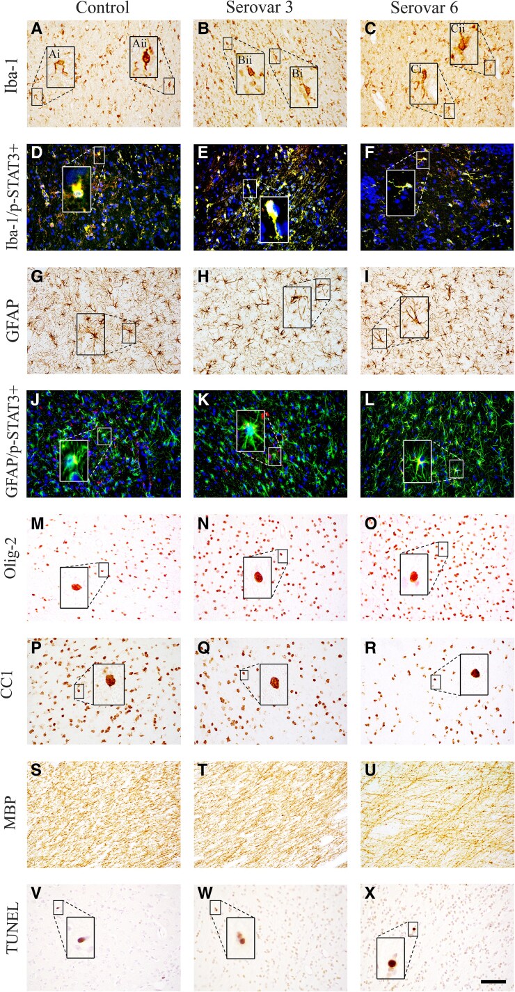
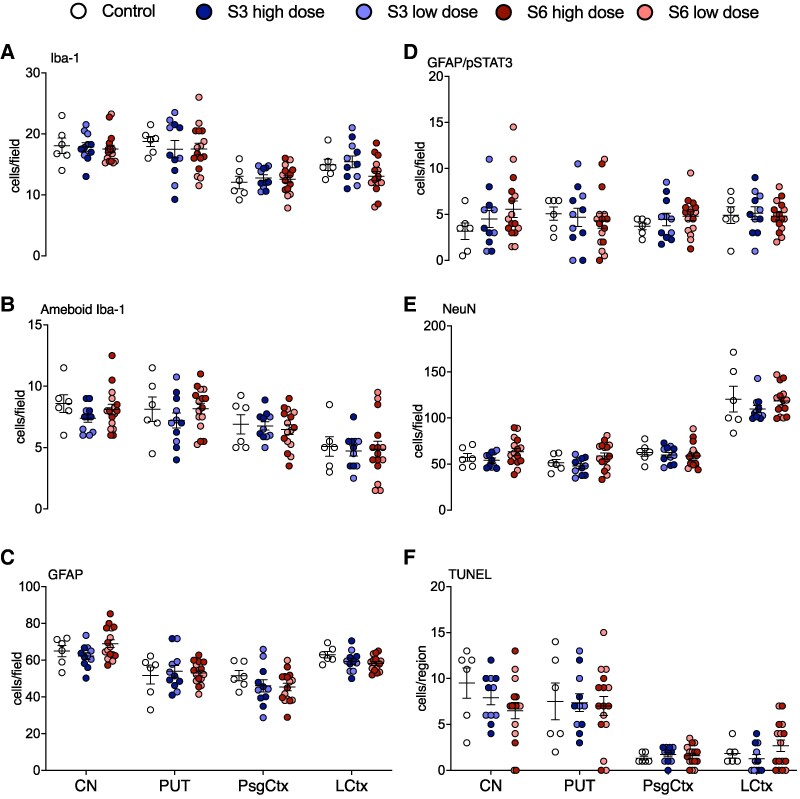
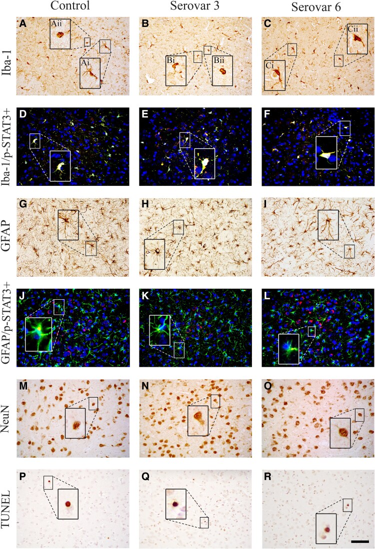
Ureaplasma parvum is commonly isolated from the amniotic fluid of pregnancies complicated by infection. While some studies have shown an association between intra-amniotic Ureaplasma species infection and brain injury and/or adverse neurodevelopment, others have not. The relationship between antenatal exposure to microbial infection and risk of poor neurological outcome is complex and multifactorial and may reflect diversities in microbial pathogenicity along with the duration and severity of the fetal inflammatory response to microbial infection. This study aimed to determine the impact of chronic intra-amniotic infection with Ureaplasma parvum serovars 3 and 6, which are among the most common serovars isolated in pregnancies complicated by infection/inflammation, on white matter development in preterm fetal sheep. Pregnant ewes carrying singleton or twin fetuses (55 days gestational age, term = 145 days) were randomly allocated to receive an ultrasound-guided intra-amniotic injection of Ureaplasma parvum serovars 3 (n = 11), 6 (n = 16) or media (control, n = 6). At 125 days of gestation, the ewe and foetus were euthanized and the fetal brain was collected for immunohistochemistry. Total numbers of oligodendrocytes (oligodendrocyte transcription factor 2-positive cells) in the periventricular white matter tract were higher in Ureaplasma parvum serovar 6-exposed fetuses than control. Numbers of mature oligodendrocytes [anti-adenomatous polyposis coli clone (CC) 1-positive cells] and myelin density (% area fraction of myelin basic protein-positive) in the periventricular and intragyral white matter tracts were lower in Ureaplasma parvum serovar 6-exposed fetuses than control. Myelin anisotropy was lower in serovar 6-exposed fetuses than control. There were no differences in numbers of total or mature oligodendrocytes, myelin density and anisotropy in Ureaplasma parvum serovar-3-exposed fetuses compared to control. Cell death, numbers of neurons, total and reactive (signal transducer and activator of transcription 3-positive) microglia and astrocytes did not differ between Ureaplasma parvum-exposed fetuses and controls within the premotor cortex and striatum. Chronic intra-amniotic infection with Ureaplasma parvum serovar 6, but not 3, impaired oligodendrocyte maturation and myelination within the large white matter tracts of the preterm sheep brain. These data suggest that the impact Ureaplasma parvum infection on white matter development may be serovar dependant, which may help to explain why some fetuses exposed to intra-amniotic Ureaplasma infection have adverse neurodevelopmental outcomes while others do not. Overall, this study demonstrates that greater emphasis needs to be placed on the taxonomy of Ureaplasma infection when designing and interpreting clinical and preclinical studies of fetal infection and neurodevelopmental outcomes.

Keywords: Ureaplasma; chorioamnionitis; infection; preterm brain injury; white matter injury.


Conflict of interest statement

The authors report no competing interests.

Figures

Graphical Abstract
Graphical Abstract
Figure 1
Figure 1
Experimental design and brain regions of interest (ROIs) for histological analysis. At 55 days (0.4) of gestation, pregnant ewes were randomly assigned to receive an ultrasound-guided intra-amniotic injection of either control media (10B broth; n = 6), Ureaplasma parvum serovar 3 [high dose: 2 × 10⁷ CFU (n = 5), low dose: 2 × 10⁴ CFU (n = 6)] or U. parvum serovar 6 [high dose: 2 × 10⁷ CFU (n = 8), low dose: 2 × 10⁴ CFU (n = 8)]. At 125 days (0.8) of gestation, umbilical arterial blood and fetal brains were collected and processed for analysis. Brain ROIs were sampled from histological sections taken 23 mm anterior to stereotaxic zero. White boxes represent the white matter ROIs that were captured within the periventricular and first and second intragyral white matter tracts. Black boxes represent grey matter ROIs that were captured within the caudate nucleus, putamen and premotor cortex. Created in BioRender. Galinsky, R. (2025) https://BioRender.com/p80o123.
Figure 2
Figure 2
White blood cell analysis in Ureaplasma parvum serovar 3 and serovar 6-exposed subjects. Total white blood cells (A), neutrophils (B), lymphocytes (C) and monocytes (D) were compared between control (white circles, n = 6), serovar 3 [low dose, light blue circles (n = 5); high-dose, dark blue circles (n = 6)] and serovar 6 [low-dose, light red circles (n = 8); high dose, dark red circles, (n = 8)]-exposed subjects. Data are individual subject means ± SEM. Statistical analysis: Kruskal–Wallis test with Benjamini–Hochberg procedure.
Figure 3
Figure 3
White matter histopathology. Numbers of total (A), amoeboid (B) Iba-1+ microglia, GFAP+ (C) and GFAP/phosphorylated signal transducer and activator of transcription (pSTAT)3+ astrocytes (D), Olig-2+ (total; E), CNPase (immature and mature; F) and CC1+ (mature; G) oligodendrocytes, proportions of mature (CC1:Olig2+; H) oligodendrocytes, TUNEL+ (apoptotic; I) cells, MBP % area fraction (density; J) and coherence (anisotropy; K) in the periventricular (PVWM) and intragyral white matter tracts (IGWM) in control (white circles, n = 6), Ureaplasma parvum serovar 3 [low dose (n = 5), light blue circles; high-dose (n = 6), dark blue circles] and serovar 6 [low-dose (n = 8), light red circles; high dose (n = 8), dark red circles]-exposed subjects. Data are individual subject means ± SEM. Statistical analysis: two-way ANOVA with Tukey’s multiple comparison test. *P < 0.05, **P < 0.01, ***P < 0.001.
Figure 4
Figure 4
Photomicrographs of white matter immunohistochemistry. Representative photomicrographs showing Iba-1 (A–C), Iba-1/phosphorylated (p)-STAT3 [activated microglia = yellow (D–F); green = Iba-1, red = p-STAT3, blue = 4′,6-diamidino-2-phenylindole (DAPI)], GFAP (G–I), GFAP/p-STAT3 (activated astrocytes = yellow, J–L; green = GFAP, red = p-STAT3, blue = DAPI), Olig-2+ (M–O), CC1 (P–R) and MBP (S–U) and TUNEL (V–X) staining in the periventricular white matter (PVWM) in control, Ureaplasma parvum serovar 3 and serovar 6-exposed subjects. Representative images of ramified (Ai, Bi, Ci) and amoeboid (Aii, Bii, Cii) microglia shown in high magnification. Scale bar is 50 μm.
Figure 5
Figure 5
Grey matter histopathology. Numbers of total (A) and amoeboid (B) Iba-1+ microglia, GFAP+ (C) and GFAP/phosphorylated (p)STAT3+ (D) astrocytes, NeuN+ neurons (E) and TUNEL+ (apoptotic; F) cells in the caudate nucleus (CN), putamen (PUT), parasagittal (PsgCtx) and lateral (LCtx) cortices in control (white circles, n = 6), Ureaplasma parvum serovar 3 [low dose (n = 5), light blue circles; high-dose (n = 6), dark blue circles] and serovar 6 [low-dose (n = 8), light red circles; high dose (n = 8), dark red circles]-exposed subjects. Data are individual subject means ± SEM. Statistical analysis: two-way ANOVA with Tukey’s multiple comparison test.
Figure 6
Figure 6
Photomicrographs of grey matter immunohistochemistry. Representative photomicrographs showing Iba-1 (A–C), Iba-1/phosphorylated (p)-STAT3 [activated microglia = yellow, D–F; green = Iba-1, red = phosphoSTAT3, blue = 4′,6-diamidino-2-phenylindole (DAPI)], GFAP (G–I), GFAP/phosphoSTAT3 (activated astrocytes = yellow, J–L; green = GFAP, red = p-STAT3, blue = DAPI), NeuN (M–O) and TUNEL (P–R) staining in the premotor cortex. Representative images of ramified (Ai, Bi, Ci) and amoeboid (Aii, Bii, Cii) microglia shown in high magnification. Areas of analysis include: caudate nucleus (CN), putamen (PUT), parasagittal (PsgCtx) and lateral (LCtx) cortices. Scale bar is 50 μm.

Similar articles

References

    1. Galinsky R, Polglase GR, Hooper SB, Black MJ, Moss TJ. The consequences of chorioamnionitis: Preterm birth and effects on development. J Pregnancy. 2013;2013:412831. - PMC - PubMed
    1. Kikhney J, von Schöning D, Steding I, et al. Is Ureaplasma spp. the leading causative agent of acute chorioamnionitis in women with preterm birth? Clin Microbiol Infect. 2017;23(2):119.e1–119.e7. - PubMed
    1. Silwedel C, Speer CP, Härtel C, Glaser K. Ureaplasma-driven neuroinflammation in neonates: Assembling the puzzle pieces. Neonatology. 2020;117(6):665–672. - PMC - PubMed
    1. Kong F, Ma Z, James G, Gordon S, Gilbert GL. Species identification and subtyping of Ureaplasma parvum and Ureaplasma urealyticum using PCR-based assays. J Clin Microbiol. 2000;38(3):1175–1179. - PMC - PubMed
    1. Leli C, Mencacci A, Latino MA, et al. Prevalence of cervical colonization by Ureaplasma parvum, Ureaplasma urealyticum, Mycoplasma hominis and Mycoplasma genitalium in childbearing age women by a commercially available multiplex real-time PCR: An Italian observational multicentre study. J Microbiol Immunol Infect. 2018;51(2):220–225. - PubMed

LinkOut - more resources