[Preprint]. 2025 May 13:2025.05.12.653516.
doi: 10.1101/2025.05.12.653516.

Recombinant protein platform for high-throughput investigation of peptide-liposome interactions via fluorescence anisotropy depolarization

Affiliations

Recombinant protein platform for high-throughput investigation of peptide-liposome interactions via fluorescence anisotropy depolarization

Antonis Margaritakis et al. bioRxiv. .

Abstract

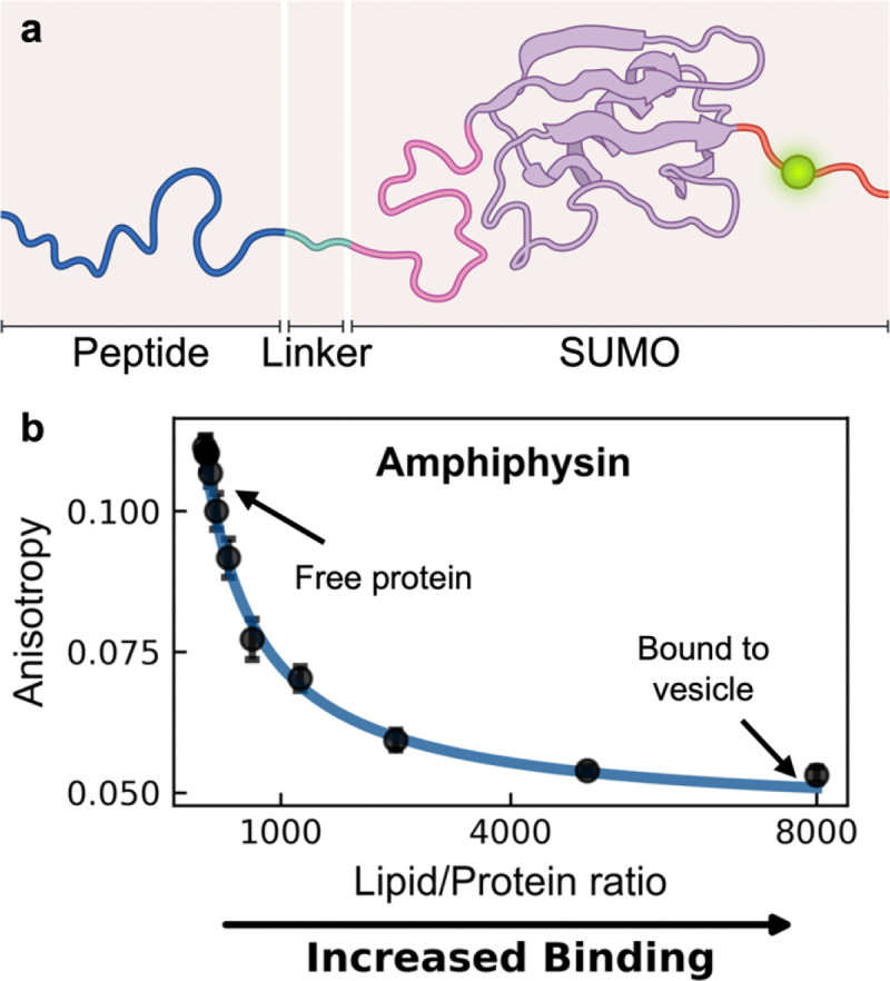
Many cytosolic proteins critical to membrane trafficking and function contain an unstructured domain that can bind to specific membranes, with a transition into an amphipathic helix induced upon membrane association. These inducible amphipathic helices often play a critical role in organelle recognition and subsequent function by these cytosolic proteins, but the tools and techniques used to characterize affinity towards specific membranes are low-throughput and highly dependent on the solubility of the inducible amphipathic helix. Here, we introduce a modular recombinant protein platform for rapidly measuring the binding affinity of inducible amphipathic helices towards a variety of membrane compositions and curvatures using high-throughput fluorescence anisotropy measurements. Inducible amphipathic helices are solubilized with a fluorescently tagged small ubiquitin-like modifier (SUMO) protein and binding to membranes quantified by leveraging the unexpected decrease in fluorescence anisotropy upon binding, a phenomenon previously observed but not well understood. By using fluorescence anisotropy decay measurements and solution NMR experiments, we deduce that this phenomenon likely occurs due to the local increase in fluorophore motion upon binding to the membrane. Altogether, this recombinant protein platform can be readily applied to any inducible amphipathic helix of interest, allowing for detailed investigation of the specific membrane biochemical parameters facilitating binding.

Keywords: Amphipathic helix; fluorescence anisotropy depolarization; high-throughput protein vesicle binding; peptide-liposome interactions; vesicles.


Conflict of interest statement

Competing Interest Statement: The authors declare no competing interest.

Figures

Figure 1 |
Figure 1 |. Our recombinant protein platform exhibits a decrease in fluorescence anisotropy upon binding to a vesicle
(a) Schematic representation of the SUMO protein platform. Peptides of interest (dark blue) are engineered at the N-terminus, linked to SUMO via soluble linker (light blue) and a mutated cysteine to facilitate fluorophore attachment (green). A cleavable C-terminal hexahistidine tag (not depicted) facilitates purification and cleaved prior to usage. (b) An unexpected decrease in fluorescence anisotropy is observed when measuring the N-terminal domain of Amphiphysin engineering to our platform upon increasing titration of 67 nm diameter DOPC/DOPS (70/30) vesicles. Data was fit using a membrane partitioning model (blue) as described in Methods.
Figure 2 |
Figure 2 |. High-throughput detection of inducible amphipathic helix binding to liposomes is possible.
(a) Schematic representation of the vesicle array used to assess protein binding in different membrane composition and curvatures. Four vesicle composition batches were prepared: DOPC/DOPS/DOPE = (70-X)/30/X with X = (0, 15, 30, 45), each extruded to three different final diameters D ≈ (94 nm, 86 nm, 68 nm). (b, c) Fluorescence anisotropy measurements of our recombinant platform engineered with the inducible amphipathic helices of (b) CHMP4B (AA:1–19, blue circles) and (c) Huntingtin (AA:1–17, red triangles) with increasing vesicle concentration. Data points show the mean of 3 technical replicate measurements for each lipid/protein ratio, with standard deviations plotted (black error bars). Data were fit using a membrane partitioning model (solid lines) with partition coefficient values shown as x105.
Figure 3 |
Figure 3 |. Binding isotherms derived from fluorescence anisotropy and tryptophan fluorescence show strong quantitative agreement.
Parallel fluorescence anisotropy and tryptophan fluorescence measurements were taken with our recombinant platform engineering with the inducible amphipathic helices of (a) Amphiphysin (AA: 1–25, F9W mutant), (b) CHMP4B (AA: 1–19, F8W mutant), (c) Endophilin-B1 (AA: 1–33, F18W mutant) and (d) Huntingtin (AA: 1–17, F11W mutant). The binding of each platform was measured against changes in a variable lipid membrane content either known or suspected to influence binding affinity: cholesterol (Chol) for Amphiphysin, cardiolipin (CL) for CHMP4B, phosphatidylethanolamine (PE) for Endophilin-B1, and cardiolipin (CL) for Huntingtin. In all cases, vesicles were measured to be ~70 nm diameter, composed of DOPC/DOPS/variable lipid at (70-X)/30/X molar ratios.
Figure 4 |
Figure 4 |. The decrease in fluorescence anisotropy is electrostatic in nature.
Fluorescence anisotropy was measured for fluorescently-tagged hexahistidine model platform binding to nickel-chelating lipid containing vesicles against different conditions: (a) decreasing vesicle size (with lipid vesicle compositions of DOPC/DGS-NTA(Ni) = 90/10 or DOPC/DOPS/DGS-NTA(Ni) = 50/40/10), (b) membrane charge (with lipid vesicle compositions of DOPC/DOPS/DGS-NTA(Ni) = (90-X)/X/10 with X shown in the legend), and (c) buffer salt concentration (with lipid vesicle compositions of DOPC/DOPS/DGS-NTA(Ni) = 50/40/10). Data points are the mean of 4 replicate measurements for each lipid/protein ratio with standard deviations plotted (black error bars). Solid lines represent fits to a depletion model as described in methods. Unless otherwise noted, all vesicles were extruded to a final diameter of ~95 nm.
Figure 5 |
Figure 5 |. Fluorescence anisotropy decay and NMR measurements support a model by which decrease in fluorescence anisotropy occurs by a local increase in fluorophore mobility.
Fluorescence anisotropy decay was measured for fluorescently-tagged hexahistidine model platform (25 nM) in 3 different states: (a) free in solution, (b) bound to neutral vesicles (DOPC/DGS-NTA(Ni) = 90/10, 25 μM) and (c) bound to negatively charged vesicles (DOPC/DOPS/DGS-NTA(Ni) = 50/40/10, 25 μM). Data were fitted as described in Methods using a two-state hindered anisotropy decay model for (a) and a hindered rotational diffusion model for (b) and (c). Insets show residuals of each fit. (d) Residual 1HN-15N signal intensities of the fluorescently-tagged hexahistidine model platform in the presence of negatively charged vesicles (DOPC/DOPS/DGS-NTA(Ni) = 50/40/10, 25 mM). For the protein remaining free in solution, three distinct areas of signal reductions were observed illustrating varying degrees of domain flexibility that are expected to be preserved in the vesicle-bound state. Vesicle-bound protein is tumbling too slowly to be observable. The differently affected areas are indicated by blue/pink, magenta and orange bars, and correspond to the protein domains shown in (e). (e) Schematic representation of our platform free in solution and bound to a vesicle, where the conformational freedom of the fluorescently-tagged domain increases.

Similar articles

References

    1. Gleisner M. et al. Epsin N-terminal Homology Domain (ENTH) Activity as a Function of Membrane Tension*. J. Biol. Chem. 291, 19953–19961 (2016). - PMC - PubMed
    1. Bussell R. & Eliezer D. A structural and functional role for 11-mer repeats in alpha-synuclein and other exchangeable lipid binding proteins. J. Mol. Biol. 329, 763–778 (2003). - PubMed
    1. Cornell R. B. Membrane lipid compositional sensing by the inducible amphipathic helix of CCT. Biochim. Biophys. Acta BBA - Mol. Cell Biol. Lipids 1861, 847–861 (2016). - PubMed
    1. Mercier V. et al. Endosomal membrane tension regulates ESCRT-III-dependent intra-lumenal vesicle formation. Nat. Cell Biol. 22, 947–959 (2020). - PMC - PubMed
    1. Puth K., Hofbauer H. F., Sáenz J. P. & Ernst R. Homeostatic control of biological membranes by dedicated lipid and membrane packing sensors. Biol. Chem. 396, 1043–1058 (2015). - PubMed

Publication types

LinkOut - more resources