[Preprint]. 2025 May 15:2025.05.10.653184.
doi: 10.1101/2025.05.10.653184.

A Transcriptomic Atlas of Healthy Human Skin Links Regional Identity to Inflammatory Disease

Affiliations

A Transcriptomic Atlas of Healthy Human Skin Links Regional Identity to Inflammatory Disease

Sahiti Marella et al. bioRxiv. .

Abstract

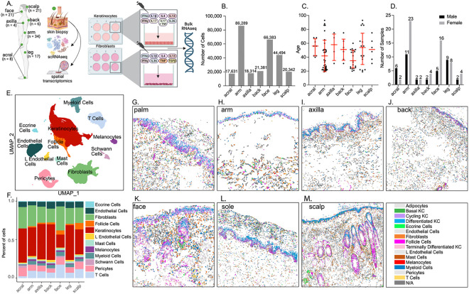
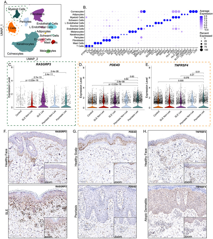
Human skin is not a uniform organ but a mosaic of anatomically distinct niches, with each site finely tuned to unique environmental demands and immune pressures. Yet, the molecular determinants that define these regional identities and their relationship to site-specific vulnerability to inflammatory disease remain poorly understood. Here, we generate a high-resolution single-cell atlas of human skin, profiling 274,834 cells from 96 healthy samples across 7 anatomically distinct sites (acral, arm, axilla, back, face, leg and scalp). Our analysis reveals striking region-specific transcriptional and cellular networks, uncovering how local immune-stromal crosstalk governs tissue homeostasis and underpins anatomical susceptibility to distinct inflammatory diseases such as such as systemic lupus erythematosus (SLE), atopic dermatitis (AD), and psoriasis. These findings illuminate the tissue-intrinsic foundations of regional immune identity and provide a blueprint/resource for the development of precision therapies tailored to the distinct immunological microenvironments of specific anatomical skin sites.

Keywords: autoimmune diseases; inflammation; regional gene expression changes; single cell; skin; transcriptomics.


Figures

Figure 1.
Figure 1.. A single-cell atlas of healthy human skin reveals anatomical site-specific differences in cellular composition.
(A) Schematic of experimental design and anatomical site sampling (B) Total number of high-quality cells captured per anatomical site following scRNA-seq and quality control (n = 17,831 cells from acral, n = 86,289 cells from arm, n = 18,314 cells from axilla, n = 21,381 cells from back, n = 66,383 cells from face, n = 44,494 cells from leg, and n = 20,342 cells from scalp) (C) Age distribution across anatomical sites (D) Sex distribution across anatomical sites (E) UMAP embedding of 274,834 cells colored by annotated cell types (F) Bar plot showing the relative abundance of major skin cell types across anatomical sites (G-M) CosMx spatial transcriptomics plots showing spatial distribution of cell types (Adipocytes, KC populations, Eccrine cells, Endothelial cells, Fibroblasts, Follicle cells, L Endothelial cells, Mast cells, Melanocytes, Myeloid cells, Pericytes and T cells) in palm, arm, axilla, back, face, sole and scalp skin.
Figure 2.
Figure 2.. Anatomical site-specific KC subtypes exhibit distinct transcriptional and cytokine response profiles in healthy human skin.
(A) UMAP visualization of KC subclusters across all anatomical sites (B) Dot plot showing marker gene expression used to define each KC subset (C) Bar plot showing the relative abundance of each KC subtype across anatomical sites (D–J) Volcano plots displaying differentially expressed genes in KCs from each anatomical site compared to all other sites (K) Dot plot showing the top 5 differentially expressed genes from KCs at each anatomical site (L) Heatmap of cytokine response module scores (IFN-α, IFN- y, IL-1β, IL-4, IL-13, IL-17, IL-17+TNF, IL-36) across KCs from different anatomical sites with data represented as a Z-score.
Figure 3.
Figure 3.. Anatomical site-specific fibroblast subtypes exhibit distinct transcriptional and cytokine response profiles in healthy human skin.
(A) UMAP visualization of fibroblast subclusters across anatomical sites (B) Dot plot showing expression of representative marker genes for each fibroblast subtype (C) Bar plot showing the relative abundance of fibroblast subtypes across anatomical sites. (D–J) Volcano plots showing differentially expressed genes in fibroblasts from each anatomical site compared to all others. (K) Dot plot showing the top differentially expressed fibroblast markers across anatomical sites (L) Heatmap of cytokine response module scores (IFN-α, IFN- y, IL-1β, IL-4, IL-13, IL-36, TNF, TGFβ) across fibroblasts from different anatomical sites.
Figure 4.
Figure 4.. Site-specific transcriptional programs in skin myeloid cells reveal regional immune states and enrichment of lupus-associated gene expression in facial skin.
(A) UMAP visualization of myeloid cell subclusters across anatomical sites (B) Dot plot showing expression of canonical marker genes across myeloid subtypes (C) Bar plot showing the relative abundance of each myeloid subset across anatomical sites (D–J) Volcano plots showing differentially expressed genes in myeloid cells from each anatomical site compared to all others. (K) Dot plot showing the top five most significant differentially expressed genes in myeloid cells from each anatomical site (L) Feature plot of RASGRP3 expression across anatomical sites (M) Hallmark 50 enrichment heatmap showing pathway activity across myeloid cells from different anatomical sites (N) CellChat of predicted incoming and outgoing signaling interaction strength across myeloid subtypes and anatomical sites.
Figure 5.
Figure 5.. Regional specialization of T cell subsets in healthy human skin reveals distinct immune activation states and scalp-specific expression of psoriasis- and AD-associated genes.
(A) UMAP visualization of T cell subclusters across anatomical sites (B) Dot plot showing expression of canonical marker genes defining each T cell subset (C) Bar plot showing the relative abundance of each T cell subset across anatomical sites (D–J) Volcano plots displaying differentially expressed genes in T cells from each anatomical site versus all others (K) Dot plot showing the top five most significant differentially expressed genes from each anatomical site (L) Feature plots showing selective enrichment of PDE4D and TNFRSF4 (OX40) in scalp CD4⁺ T cells and Tregs (M) Hallmark 50 pathway enrichment heatmap demonstrating site-specific pathway activation in T cells (N) CellChat analysis showing incoming and outgoing signaling interaction strengths across anatomical sites.
Figure 6.
Figure 6.. Disease-specific upregulation of RASGRP3, PDE4D, and TNFRSF4 in SLE, psoriasis, and AD highlights their roles in anatomical site-specific skin inflammation.
(A) UMAP visualization of major skin cell types from an independent scRNA-seq dataset including control, SLE lesional and non-lesional skin, and psoriasis lesional and non-lesional skin (B) Dot plot showing expression of key marker genes across cell types in the inflammatory skin dataset (C–E) Violin plots of RASGRP3 (C), PDE4D (D), and TNFRSF4 (OX40) (E) expression across disease groups (F–H) Immunohistochemistry (IHC) validation (F) RASGRP3 protein expression in SLE lesional facial skin compared to healthy facial skin (G) PDE4D protein expression in psoriatic skin compared with healthy scalp skin (H) TNFRSF4 protein expression in AD facial skin compared with healthy facial skin.
Figure 7.
Figure 7.. Genetic risk loci for inflammatory skin diseases align with site-specific gene expression programs, revealing potential mechanisms of regional disease susceptibility.
(A–D) Chromosomal distribution of GWAS loci mapped to genes within ±200 kb of lead SNPs for (A) acne, (B) AD, (C) SLE and (D) psoriasis (E) Bar plot showing the number of GWAS-mapped genes overlapping with anatomical site-specific differentially expressed genes (F) Table displaying the disease-associated genes overlapping with anatomical site-specific DEGs across all anatomical sites (G) Dot plot showing the expression of selected GWAS-mapped genes across anatomical sites. Color indicates average scaled expression (z-score), and dot size represents the percentage of expressing cells.

Similar articles

References

    1. Damiani G. et al. The Global, Regional, and National Burden of Psoriasis: Results and Insights From the Global Burden of Disease 2019 Study. Frontiers in Medicine 8 (2021). 10.3389/fmed.2021.743180 - DOI - PMC - PubMed
    1. Izmirly P. M. et al. Prevalence of Systemic Lupus Erythematosus in the United States: Estimates From a Meta-Analysis of the Centers for Disease Control and Prevention National Lupus Registries. Arthritis & Rheumatology 73, 991–996 (2021). 10.1002/art.41632 - DOI - PMC - PubMed
    1. Lee H. J., Hong Y. J., Han K. D. & Lee J. H. Atopic Dermatitis Severity and Risk for Psoriasis: A Nationwide Population-Based Study. Dermatology 240, 262–270 (2024). 10.1159/000536143 - DOI - PMC - PubMed
    1. Tian J. et al. Global epidemiology of atopic dermatitis: a comprehensive systematic analysis and modelling study. Br J Dermatol 190, 55–61 (2023). 10.1093/bjd/ljad339 - DOI - PubMed
    1. Ali Z. et al. Assessing anatomical distribution of atopic dermatitis identifies a cluster of patients with late onset and low risk of asthma and allergy: An observational study. Health Sci Rep 6, e1219 (2023). 10.1002/hsr2.1219 - DOI - PMC - PubMed

Publication types

LinkOut - more resources